INTRODUCTION
Microglia are cells of the myeloid lineage that are found dispersed throughout the brain and spinal cord and make up between 5 and 20% of central nervous system cells depending on the brain structure (Lawson et al., Reference Lawson, Perry, Dri and Gordon1990; Lyck et al., Reference Lyck, Santamaria, Pakkenberg, Chemnitz, Schrøder and Finsen2009). They differ from other glia by virtue of their non-neuronal origin and are thought to infiltrate the brain during development following their birth from hematopoieitic precusors in the periphery. Although their precise origin and the timing and avenue of their migration into the brain are still debated, it is now agreed that a significant fraction of microglia derive from the embryonic yolk sac which is the major site of hematopoiesis in the early embryo (Alliot et al., Reference Alliot, Godin and Pessac1999; Herbomel et al., Reference Herbomel, Thisse and Thisse2001; Chan et al., Reference Chan, Kohsaka and Rezaie2007; Ginhoux et al., Reference Ginhoux, Greter, Leboeuf, Nandi, See and Gokhan2010). Myeloid cells from the yolk sac rapidly migrate to colonise embryonic organs, including the brain. During later development there is a sharp increment in microglia density in the brain (Dalmau et al., Reference Dalmau, Finsen, Zimmer, González and Castellano1998; Paolicelli et al., Reference Paolicelli, Bolasco, Pagani, Maggi, Scianni and Panzanelli2011), but to what extent this is driven by local proliferation or new recruitment of circulating myeloid cells remains unclear. Experiments based on parabiosis (when two animals are joined surgically to create a common vascular compartment) suggest that infiltration of circulating monocytes into the adult brain is minimal under non-traumatic conditions and that bone marrow-derived cells are recruited only upon brain injury (Ajami et al., Reference Ajami, Bennett, Krieger, Tetzlaff and Rossi2007; Ginhoux et al., Reference Ginhoux, Greter, Leboeuf, Nandi, See and Gokhan2010). Other studies, however, provide evidence for the recruitment of circulating monocytes into the unperturbed brain during late developmental stages, leaving open the possibility that a window of opportunity exists during which monocytes can be recruited from the periphery, possibly at a point when the blood–brain barrier is immature and permeable to such cells (Hristova et al., Reference Hristova, Cuthill, Zbarsky, Acosta-Saltos, Wallace and Blight2010). Lineage tracing studies supporting this idea show that up to 40% of resident microglia in the adult brain are positive for the bone marrow hematopoieitic marker Hoxb8 (Chen et al., Reference Chen, Tvrdik, Peden, Cho, Wu and Spangrude2010). However, these data are at odds with other lineage tracing studies examining the contribution of yolk sack hematopoieitic cells to adult microglia that suggest that the vast majority of microglia have an embryonic origin with minimal replenishment from circulating monocytes (Ginhoux et al., Reference Ginhoux, Greter, Leboeuf, Nandi, See and Gokhan2010). Thus, microglia remain enigmatic brain cells whose infiltration in the brain appears to be carefully orchestrated with its development and whose function, therefore, is likely to be critically linked to brain maturation as well as adult function.
MICROGLIA – SURVEYORS OF THE HEALTHY BRAIN
Since they were first described almost one century ago (del Rio Hortega, Reference Del Rio-Hortega and Penfield1932), microglia have been appointed as the scavengers of the brain due to their peculiar and unique phagocytic function. According to the early view, microglia were thought to remain for the most part in a ‘resting’ state, waiting for a danger stimulus that would transform them into an ‘activated’ state in which they were able to phagocytose neuronal debris and dead cells. This view prevailed until modern microscopy techniques made it possible to view microglia in their ‘resting’ state in the living animal, at which point it was discovered that microglia are in fact highly motile in their baseline state (Davalos et al., Reference Davalos, Grutzendler, Yang, Kim, Zuo and Jung2005; Nimmerjahn et al., Reference Nimmerjahn, Kirchhoff and Helmchen2005; Peri and Nüsslein-Volhard, Reference Peri and Nüsslein-Volhard2008). By using in vivo imaging, Wake and co-workers showed that microglia processes make brief contacts (~5 min) with neuronal processes, at a frequency of about once per hour, suggesting a direct role for microglia in monitoring the functional state of neurons (Wake et al., Reference Wake, Moorhouse, Jinno, Kohsaka and Nabekura2009). This led Hanisch and Kettenmann to suggest that the baseline state of microglia is better termed, ‘surveying’ rather than ‘resting’, to highlight their constant surveillance of the brain parenchyma (Hanisch and Kettenmann, Reference Hanisch and Kettenmann2007). Nevertheless, the focus of early microglia research on the activated state and stimuli and conditions that underlie its induction, still pervades the field and it is not until recently that researchers have shown renewed interest in the function of ‘resting’ microglia, and therefore also their function in normal brain development.
Several studies support a role for microglia in neuronal elimination in the uninjured brain. For example, during hippocampal neurogenesis in the adult brain, newborn granule cells are produced in the subgranular zone (SGZ) with a small percentage of them maturating and becoming part of the mossy fiber circuitry, while the majority undergo apoptosis (Ming and Song, Reference Ming and Song2011). Surveying microglia, in their basal and ramified status, are responsible for the phagocytosis of these apoptotic bodies, engulfing them to produce ‘ball-and-chain’ structures at their terminal process (Sierra et al., Reference Sierra, Encinas, Deudero, Chancey, Enikolopov and Overstreet-Wadiche2010). Importantly these data show that phagocytosis of neuronal material by microglia do not require the induction of the activated state seen during brain injury and show that surveying microglia can have a phagocytic function. Similarly, in the developing zebrafish brain microglia actively contribute to phagocytosis of apoptotic neurons (Peri and Nüsslein-Vollhard, Reference Peri and Nüsslein-Volhard2008). Their observation that neurons expressing apoptotic markers are always found engulfed within microglia suggests, furthermore, that microglia are actively involved in the induction of neuronal death, rather than being passive ‘garbage collectors’ that clean up cellular debris after apoptosis has been completed (Peri and Nüsslein-Vollhard, Reference Peri and Nüsslein-Volhard2008). These observations point to an intimately orchestrated reciprocal signaling between neurons and microglia.
Finally, in vivo imaging studies suggest that microglia have a specific affinity for synaptic structures. A recent study by Tremblay et al. used transgenic mouse lines expressing fluorescent proteins in neurons and microglia (Thy1::YFP;Cx3cr1::GFP) to monitor their contacts in the living brain using time-lapse imaging (Tremblay et al., Reference Tremblay, Lowery and Majewska2010). Looking at the visual cortex, they found that neuronal spines, protruberances from neuronal dendrites that harbour postsynaptic sites, are frequently contacted by distal microglial processes and that, occasionally, with a frequency increased by visual deprivation, spines disappeared after being contacted by microglia (Tremblay et al., Reference Tremblay, Lowery and Majewska2010). Their data suggested for the first time that microglia might have a role in the selective elimination of synapses in the uninjured brain, a function also supported by observations that dysfunction in microglial activity can lead to synaptic impairment (Graeber, Reference Graeber2010).
MICROGLIA MEDIATE NEURON AND SYNAPSE ELIMINATION DURING DEVELOPMENT
The massive invasion and proliferation of microglia during brain development and the highly conserved feature of microglia colonisation of the brain across species (Pont-Lezica et al., Reference Pont-Lezica, Béchade, Belarif-Cantaut, Pascual and Bessis2011) suggest a critical role for these cells in brain maturation. Studies across several species have highlighted the instructive role of microglia in promoting neuronal cell death during development (Marín-Teva et al., Reference Marín-Teva, Dusart, Colin, Gervais, van Rooijen and Mallat2004; Mallat et al., Reference Mallat, Marín-Teva and Chéret2005; Peri and Nüsslein-Vollhard, Reference Peri and Nüsslein-Volhard2008; Wakselman et al., Reference Wakselman, Béchade, Roumier, Bernard, Triller and Bessis2008). In several species microglia have been closely associated with developing axon bundles (Cuadros et al., Reference Cuadros, Martin, Coltey, Almendros and Navascués1993; Dalmau et al., Reference Dalmau, Finsen, Zimmer, González and Castellano1998; Herbomel et al., Reference Herbomel, Thisse and Thisse2001; Santos et al., Reference Santos, Calvente, Tassi, Carrasco, Martín-Oliva and Marín-Teva2008) and post-mortem histological studies in humans describe a prominent accumulation of microglia at 10–12 weeks of gestation at the cortical plate–subplate junction, a position where the first synapses are detected, and later at 19–30 weeks of gestation, at axonal crossroads in white matter. This localised accumulation of microglia at points of axonal and synaptic development suggests that microglia are actively involved in the refinement of neuronal connections, including axonal guidance and synaptogenesis (Verney et al., Reference Verney, Monier, Fallet-Bianco and Gressens2010).
We have recently followed up on these observations with the aim to find evidence for a role of microglia in synapse elimination. This work was based on the observation that the expression of the chemokine fractalkine, Cx3cl1, becomes strongly upregulated in the mouse forebrain during the third and fourth postnatal week (Mody et al., 1999), the major period of synapse formation and maturation, and that its receptor is exclusively expressed by microglia (Jung et al., Reference Jung, Aliberti, Graemmel, Sunshine, Kreutzberg and Sher2000). This chemokine signaling system appeared to be well placed to serve as a neuron-microglia signal related to synaptogenesis. In fact, we found that mice lacking the fractalkine receptor, Cx3cr1, showed a ~30% decrease in the density of microglia in the brain during the second, third and fourth postnatal week, confirming a critical role of this chemokine in either attracting or promoting the proliferation of microglia during synaptogenesis (Paolicelli et al., Reference Paolicelli, Bolasco, Pagani, Maggi, Scianni and Panzanelli2011). The initial stages of synaptogenesis in cortical structures are associated with a maximal level of synapse formation and elimination (Cruz-Martin et al., Reference Cruz-Martin, Crespo and Portera-Cailliau2010) and it is notable that at precisely this point microglia arborisation and density increase dramatically in the forebrain (Dalmau et al., Reference Dalmau, Finsen, Zimmer, González and Castellano1998; Paolicelli et al., Reference Paolicelli, Bolasco, Pagani, Maggi, Scianni and Panzanelli2011).
Next, we used immunohistological measures to measure synapse maturation in Cx3cr1 knockout mice and found an increase in the number of synapses. Electrophysiological assessment of synaptic maturation in the Cx3cr1 knockout mice confirmed that excitatory synapses were more abundant, but also revealed that they were weaker, with a failure to develop efficient action-potential stimulated synaptic release as measured by the ratio of spontaneous (sEPSC) and mini (mEPSC) excitatory postsynaptic currents (Paolicelli et al., Reference Paolicelli, Bolasco, Pagani, Maggi, Scianni and Panzanelli2011). This second finding has important implications because it argues that microglia-mediated synapse elimination is required for the strengthening of surviving synapses and suggests the presence of reciprocal signaling between neurons and microglia to orchestrate synaptic pruning. We hypothesised that the combination of increased synapses, but decreased maturation of synapses could be caused by a deficit in synaptic pruning as a result of decreased microglia surveillance. More direct evidence for synaptic pruning by microglia was provided by imaging experiments in fixed tissue that captured examples of both pre and post-synaptic proteins engulfed by microglia in the developing forebrain. Higher-resolution imaging using electron microscopy uncovered evidence for engulfment of synaptic material via a clathrin-coated vesicle endocytosis pathway.
Similar engulfment and deficient pruning of presynaptic structures were recently reported in mice lacking the complement factors C1q and C3 and the C3 receptor (CR3) in the retinothalamic system (Stevens et al., Reference Stevens, Allen, Vazquez, Howell, Christopherson and Nouri2007; Schafer and Stevens, 2011) confirming the hypothesis that the classical complement system is a critical signal for microglia-mediated synapse elimination. Neuronal engulfment via the classical complement pathway is initiated by binding of C1q to desialylated sugar residues on neuronal plasma membrane proteins and the subsequent induction of C2 and C4 cleavage and C3 binding (Linnartz et al., Reference Linnartz, Kopatz, Tenner and Neumann2012). Engulfment of the complement complex is then mediated by CR3 receptors located on microglia. Intriguingly, both C1q and C3 are expressed transiently in a punctate, synapse-like pattern during the synaptogenetic period in mouse (Stevens et al., Reference Stevens, Allen, Vazquez, Howell, Christopherson and Nouri2007) and high-resolution immunofluorescence microscopy revealed these puncta to be directly adjacent to many, but not all pre- and postsynaptic structures. We have recently confirmed these localisation studies, showing that C1q immunoreactive puncta do not overlap with, but are located close to postsynaptic sites during the early postnatal period in mouse (Fig. 1). These C1q immunoreactive puncta do not overlap with microglia processes, and instead appear to colocalise with the astrocytic marker GFAP, suggesting that astrocytes could represent a source for C1q during this development stage. Expression of C1q in astrocytes during this period is supported by mRNA in situ hybridisation data that show a distinctive non-neuronal pattern of C1q expression in the hippocampus during the early postnatal stage (Allen Brain Atlas). The precise localisation of C3 at this developmental stage remains unclear, although in vitro studies show that it is expressed on neurons and possibly also released by microglia (Walker et al., Reference Walker, Kim and McGeer1995; Thomas et al., Reference Thomas, Gasque, Vaudry, Gonzalez and Fontaine2000). The CR3 receptor, on the other hand, is exclusively expressed by microglia in the brain (Ling et al., Reference Ling, Kaur, Yick and Wong1990; Stevens et al., Reference Stevens, Allen, Vazquez, Howell, Christopherson and Nouri2007).
C1q expression does not overlap with synapses or microglia. (Upper panel) Representative high-resolution confocal single-plane images of CA1 stratum radiatum taken from the hippocampus of a Cx3cr1::GFP/ + reporter mouse (KO/+) at postnatal day 15 revealed apposed, non-overlapping localisation of C1q (indicated by arrowheads) and PSD95-immunoreactive puncta and GFP-positive microglia processes (scale bar: 1 µm). (Lower panel) Representative low-resolution confocal single-plane images showed C1q staining overlapping with the astrocytic marker GFAP, but not with GFP-positive microglia processes (scale bar: 10 µm).

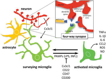
Although the precise order and timing of events leading from the apposition of C1q to synapses, to its presumed release and binding to neuronal membranes, and the assembly of the remaining complement proteins in preparation for phagocytosis remains to be worked out, these data allow us to propose a four-way synapse model of synaptic pruning. In this model pre- and post-synaptic specialisations are joined by astrocyte and microglia processes, each contributing essential classical complement pathway components to promote microglia-mediated synaptic engulfment and elimination (Fig. 2). Several questions are raised by this model. What are the neuronal signals that initiate the cascade? How important is neuronal activity and/or plasticity in marking synapses for elimination? What is the initiating ‘eat me’ signal? How does synaptic pruning work in adulthood, when the classical complement factors no longer show punctate expression patterns?
Role of microglia in synapse elimination during development. Under normal conditions microglia are in a ‘surveying‘ state in which their highly motile processes extend and retract to interrogate the neuronal environment. Together with the more stationary processes of astrocytes, and the pre- and postsynaptic specialisations (see inset) they form a ‘four-way’ synapse. Neurons produce the chemokine fractalkine (Cx3cl1) to attract and/or promote the proliferation of microglia during synaptogenesis. Critical components of the complement system are produced by each branch of the four-way synapses and are critical for microglia-mediated phagocytosis of unwanted pre- and post-synaptic structures. C1q is expressed by astrocytes, the complement receptor 3 (CR3) by microglia, and neurons express C3 (see inset). Under conditions of pathogenic insult or neurodegeneration, pathogen-associated molecular patterns (PAMPs, e.g. LPS, IFN-γ) can drive microglia into an ‘activated’ state characterised by the secretion of pro-inflammatory factors, including TNF-α, IL-1β, IL-6, ROS, NO. Induction of the activated state is inhibited by fractalkine, Cx3cl1, as well as CD22, CD47, CD200.

WHEN DANGER CALLS
As mentioned above, a large body of research has documented the role of microglia in responding to pathogenic signals that reach or originate in the brain and in resolving injury in the adult brain. Less is known, however, about the impact of pathogenic signals on microglia function during development. Could the activation of microglia during a time when they are critically involved in synaptic maturation has a detrimental and potentially long-term impact on brain wiring? Under normal conditions microglia activation is suppressed by ‘off’ signals (e.g. Cx3Cl1, CD22, CD47 and CD200) that are expressed by neurons and maintain microglia in the default, surveying state. ‘Off’ signals also suppress the expression of pro-inflammatory factors. In contrast, ‘on’ signals emanating either from neurons or other members of the local brain environment (e.g. glutamate, purines and chemokines) or from exogenous factors that manage to infiltrate the brain (e.g. pathogen-associated molecular patterns or PAMPs) can trigger microglia activation, promoting motility and the release of pro-inflammatory molecules (Fig. 2; Biber et al., Reference Biber, Neumann, Inoue and Boddeke2007; Ransohoff and Cardona, Reference Ransohoff and Cardona2010; Saijo and Glass, Reference Saijo and Glass2011). Activation of microglia involves dramatic changes in cell shape and motility, including retraction of long processes, transformation into an amoeboid shape, and rapid migration to the site of injury. It is well recognised that microglia can sense and rapidly respond to threats to CNS integrity by expressing a broad pattern of cytokines, either pro- or anti-inflammatory, and by changing their MHC expression profile. Nevertheless, it is important to keep in mind that, despite a common morphological transformation during activation, the final state of microglia upon activation is not an all-or-none phenomenon and different activation states can be distinguished by their gene expression signatures.
According to the type and concentration of ‘on’ signals, activated microglia can express different spectra of pro- or anti-inflammatory cytokines and under certain circumstances, for example, microglia can express an activated pro-inflammatory phenotype without being able to carry out a phagocytic response (Koenigsknecht-Talboo and Landreth, Reference Koenigsknecht-Talboo and Landreth2005). Microglia can exacerbate inflammatory processes by releasing reactive oxygen species (ROS), nitric oxide (NO) or tumor necrosis factor (TNF)-α, or, on the other hand, can confer neuroprotection and promote neurogenesis (Fig. 2). In some cases fully activated microglia can have neurotoxic effects. In models of neurodegenerative disorders such as Parkinson's disease, microglia activation mediated by Inteferon (INF)-γ has been clearly shown to induce neuronal death (Block et al., Reference Block, Zecca and Hong2007; Mount et al., Reference Mount, Lira, Grimes, Smith, Faucher and Slack2007). At the same time, other studies demonstrate that microglia can release neurotrophic factors favoring neuroprotection, neurogenesis, and glia development (Monje et al., Reference Monje, Toda and Palmer2003; Kitamura et al., Reference Kitamura, Takata, Inden, Tsuchiya, Yanagisawa and Nakata2004). This dual aspect of microglia make understanding their function in a particular neuronal process not straightforward. Cataloguing different activated repertoires and understanding their impact on synaptic pruning and maturation will be critical to understanding the effects of pathological conditions on brain maturation.
However, several external signals have been clearly shown to affect the phagocytic activity of microglia, and thus are likely to impact synaptic pruning. Trophic factors, such as glial-derived neurotrophic factor, M-CSF or the ciliary neurotrophic factor have been shown to promote phagocytic activity (Mitrasinovic et al., Reference Mitrasinovic, Vincent, Simsek and Murphy2003; Chang et al., Reference Chang, Fang, Lee and Tzeng2006; Lee et al., Reference Lee, Yang, Fang and Tzeng2009), while prostaglandin E2 attenuates phagocytosis through the E-prostanoid receptor 2 (Nagano et al., Reference Nagano, Kimura and Takemura2010). Kettenmann and co-workers showed that the neurotransmitter serotonin reduces the phagocytic efficiency of microglia while simultaneously promoting their motility towards the injury site (Krabbe et al., Reference Krabbe, Matyash, Pannasch, Mamer, Boddeke and Kettenmann2012). Evidence suggests that an inflammatory and oxidative milieu, moreover, can negatively regulate microglial phagocytic function. Microglia isolated following IFN-γ/LPS stimulation in vivo, for example, exhibited loss of phagocytic and migratory activity (Broderick et al., Reference Broderick, Duncan, Taylor and Dick2000). Similarly, recent studies showed that stimulation of microglia with IL-1β, LPS or t-BHP (a strong oxidant) significantly reduced phagocytosis in a nuclear factor kappa-light-chain-enhancer of activated B cells (NF-κB)-mediated manner (Pan et al., Reference Pan, Zhu, Lin, Zhang, Ye and Huang2011). Moreover, in the presence of high levels of the pro-inflammatory factors IL-1β and TNF-α, C3 receptor-mediated phagocytic activity is reduced, whereas FcR-mediated phagocytosis is not affected, suggesting that more than one distinct signaling pathway controls phagocytic activity (Koenigsknecht-Talboo and Landreth, Reference Koenigsknecht-Talboo and Landreth2005). As discussed above, C3 is likely to be a critical ‘eat me’ signal that instructs microglia for removing synapses (Stevens et al., Reference Stevens, Allen, Vazquez, Howell, Christopherson and Nouri2007; Linnartz et al., Reference Linnartz, Kopatz, Tenner and Neumann2012). Thus, in general, pro-inflammatory factors are likely to impact negatively on synaptic pruning by blunting C3-mediated phagocytic activity associated with the non-activated, surveying state.
CONCLUSION
Evidence is mounting that microglia have a special role during brain development in sculpting neuronal circuits. Early during development they are critical for the elimination of unneeded neurons and later they are involved in removing unwanted synapses. Moreover, multiple data suggest that neurons and microglia have evolved recurrent signaling mechanisms to orchestrate these functions, and that microglia do not just arrive after-the-fact to scavenge neuronal debris, but rather are intimately involved in the selection and elimination process. Finally, data suggest that microglia have a particular crucial role during development in interpreting changes in the brain environment and translating them to modulate neuronal circuit maturation. Any reduction in phagocytic capacity due to increased activation of microglia will have grave consequences for synaptic maturation and potentially irreversible consequences on brain wiring.
Some data suggest that microglia may be more prone to pathogenic stimuli during development, possibly in order to better react to and resolve injury during such a critical period of brain maturation. Such a hypersensitivity to environmental pathogens might explain why bacterial infections at P4, but not at P30, in the rat have been shown to cause significant long-term changes in the cytokine profile of microglia and behavior (Bilbo et al., Reference Bilbo, Biedenkapp, Der-Avakian, Watkins, Rudy and Maier2005, Reference Bilbo, Rudy, Watkins and Maier2006, Reference Bilbo, Barrientos, Eads, Northcutt, Watkins and Rudy2008; Meyer et al., Reference Meyer, Nyffeler, Engler, Urwyler, Schedlowski and Knuesel2006, Reference Meyer, Murray, Urwyler, Yee, Schedlowski and Feldon2008). Early hypersensitivity of the microglia system coupled to its critical role in neuron elimination and synaptic pruning, means that disturbances in microglia homeostasis could be a route to mental illness with a developmental onset. The concept of prenatal inflammation as a risk factor for neuropsychiatric disorders such as autism and schizophrenia has remained current and epidemiological evidence confirms that prenatal exposure to immune challenge can impair later brain maturation (Ciaranello and Ciaranello, Reference Ciaranello and Ciaranello1995; Meyer et al., Reference Meyer, Feldon and Fatemi2009, Reference Meyer, Feldon and Dammann2011; Brown and Derkits, Reference Brown and Derkits2010; McAlonan et al., Reference McAlonan, Li and Cheung2010; Buehler, Reference Buehler2011; Patterson, Reference Patterson2011; Vuillermot et al., Reference Vuillermot, Joodmardi, Perlmann, Ove Ögren, Feldon and Meyer2012). For example, cytokine-associated inflammatory events can be critical mediators of the neurodevelopmental effects of prenatal infection (Gilmore and Jarskog, Reference Gilmore and Jarskog1997; Meyer et al., Reference Meyer, Murray, Urwyler, Yee, Schedlowski and Feldon2008) and interleukins such as IL-6 can induce long-term neural and behavioral deficits (Smith et al., Reference Smith, Li, Garbett, Mirnics and Patterson2007). It remains to be determined what role, if any, microglia-mediated neuron elimination and synaptic pruning plays in such developmental programming phenomena.
Statement of interest
None.