Introduction
The dramatic changes in the market forms for poultry in recent years, from a predominantly whole bird commodity to a modern, highly diversified industry focused on cut up, deboned, and ready-to-eat further processed products, has resulted in a change of quality expectation for poultry products. The major poultry meat quality attributes include appearance, texture, juiciness, flavour, and functionality. With increasing trends in further processing, meat functionality has increased in relative importance, especially because of its key role in determining the sensory quality of complex ready-to-eat products (Fletcher, Reference FLETCHER2002).
Many different methods for the measurement of meat quality traits are available which are based on different principles, and instruments and/or probes. Close attention is needed when using such measurements in order to standardise meat sample preparation and handling before and during analysis. In view of the complexity of meat processes during post mortem time and quality trait determination, it is not surprising, therefore, that the results obtained in different studies and laboratories are not always in agreement. For comparison of the results from different studies therefore, it is extremely important that all investigators adhere strictly to the same measuring specifications. That is why the development of a set of standardised methods for meat quality measurement is indispensable.
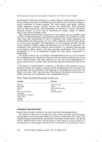
The purpose of this review is to produce a document that can serve as a set of common base methodologies that will permit comparison between research studies that are carried out by different groups. This paper represents the first step in the standardisation of quality measurements in poultry meat, and includes chemical and physical traits (Table 1).
Development of methodologies is continuing, so the input of all researchers active in the field of poultry meat quality is necessary for this standardisation to be successful. The present paper must be considered as the beginning of a standardisation process that will improve over time. Table 1 provides lists of the chemical and physical characteristics of poultry meat that will be addressed in this standardisation process.
Chemical and physical characteristics of poultry meat.

Chemical characteristics
moisture (water content or dry matter determination)
The objective of this test is to measure the water content of poultry meat and poultry meat products as part of proximate analysis; its content is complement of dry matter. The standard reference method for the measurement of moisture in meat utilises oven drying (AOAC Method 950.46, 1990). Ground muscle or meat (about 4 g) may be dried in a conventional oven (air drying) at 100-102°C for 16-18 hours or in a convection oven at about 125°C for 2-4 hours. A vacuum oven (≤100 mm Hg) at 95-100°C for about 5 hours may also be used. The residue is then weighed. Use of the high-temperature ovens and/or use of vacuum serve to shorten drying time, but may be not suitable for samples with high fat content. In all cases, it is necessary to hold the samples in desiccators during cooling to prevent water absorption from the air prior to the weighing of the dry matter residue. If high-fat content samples are dried in cellulose thimbles for subsequent fat extraction, some melted fat may soak through the thimble and may be lost, resulting in erroneously high values for moisture. The use of aluminium weighing dishes alleviates this concern. It is important to keep in mind that oven drying does not determine moisture alone, but rather measures a mixture of volatile materials that are driven off by the temperature used. To achieve consistency, all drying methods must follow the prescribed drying conditions carefully (Sebranek, Reference SEBRANEK2004).
Because all air-drying methods are relatively time-consuming, the use of microwave ovens has been thoroughly investigated for rapid drying. The most successful approach has been that of CEM, who designed a microwave oven specifically for measurements of moisture content of meat and poultry products. Samples are placed between glass fibre pads, weighted, dried for 3-5 minutes and the dry matter is then reweighed to determine moisture loss (AOAC Method 985.14, 1990).
In summary, all official methods recommended by AOAC can be used to measure moisture content of meat without any preference. However, use of high temperature and/or vacuum may be not suitable for samples with high fat content. It is recommended that duplicate samples be measured per muscle sampled from each bird.
total lipids
The objective of these analyses is to assess the lipid content of poultry meat and poultry meat products as part of proximate analysis. The method of choice for the measurement of the total fat content of meat has long been through the use of several solvent-based procedures. These procedures include ether extraction followed by gravimetric measurement (AOAC Method 960.39, 1990), tetrachloroethylene extraction followed by specific gravity measurements (Foss-let, AOAC Method 976.21, 1990), and methylen chloride extraction followed by gravimetric measurements (CEM, AOAC Method 985.15, 1990).
Conventional fat extraction with ether typically requires several hours, while the Foss-let and CEM require specialised equipment that is relatively expensive. While these two methods are still being used in some laboratories, both the Foss-let and the CEM extraction units have been discontinued by the manufacturers (and in some countries are no longer allowed) because of concerns for toxicity of waste organic solvents. Rapid solvent extraction can be achieved by Soxtec units, which have been approved by the AOAC for meat analysis. The amount of sample required is generally about 20 g of ground and homogenized meat (Sebranek, Reference SEBRANEK2004).
Solvent extraction methods with conventional organic solvents such as ether are utilised to measure total fat content as triglycerides. However, other lipid components such as phospholipids and free fatty acids are not included in this extraction. These lipid components are relatively low in quantity in meat products (1% or less), but in some cases it is important to include them in order to get a true total fat measurement. To include phospholipids and other minor lipids in extraction procedures, a modified solvent (chloroform-methanol, Folch method) or a pre-extraction acid hydrolysis treatment (Schmid-Bondzynski-Ratzlaff method or Weibull-Stoldt method) can be used (Sebranek, Reference SEBRANEK2004).
Due to general low fat content of poultry meat, modified solvent (chloroform-methanol, Folch method) or a pre-extraction acid hydrolysis treatment (Schmid-Bondzynski-Ratzlaff or Weibull-Stoldt method) methods should be preferred. Because of the use of solvents, generally the measurement is not repeated, but in this case it is strongly recommended that a minimum of 10 repetitions per treatment be used in order to obtain a good reliability of the treatment averages.
proteins
The protein content of poultry meat and poultry meat products is included generally as part of proximate analysis. The long-time standard for protein analysis has been the Kjeldahl method (AOAC Method 976.05, 1990). This method includes two phases:
i. a catalysed mineralization of nitrogen by heating in concentrated sulphuric acid;
ii. an alkaline treatment followed by a distillation and dosage of the produced free NH3.
The content of total nitrogen is included in the proteins, peptides and in other non-peptidic compounds. This content may be related to the total protein content using a general coefficient (6.25). Generally the sample required must weight approximately 2 g.
The heavy-metal catalyst used plus the concentrated acid and alkali waste generated by conventional Kjeldahl procedures have become an increasing disposal concern for laboratories. Improvements in instruments for providing automated, relatively rapid Kjeldahl analyses have included accelerated digestion units (Labconco) and automated, rapid distillation units (Kjeltec). These instruments are based on the Kjeldahl method but provide far easier and more rapid analysis than the traditional Kjeldahl method (Sebranek, Reference SEBRANEK2004).
A combustion method has also received AOAC approval (AOAC Method 992.15, 1990). This method is based on complete sample combustion (at about 850°C), followed by the quantification of released nitrogen. A major advantage of the combustion method is that no toxic or hazardous wastes are produced. The combustion procedure is also relatively fast, requiring about five minutes per sample. Repeatability has been reported as 0.12-0.41%. One concern for the combustion method has been that a relatively small sample size is required, making sample preparation critical to the method's success (Sebranek, Reference SEBRANEK2004). It should be remembered that automated instruments based on Kjeldahl method are preferred and at least two duplicate samples per muscle sampled from each bird are recommended.
ash
The determination of ash indicates the mineral content of poultry meat and poultry meat products as part of proximate analysis. The method for measuring the ash content of meat is based on a gravimetric measurement and it involves two steps:
i. addition of a well known quantity of magnesium acetate solution to the muscle sample (about 3 g), drying at 100°C followed by incineration at 550-600°C (the weight of the residue minus the magnesium oxide derived from the magnesium acetate represents the ash content);
ii. the sample's weight difference after incineration at 550°C, without addition of magnesium acetate solution (AOAC 920.153, 1990).
It is recommended at least to duplicate muscle samples from each bird.
fatty acid composition
The fatty acids are isolated either as total fat or as free fatty acids and are then converted to fatty acid methyl esters (FAMEs), which are extracted with a non-polar solvent, and a small proportion is then injected into the gas-liquid chromatography (GLC). The results are calculated either per 100 g sample or per 100 g fat or as a percentage of the total fatty acids.
Isolation
Isolation of fat or free fatty acids: fat can be obtained from Folch extraction (Folch et al., Reference FOLCH, LEES and SOLANE-STANLEY1957) or by using acid hydrolysis (see total lipid analysis). The Soxhlet extraction method should be avoided because this method does not extract the phospholipids.
Methylation of fatty acids
This is necessary to make fatty acids volatile enough for GLC. There are three main methods (Leth, Reference LETH2004):
i. boiling in methanol for 2 h with a strong acid such as hydrochloric acid or sulphuric acid;
ii. basic catalysed trans-esterification is even more rapid; when using sodium methoxide in methanol as the catalyst, the methylation takes only a few minutes (take care to avoid water);
iii. sample is boiled for a few minutes in methanol and potassium hydroxide, cooled, boron trifluoride in methanol (as catalyst) is added and the mixture is boiled again for a few minutes in a stoppered glass tube.
The FAMEs are extracted with n-hexane or isooctane, and they are then ready for GLC.
GLC of fatty acids
The FAMEs are injected on a capillary column, usually 50-100 m long, with internal diameter 0.25-0.32 µm. The capillary column is coated with a highly polar material to achieve the necessary separation of FAMEs (100% biscyanopropyl polysiloxane or 90% biscyanopropyl polysiloxane 10-cyanopropylphenyl polysiloxane). Highly polar column material is necessary in order to separate the cis-trans and positional isomers. To achieve sufficient separation, a carefully designed temperature programme is necessary. A flame ionisation detector (FID) is usually used to detect the eluted peaks. An internal standard is often used for quantification of the fatty acids. The odd numbered fatty acids, most commonly C17, but also C13 and C23, can be used as the internal standard, since they occur in nature in very small amounts. Detection can be also achieved using mass spectrometry (MS) with electron impact ionization. For FAMEs, only the molecular weight can be ascertained. Other structural information cannot be obtained, because the double bonds shift back and forth during ionization. However, if fatty acids are esterified with nitrogen-containing heterocyclic compounds like picolinyl esters or dimethyloxazoline (DMOX) esters, the double bonds will be stabilised, and their position in the molecule can be seen, although the difference between cis and trans mass spectra is not visible (Leth, Reference LETH2004).
Although methods for FA analysis rely on GLC capillary columns of FAMEs, there are many variations. This implies that all isolation, methylation and chromatographic conditions should be specified to have a better possibility to compare results obtained in different laboratories/studies. A minimum of 10 repetitions per treatment is required in order to have reliable results.
cholesterol
Saponification and extraction are applied for cholesterol as for other fat-soluble compounds. The unsaponifiable matter is purified by thin-layer chromatography on silica or by column chromatography on alumina to separate tocopherols and triterpenes, and the purified cholesterol is silylated and injected into a gas chromatograph equipped with a 50 m semi-polar capillary column. Betulin, a dihydroxysterol, can be added as the internal standard before saponification, thus correcting for loss of cholesterol during the analysis. 5-alfa-cholestane is also often used as internal standard. However, it will separate from cholesterol during purification, and it cannot be silylated and so must be added after silylation, thus being an internal standard only for the GLC. The compound 5-alfa-cholestanol behaves like cholesterol during the analysis and is used as an internal standard, which can be added before saponification. Several silylating reagents have been used to form trimethylsilyl ethers with hydroxy groups, for instance N-methyl-N-(trimethylsilyl) trifluoroacetamide (MSTFA) or N,O-bis(trimethylsilyl) trifluor-acetamide (BSTFA) in pyridine. All these seem to work well if the silylating conditions are arranged properly. Methods also exist without saponification, and with purification just by isolation of the fat and injection of the fat solution, or with saponification but no purification, and in both cases without silylation. In that case, 5-alfa-cholestane is a suitable internal standard (Leth and Ertbjerg, Reference LETH and ERTBJERG2004).
The determination of cholesterol is straightforward with GLC on capillary columns, but many variations on this trait are described in literature. The use of an internal standard before saponification to correct the loss of cholesterol during the analysis is recommended. There are also methods without saponification or purification. However, the best separation and the most precise results are found when the full method is used.
lipid oxidation
This is required to estimate the degree of rancidity of poultry meat to lipid oxidation. The degree of rancidity in fats and meats has traditionally been measured using an assay for the determination of substances which react with thiobarbituric acid (TBA) to give an absorption peak at 532-535 nm (TBARS test). The major reactant is malonic dialdehyde and the results are expressed as mg malonic dialdehyde per kg of meat (Baeza, Reference BAEZA2004). The most generally accepted method has been described by Lynch and Frei (Reference LYNCH and FREI1993). Samples (0.5 ml: 1 g of muscle minced in 10 ml of KCl 0.15 M + butyl-hydroytoluene 0.1 mM) are held in boiling water for 10 min with 1% (w/v) 2-thiobarbituric acid in 50 nM NaOH (0.25 ml) and 2.8% (w/v) trichloroacetic acid (0.25 ml). After cooling at room temperature, the pink chromogen is extracted into n-butanol (2.0 ml) and its absorbance is measured at 535 nm. TBARS concentration is calculated using 1,1,3,3-tetramethoxypropane. It is also possible to measure TBARS after oxidation with FeSO4 (Kornbust and Mavis, 1980). If samples are very different in lipid content, TBARS can be expressed as nmoles malonaldehyde (MDA)/mg protein content of the meat determined according to the Lowry procedure (Lowry et al., Reference LOWRY, ROSEBROUGH, FARR and RANDALL1951).
Fluorimetric TBARS methods, which have much greater sensitivity than spectrophotometric methods, have also been developed for samples with low lipid-oxidation products such as fresh, raw chicken and turkey meat (Draper et al., Reference DRAPER, SQUIRES, MAHMOODI, WU, AGARWAL and HADLEY1993; Jo and Ahn, Reference JO and AHN1998). Methods to measure susceptibility to lipid oxidation are based on different determination principles so the absolute results between studies are difficult to compare. This determination needs to be investigated to define standard measurement conditions.
amino acids
Amino acids are a very diverse group of chemical compounds with a wide variety of side chains so this diversity gives them very different behaviour in terms of oxidative stability, hydrolysis, solubility in water and chromatographic separation. Thus simultaneous chemical analysis of all of the naturally occurring amino acids is very complicated. Many methods have been proposed examining all of the aspects of amino acid analysis from hydrolysis to chromatographic separation and detection, with many variations and special procedures for some of the amino acids.
Hydrolysis
Hydrolysis is carried out by boiling the sample in strong hydrochloric acid for close to 24 h either with a reflux condenser or in sealed ampoules. Other possibilities are microwave systems with the use of special vessels, which can withstand high pressure. This allows the use of temperatures as high as 180°C and reduces the hydrolysis time to 10 min. It has been observed that tryptophan is totally destroyed in the hydrolysis process, and so a special procedure with basic hydrolysis is necessary (see below). Cysteine and methionine are not stable and form several intermediary oxidation products. This problem can be overcome by driving the oxidation to completion with phenol-containing performic acid to cysteic acid and methionine sulphone. However, this process destroys tyrosine, which then must be determined in an unoxidized sample. Recoveries for serine, threonine, valine, leucine and isoleucine tend to be a bit low (85-95%), which is usually considered tolerable or can be rectified with the use of a correction factor. Glutamine and glutamic acid are determined together, as are aspargine and aspartic acid. Thus the hydrolysis procedure is a series of compromises with many parameter sets described in the literature to obtain reasonable results for most of the amino acids. It is necessary to hydrolyse both an oxidized and an unoxidized sample and to carry out a special procedure for tryptophan, when analysis for total amino acids is the goal (Leth, Reference LETH2004).
Chromatographic Separation
The chromatographic separation of amino acids can be carried out with either ion-exchange chromatography or reversed-phase high performance liquid chromatography (HPLC). Ion-exchange chromatography, which requires expensive automated amino acid analysers, has prevailed for many years, but is now gradually being replaced by reverse-phase HPLC carried out on standard equipment but with commercially available kits for derivatisation and detection. It is common to use external standardisation with chromatography of a mix of standard amino acids instead of the use of some internal standards.
Cation exchange chromatography separates amino acids in their free form by gradient elution with a row of sodium-based buffers, giving a typical run time of about 1.5 h. Detection of the amino acids is typically performed by light absorption after post-column reaction with ninhydrin, which forms a purple complex with all primary amino acids with a maximum at 570 nm. The secondary amino acids proline forms an intermediate reaction product with a maximum at 440 nm.
Reverse-phase HPLC is usually carried out on C18 columns with particle size 5 or 3 mm, and gradient elution is common with a mixture of acetonitrile, methanol and acetate or phosphate buffers. However, a great many variations on this theme are described in literature.
Derivatisation is necessary either pre- or post-column to separate and detect amino acids. A huge number of reagents are commercially available as kits and adapted especially to their equipment. Some of the more common reagents are phenylisothiocyanate (PITC), o-phthalaldehyde (OPA), 9-fluorenylmethylchloroformate (FMOC), 6-aminoquinolyl-N-hydrxysuccinimidyl carbamate (AQC) and dabsyl chloride. They make ultraviolet (UV) of fluorimetric detection possible and impart a higher degree of hydrophobicity to the amino acids, which allows sufficient retention on a reversed-phase HPLC column, and elution times are faster than with ion-exchange chromatography, usually from 15-40 min. An excess of the reagent must be added to the free amino acids and heated at 50-70°C for about 10 min. It is often necessary to remove the excess reagent to avoid false peaks in the separation of the amino acids. However, FMOC reacts very quickly, in about 90 sec at room temperature. The reagents usually react with both primary and secondary amino acids and derivatives with either fluorescence or UV absorbance are formed. OPA reacts only with primary amines and forms unstable derivatives, which make it more suited for post-column derivatisation. Since tryptophan, as mentioned earlier, is destroyed by acid hydrolysis, it is necessary to perform a basic hydrolysis with sodium, barium or lithium hydroxide by prolonged heating for about 16 h at 110°C. However, tryptophan is also somewhat unstable by basic hydrolysis, which can be compensated by adding an internal standard, for instance 5-methyltryptophan or ɑ-methyltryptophan, before hydrolysis. Tryptophan is then determined by reverse-phase HPLC with fluorescence detection, as tryptophan is one of the few amino acids with native fluorescence (Leth, Reference LETH2004).
Many methods have been proposed examining all the aspects of amino acid analysis from hydrolysis to chromatographic separation and detection, with many variations and special procedures for some of the amino acids. This determination needs to be investigated to define standard measurement conditions.
collagen
Measuring the collagen content and solubility is important as it is related to meat tenderness. Heat-solubility of collagen is generally determined according to the method of Hill (Reference HILL1966) and collagen content is estimated by measuring total hydroxyproline content according to Bergman and Loxley (Reference BERGMAN and LOXLEY1963) of the procedure of Woessner (Reference WOESSNER1961), using standard curve. A factor of 7.14 (Listrat et al., Reference LISTRAT, RAKADJIYSKI, JURIE, PICARD, TOURAILLE and GEAY1999), 7.25 (Smith et al., Reference SMITH, FLETCHER, BUHR and BEYER1993; Liu et al., Reference LIU, NISHIMURA and TAKAHASHI1996; Silva et al., Reference SILVA, PATARATA and MARTINS1999), or 7.52 (Silva et al., Reference SILVA, PATARATA and MARTINS1999), is used to convert hydroxyproline content to collagen content. The amount of heat-soluble collagen is expressed as a percentage of the total amount of collagen, and collagen content is expressed as mg of collagen per g of skeletal muscle. Hydroxyproline and L-hydroxyproline content can be determined by a colorimetric procedure in which the sample is hydrolysed in sulphuric acid at 103°C, filtered and diluted. Hydroxyproline is oxidized with cholaramine-T. After dilution of 4-dimethylaminobenzaldehyde a red-purple colour is developed, which is measured photometrically at 538 nm.
Although methods to measure collagen content are based on the determination of hydroxyproline by colorimetric method, different conversion factors are present in published literature.
pigments
The major pigments responsible for colour in meat are myoglobin, haemoglobin and cytochrome c. The concentration of haem pigments in poultry meat can be determined indirectly but rapidly by extracting and measuring the iron concentration in meat as described by Hornsey (Reference HORNSEY1956). The colour of meat can change during storage, according to the status of the myoglobin, which may change from the purplish red of deoxygenated myoglobin to the bright red of oxymyoglobin or brown/grey of metmyoglobin. It is possible to follow the amount of metmyoglobin present on the meat surface, by using one of two methods. One involves calculating the difference between meat-reflectance values at two separate wave length, 580 and 630 nm (%R630-%R580) as described by Van Den Oords and Wesdorp (Reference VAN DEN OORDS and WESDORP1971), when measured with a spectrophotometer equipped with an integration sphere. The second method requires the determination of the ratio between the absorption (K) and diffusion (S) of light coefficients at two separate wave lengths, 525 and 572 nm (K/S 525 and K/S 572) according to Stewart et al. (Reference STEWART, ZIPSER and WATTS1965). The porphyrin ring of haem absorbs visible light in the Soret region (400-440 nm). Haem iron interactions with ligands (oxygen, CO, cyanide, etc.) are responsible for the beta and alpha absorption peaks. Deoxymyoglobin exhibits a strong absorption maximum at 555 nm. Oxymyoglobin absorbs at 420 nm in the Soret region, and has large twin maxima at 544 and 582 nm. Metmyoglobin exhibits peaks at 409 and 500 nm, with a small peak at 630 nm. At 525, deoxy-, oxy- and met-myoglobin absorption spectra intersect (isobestic point). Thus, spectrophotometric absorbance at 525 nm is a measure of total myoglobin concentration. Treatment of meat samples with potassium ferricyanide plus cyanide converts all myoglobin forms to cyanometmyoglobin, allowing quantization by spectrophotometric measurement of absorbance at 540 nm. Cytochrome C may be spectrophotometrically distinguished from myoglobin by its absorption peaks at 521 and 550 nm. The different methods to measure meat pigment content do not produce comparable results, so currently it is not possible to indicate a standard method. However, the method proposed by Hornsey (Reference HORNSEY1956) can be preferentially used as the current standard to measure the total pigment content.
Physical traits
pH and R-value
These measurements indicate the rate and the intensity of the muscle acidification during post mortem time. pH is measured with a pH meter equipped with an electrode calibrated prior to its use at pH 4.0 and 7.0. The probe has to be stored in 3 M KCl solution. In poultry, in order to estimate the rate of muscle acidification during post mortem time, pH is conventionally measured about 15 min after the bird's death because of the higher rate of pH drop in poultry in comparison to what takes place with other species. However, more measurements can be taken during the initial phase of decline, such as at 5, 15, 30, 60, 120 min or more. To estimate the intensity of acidification fall, pH is measured at 24 h post mortem and this is called ultimate pH (pHu). However, pHu can be attained in poultry breast meat after 6-8 h post mortem under commercial chilling conditions. In most cases the pH is measured on chilled muscle so it is important that the pH meter is provided with a temperature control system. The pH is more easily measured by using a probe method that inserts a thin electrode directly into the muscle after incision of the muscle. One should use a minimum depth of 1 cm. The pH can also be measured by the sodium iodoacetate, which is a modification of the method initially described by Jeacocke (Reference JEACOCKE1977). The pH is measured using a pH meter on a meat homogenate (about 2 g) in a 5 mM sodium iodoacetate solution with 150 mM of potassium chloride (18 ml). This method can be suitable for the determination of early post mortem time pH because iodoacetate stops the acidification in the meat sample, and pH can be measured after collecting and homogenizing several samples. Meat samples can also be stored at –40°C, and after they are powdered by immersion in liquid N2, they can be used to determine pH.
R-value determination can be done at the same time as the pH is measured in order to assess the depletion of ATP within the muscle during the post mortem time. R-value is an indicator of ATP depletion within a muscle, and it can be measured by spectrophotometer using a filtered meat homogenate in a perchloric acid solution, added with sodium phosphate buffer (pH = 7.0) (Honikel and Fischer, Reference HONIKEL and FISCHER1977). The R-value is calculated as the ratio of absorbance between adenine nucleotides (260 nm, ATP + ADP + AMP) and inosine (250 nm, inosine + inosine monophosphate + hypoxanthine metabolites of AMP). R-values from 1.07 to 1.26 indicate approximate time of rigor onset and full rigor, respectively. Samples have to be transferred immediately after collection to liquid nitrogen to stop metabolic processes.
The iodoacetate method should be preferred at least for the determination of early post mortem time pH. When using probe method, it is recommended to carry out the calibration after each set of 30-50 measurements, and at least once each day. The R-value measurements during the post mortem period can be carried out at the same times adopted for pH analysis. It is recommended at least two duplicate samples from each muscle sampled be used from each bird.
colour
Colour is an appearance property that is important to consumers and is associated with the functional properties of meat, and methods for measuring colour can be used to determine colour changes during and following processing (Froning, Reference FRONING1995). Colour measurement can be performed using a reflectance colorimeter (several models are present on the market). Different light sources can be utilised for measuring colour. The light source is characterised by the light energy distribution it can emit according to visible part of the electromagnetic spectrum. This characteristic can be measured very accurately and enables the colourimetry to define an illuminant which is the curve of light energy emission. This illuminant corresponds to a reference source (for example: ‘D65’ for daylight, ‘A’ for a bulb with tungsten filament, or ‘CWF’ for a fluorescent tube) and its definition is standardised by the CIE (Commission Internationale de l'Eclairage). The use of different illuminants consistently affects the absolute colour values even if the colour differences do not vary. So it is extremely important to indicate which illuminant has been used into the experimental procedures (Barbut, Reference BARBUT2002a; Baeza, Reference BAEZA2004).
Different numerical systems for colour measurements have been developed. The CIE in 1931 incorporated the spectral aspect of illuminating with three different colours into so-called tristimulus values known as the X, Y, Z. The CIE X, Y, Z system defines a colour by the additive mixture of three primary colours, X (red), Y (green) and Z (blue), parameters that are required to match colour of a mixture as viewed by ‘a standard observer’ (human) under defined illumination and viewing conditions. This system is useful for defining colours, but the results are not easily visualized by other readers of scientific papers. Because of this, colour was positioned in the CIE (Reference CIE1978) colour space according to its coordinates. As with any three dimensional space these coordinates can be either Cartesian (L*a*b*) or Polar (L*C*h°). The L*a*b* system considers Lightness (L*), Red/Green (a*) and Blue/Yellow (b*) colour deviations while the L*C*h° system considers Lightness (L*), Saturation or Chroma (C*) and Hue (h°). Hue [h° = tg-1 (b*/a*)] and Chroma [C* = a*2 + b*2)0.5] can be calculated from the a* and b* coordinates. Another frequently used method for food applications is the Hunter L, a, b solid scale. The relationship between the CIE and other colour scales has been discussed by Hunter (Reference HUNTER1975), and Acton and Dawson (Reference ACTON and DAWSON1994). There are available equations that allow one to transform the data obtained by the different systems. The most common poultry muscle used for colour measurement is the breast. It is preferable measured for its colour on the medial surface of the bone side to avoid breast fillet surface discolorations due to possible over scalding during slaughtering. An area of measurement should be selected that is free from obvious defects (bruises, discolorations, haemorrhages, full blood vessels, picking damage) or any other condition which may prevent the obtainment of a uniform colour reading.
For the evaluation of colour changes over time, the colour difference (ΔE*) can be calculated as follows: ΔE* = [(L1*-L2*)2+(a1*-a2*)2+(b1*-b2*)2]1/2, where L1*, a1*, b1*, represent the initial colour readings measured at time ‘0’, and L2*, a2*, b2* refers to each subsequent measurement time. When the meat sample is packaged, it is useful to perform a colorimeter calibration by placing the standard white tile inside the plastic bag that is used to store the meat in order to negate the colour and light reflectance properties of the packaging material. The colour difference (ΔE*) is then calculated by the colorimeter as follows: ΔE* = [(ΔL*)2 + (Δa*)2 + (Δb*)2]1/2; the values of ΔL*, Δa*, and Δb* are the differences in colour between the colour of the calibration tile and the sample.
Recommended parameters are a light sources of D65 (corresponding to daylight), a standard observer at 10° and CIE L*a*b* colour scale. It is important to define the post mortem time, the location of the measure, the muscle chilling and the cutting conditions (Petracci and Fletcher, Reference FLETCHER2002). Attention should be also placed to standardise the thickness (> 1 cm) of the sample and the background where the sample is placed for colour measuring (Bianchi and Fletcher, Reference FLETCHER2002).
Water Holding Capacity (WHC)
WHC measures the ability of meat to retain its liquid portion during storage, processing and cooking. The major methods used to estimate WHC in fresh poultry and poultry products include gravimetric, press, centrifugation and cooking methods.
Gravimetric method (drip loss method)
WHC is determined by measuring a standard-sized sample of muscle sampled at 24 h post mortem, and then suspending it in a plastic bag or glass/plastic box at 2-4°C for at least 24 hours, before removing it and blotting the excess surface fluids followed by measuring the weight loss gravimetrically (Honikel, Reference HONIKEL1998). WHC measurements are commonly carried out on breast, thigh and drumstick meat. As discussed by Van Laack et al. (Reference VAN LAACK, LIU, SMITH and LOVEDAY2000), the drip loss method was initially developed for PSE pork in which losses are abundant (2-5%). In comparison, poultry meat tends to lose less of its moisture, so it is important to store the samples for longer than 24 h before the measurements are taken.
Press methods
These methods have been used to evaluate the amount of ‘squeezable’ water that is present in poultry meat (Kauffman et al., Reference KAUFFMAN, EIKELENBOOM, VAN DER WAL, MERKUS and ZAAR1986a,b). The sample is compressed between two parallel plates using a hydraulic press or a texture analyzer. The released moisture is collected on a pre-weighed filter paper (Trout, Reference TROUT1988; Zhang et al., Reference ZHANG, MITTAL and BARBUT1993). During the test, the sample is usually compressed into a thin film allowing the free water to squeeze out of the sample. Over the years, a wide range of conditions have been reported for the press method of meat sample evaluation. They range from force of 0.01-44 kN, sample size of 0.3-1.5 g, temperature of 4-23°C and compression time from 1-20 minutes. In addition, different filter papers have been used.
Centrifugation
High-speed centrifugation has been applied to meat samples placed in test tubes. Reported conditions varied in sample size (1.5-20 grams) and force applied (1,500-190,000 g). Such large differences in test procedures make it very difficult to compare different studies. If extra moisture is added to the meat sample, at high g force water is expelled from the sample, while at low g force, it is retained (Barbut, Reference BARBUT2002b).
Cooking loss
Meat samples should be standardised for thickness and weight, and should be freshly cut, weighed, and should be represented by several individual slices. Precautions should be taken regarding the geometry of the sample to make them all consistent (i.e. in muscle fibre orientation, surface area to weight ratio, etc.) for the measurement of cooking loss. Different methods of cooking can be used (AMSA, Reference AMSA1995), such as:
i. Roasting – this method uses heat that is transmitted to the meat by convection, either by normal or forced air, in a closed preheated oven. It is recommended that investigators do not use high-velocity or forced convection oven cookery. The meat samples should be placed on a rack either in or over a shallow pan to catch drippings;
ii. Steam cooking – this method uses meat samples placed on metallic trays in a preheated steam oven;
iii. Water Bath method – this method uses meat samples that are placed in thin-walled plastic bags where the samples are immersed in a continuously boiling water-bath for cooking, with the bag opening extending above the water surface.
Irrespective of the adopted cooking method, cooking conditions (heating rate and end-point temperature at the thermal centre) must be defined, controlled, and recorded. In order to monitor temperature changes of meat samples during cooking, thermocouples connected to recorders or hand-held digital read-out thermocouple thermometers should be used.
With regard to drip and cooking loss methods, it is important that the investigator defines exactly where the sample has been taken and the cutting conditions at the time of sampling. Meat samples should be represented by individual slices standardised for thickness, weight and geometry (muscle fibre orientation, surface area to weight ratio). It is recommended to keep the samples stored at least for 48 h to measure drip loss. As far as cooking methods are concerned, the internal temperature of the samples should be standardised at 2-4°C (chilling conditions) just prior to their placement in the cooking environment. Recommended final internal temperature is from 75 to 80°C. However, as different measurement conditions have been used for determining WHC by press and centrifugation methods, additional research needs to be done in order to define the optimum measurement conditions.
texture
Texture is a useful measurement related to tenderness or toughness of meat. Texture is considered to be the most important sensory characteristic of poultry products, and thus a great deal of emphasis has been placed on instrumental procedures to evaluate the structure of muscle fibres. Instrumental determination of texture, or tenderness, is usually evaluated on intact pieces or cores of meat that are large enough to ensure representative sampling of the muscle. This insures that treatment effects can be accurately measured. The most common poultry meat used for the measurement of texture is the breast. Texture should be evaluated on meat cuts after cooking. For shear force evaluation, strips or core samples should be prepared from each meat cut to be evaluated. Strips/cores should be removed parallel to the longitudinal orientation of the muscle fibres (for example, three samples that are 1 cm2 x 3 cm should be taken for each breast muscle used). The breasts should be sampled by making a single blade cut perpendicular to the fibres. Shear forces have been historically recorded in weight measurements (i.e., lb, kg), but they can also be converted to Newton force units, if appropriate. Most of the data that has been used to determine tenderness in cooked intact poultry in the past has been generated using a Warner-Bratzler instrument, tensile tests, or with the Kramer Shear Press (Lyon and Lyon, Reference LYON and LYON2001).
Warner-Bratzler
The Warner-Bratzler shear device is small and portable, consisting of a rectangular blade with a triangular hole cut from the centre. The blade is attached to a circular fan scale. The sample of known dimensions, usually a rectangular strip, is placed in the triangular notch of the single blade. Two bars are lowered by a hydraulic motor and the sample is pushed across the apex of the triangular notch. As the bars are lowered across the sample, the peak force to shear across the fibre is recorded as kilograms on a circular fan scale. Usually, strips of parallelepiped shape with a square base are sheared using this device and shear force is expressed in force unit/shear area which corresponds to the area of square base of the parallelepiped-shaped strip.
The benefits of the Warner-Bratzler device are its reliability, ruggedness, ease of use, portability, and low cost. The device lends itself to on-site quality control work. The limiting factor is that only peak load or peak shear force is generated during the test, so the researcher or quality control personnel must have sufficient experience with sensory panel data to add validity to the shear values.
Kramer shear press
The other shear test that has been extensively used for red meat and poultry texture studies has been conducted using a Kramer shear press. The shear test cell is composed of two main parts, a metal box with slots which hold the sample and a top part with five or ten blades spaced to fit into the slots. This device is attached to a system designed to move the multiple blades down and trough a rectangular sample placed in the cell. The multiple blades are lowered across the sample. The sample is initially compressed and the shear across the fibres forces the resulting strips to come out of the bottom of the slotted cell. Usually, core samples of cylindrical or parallelepiped shapes are sheared using this device and shear force is expressed as force unit/grams of sample weight. In the same way small meat blocks or ground meat may be used. The Kramer shear press is rugged, but it is much heavier, less portable, and more expensive than the Warner-Bratzler machine.
Warner-Bratzler, Kramer shear press and Tensile tests using an Instron-type machine
The blade designs of the original Warner-Bratzler and Kramer shear press systems have been reproduced on other instruments such as the Instron Universal Testing Machine and the Texture Technology Texture Analyzer. The multiple blade cell is also referred to as the Allo-Kramer shear cell. The newer systems are accompanied by software to program the machines and to record more dimensions of the force/distance or force/time curves. Tensile tests can be used for measurement of the biomechanical properties of the meat. Sample preparation for these machines is the same as recommended for the Warner-Bratzler shear test. The parameters for the tensile tests are force (N) recorded at 20% (K20) and 80% (K80) of strip height, specifically expressing the mechanical resistance of muscle fibre and of muscle fibre plus collagen (Culioli et al., Reference CULIOLI, TOURAILLE, BORDES and GIRARD1990). Recently razor blade method was proposed as alternative to Warner-Bratzler and Kramer shear press methods (Cavitt et al., Reference CAVITT, YOUM, MEULLENET, OWENS and XIONG2004). This method allows to determine shear energy (N*mm) and shear force by penetrating (depth of 20 mm) by razor blade (height 24 mm; width 8 mm) on intact fillets without sample preparation.
Evaluation of ground poultry meat almost always requires more than just an evaluation of overall tenderness. Thus, some type of Universal Testing Machine is essential. At a minimum the measurements include: shear force (by Warner-Bratzler or Allo-Kramer), hardness, springiness, cohesiveness, gumminess, and chewiness. For shear force evaluation, one strip (2.5 cm wide) should be cut from the centre of each of 10 patties per formulation. Each strip should be:
i. sheared once with a multi-bladed Allo-Kramer shearing device;
ii. sheared three times with either a single-blade straight-edge Allo-Kramer shearing device or a straight-edge Warner-Bratzler shear attachment.
Hardness, springiness, cohesiveness, gumminess, and chewiness should be determined according to methods published by Bourne (Reference BOURNE1978). One core should be removed from the centre of 10 cooked patties and compressed twice to 70 percent of their original height. Hardness is the peak force during the first compression cycle (‘first bite’). Cohesiveness is the ratio of the peak force during the first compression (Area2/Area1). Springiness (originally called elasticity) is the height that the food recovers during the time elapsed between the end of the first bite and the start of the second bite. Gumminess is the product of hardness and cohesiveness. Chewiness is the product of gumminess and springiness. A crosshead speed of 100 mm/min should be used (Lyon and Lyon, Reference LYON and LYON2001).
All shear tests can be profitably used to measure texture of poultry meat. However particular attention should be placed in sample preparation. It has to be considered that post mortem deboning time, sample size, location within the muscle, orientation of the fibres to shearing blade, and the presence of connective tissue, are all critical factors that must be controlled if the investigator is to ensure reliable results. Also cooking method and cooking conditions should be carefully controlled and reported and recommended internal meat temperature at the end of the cooking process for poultry is between 75 and 80°C. After cooking, before strip/core preparation, cooling time and temperature should be standardised. Two different cooling times and temperatures are acceptable. Strips/cores are easier to obtain and more uniform in diameter if obtained from chilled meat. One suggested method is to chill the samples overnight at 2-4°C before sampling. This procedure reduces the variation in shear force due to sample temperature at shearing. If samples are not chilled before sampling, they should be cooled to a temperature between 24-28°C throughout the sample before the sample is taken. Strip/core samples should be removed parallel to the longitudinal orientation of the muscle fibres. When using an Instron Universal Testing Machine or similar instrument, a crosshead speed between 200-250 mm/min is recommended because of the strong indication that differences in crosshead speed can influence shear force results. It is also strongly recommended that load cells and full scale load range be selected from which sample loads will be between 20% and 80% of the cell or range.
sarcomere length
This is the measurement of the contractile muscle status during post mortem time. Sarcomere length (distance between adjacent Z-discs) is highly correlated with tenderness of pre-rigor and rigor meat. It is recommended sarcomere length measurements be carried out at the same post mortem times adopted for pH analysis. Different methods for measuring sarcomere length have been proposed based on a diffraction method using a helium-neon laser (Cross et al., Reference CROSS, WEST and DUTSON1980; Koolmees et al., Reference KOOLMEES, KORTEKNIE and SMULDERS1986; Gif et al., Reference GIF, TOURNAYRE and CULIOLI1995), a phase-contrast light microscope (Silva et al., Reference SILVA, PATARATA and MARTINS1999), or an electron microscope (Kuber et al., Reference KUBER, DUCKETT, BUSBOOM, SNOWDER, DODSON, VIERCK and BAILEY2003; Kolczak et al., Reference KOLCZAK, POSPIECH, PALKA and LACKI2003).
Sarcomere length in poultry meat is usually measured by diffraction methods using a helium-neon laser. The method proposed by Cross et al. (Reference CROSS, WEST and DUTSON1980) uses small meat samples (3.0 x 3.0 x 2.0 cm) prepared with the muscle fibres running longitudinally. The samples are placed in scintillation vials, with 5% glutaraldehyde solution added, and are then held for 4 h at 4°C (fixation). The glutaraldehyde solution is then replaced with the 0.2 M sucrose solution and fixed again overnight at 4°C. The meat samples can be stored at 4°C for up to 7 days in this solution. Similar methods were proposed by Koolmees et al. (Reference KOOLMEES, KORTEKNIE and SMULDERS1986) and Gil et al. (Reference GIL, HOURTOS and SARRAGA1998). Sarcomere length can be also determined using a modification of the laser diffraction method described by Cross et al. (Reference CROSS, WEST and DUTSON1980). Using their method, crude homogenates of the breast samples are prepared by blending 2.5 g of muscle with a solution containing 150 mM KCl and 5 mM sodium iodoacetate. Using this method, at least 15 readings are recommended to be taken and averaged for each breast sample.
Another method has been proposed by Kuber et al. (Reference KUBER, DUCKETT, BUSBOOM, SNOWDER, DODSON, VIERCK and BAILEY2003) and Kolczak et al. (Reference KOLCZAK, POSPIECH, PALKA and LACKI2003), in which the muscle samples are fixed in two steps (glutaraldehyde and osmium tetroxide), dehydrated, polymerised with resin, sliced into blocks and thin longitudinal sections are then obtained using a ultramicrotome. The sections are then stained with lead citrate and uranyl acetate before viewing with a transmission electron microscope. The values for sarcomere length in breast muscle vary between 1.53 and 1.84 µm.
Methods to measure sarcomere length are based on different determination principles so the results between studies are not directly comparable, and hence this determination needs to be investigated further to define standard measurement conditions.
Conclusions
This paper provides the first comprehensive attempt to pull together and standardise the common methods that can be used for the measurement of the physical and chemical properties of poultry meat for quality evaluation. For the evaluation of chemical composition, there are standard methods that have been largely adopted in the majority of published papers, and thus, do not need further harmonization. It must be remembered that there is still a need for standardised methods to determine physical traits of poultry meat that will facilitate comparisons between studies and to provide reference values. Therefore, the present paper is designed rather as a working paper which can be improved over time.