I. INTRODUCTION
The luminescent properties of lanthanide ions have been extensively studied because of their several useful applications in the areas of medicine,Reference Katelnikovas, Barkauskas, Ivanauskas, Beganskiene and Kareiva1 solid state lasers, color TV monitors, fluorescent lamps,Reference Wei and Chen2 and phosphors,Reference Oliveira, Cebim, da Silva and Davolos3–Reference Matos, Pereira, Calefi, Ciuffi and Nassar5 among others.Reference Thirumalai, Chandramohan and Vijayan6 The spectroscopic properties of lanthanide ions, especially europium (III), have prompted their use as luminescent probes. This is due to the facile interpretation of the europium (III) emission spectra, which depend on the surroundings of the ion.Reference Matos, Calefi, Ciuffi and Nassar4, Reference Zevin and Reisfeld7–Reference Pereira, Matos, Ávila, Nassor, Cestari, Ciuffi, Calefi and Nassar11
Although the low absorptive coefficient of lanthanide ions has limited their utilization, they form stable coordination compounds in which the lanthanide preferentially binds to oxygen or nitrogen atoms on the ligand. This fact, combined with the resulting ligand-to-metal energy transfer (LMCT) has led to their increasing use in countless fields. In this context, dipicolinic acid (2,6-pyridinecarboxylate) is a tridentate ligand that is able to coordinate to metal ions via its oxygen and/or nitrogen atoms. There are various literature works on the spectroscopic characterization of complexes of lanthanide ions with dipicolinic acid (dpa), and many of their applications have been described.Reference Matos, Calefi, Ciuffi and Nassar4, Reference Reinhard and Gudel12–Reference Hamacek, Zebret and Bernardinelli18 The dpa ligand presents high absorptivity coefficient, and its coordination with lanthanide ions via its oxygen and nitrogen atoms culminates in luminescent compounds.
Rapid Prototyping (RP) is a technology through which a virtual image is transformed into a solid 3D piece.Reference Chua, Leong and Lim19 The Fused Deposition Modeling (FDM) makes use of an ABS polymer to obtain such a piece.Reference Kruth, Leu and Nakagawa20 This technique utilizes the fusion temperature of the ABS polymer to prompt the formation of a layer. In a further step, another layer is deposited on the first, and so on. At the end of this layer-by-layer deposition, a 3D piece is achieved. Depending on the capacity and the thermal properties of the initial polymer filament, the FDM process can generate an ABS polymer with distinct properties in terms of impact strength, tensile strength, and processability.Reference Bellehumeur, Li, Sun and Gu21,Reference Li, Lu, Pang, Bai, Zhang and Guo22
The RP procedure culminates in complex forms that can be applied in a number of areas, including aircrafts, automobiles, medicine, molding prototypes, and architectural models, among others. Depending on the application, it is necessary to carry out modifications to the surface or to the intrinsic pores of the polymer, which can be attained via functionalization. The sol-gel methodology is the technology recommended for the accomplishment of such functionalization because it is a process that takes place in colloidal solution. In this way, the reagent has easy access to the internal part of the materials containing different pore sizes. Functionalization by the sol-gel process can be used with a view to increasing the thermal stabilityReference Bandeira, De Campos, De Faria, Ciuffi, Calefi, Nassar, Silva, Oliveira and Maia23 of the protective coatingReference De Campos, Bandeira, Calefi, Ciuffi, Nassar, Silva, Oliveira and Maia24 for bioapplications.Reference Bandeira, de Campos, Calefi, Ciuffi, Nassar, Silva, Oliveira and Maia25
In this work, a piece composed of ABS polymer prepared based on RP procedure was used as substrate for a europium (III) luminescent complex. The sol-gel methodology was used for the preparation of the colloidal europium (III) solution, and the dip-coating technique was utilized for coating ABS polymer with colloid containing the lanthanide. Characterization of the as-prepared material was performed by thermal analyses, x-ray diffraction (XRD), scanning electron microscopy (SEM), and luminescence spectroscopy.
II. EXPERIMENTAL
A. Materials and methods
1. Sample preparation
The sols were prepared using tetraethyl orthosilicate (TEOS), ethanol as solvent, and water, under stirring. The europium (III) complex with dpa (Na3[Eu(dpa)3]) hereafter referred to as Eu:dpa complex or Na3[Eu(dpa)3].2H2O was prepared as described in the literatureReference Reinhard and Gudel12 and added to the colloidal solution. The ABS substrate was dipped into the resulting lanthanide colloidal solution for 1 h, washed in distilled water and ethanol, and dried at 50 °C for 1 day. Before coating with the gel containing the europium (III) complex, the ABS polymer had been treated with either diluted acid (acetic acid 1.0 mol/L) or base (sodium hydroxide 1.0 mol/L) for 1 h, followed by washing and drying. This procedure was necessary for activation of the ABS surface.
The ABS substrates used in this work consisted of square slides with the following dimensions: 20 x 20 x 1 mm. They were built on the FDM equipment provided by the Research Center of the Brazilian government (CTI).
2. Thermal analysis
Thermogravimetry (TG) curves and their derivatives (DTG) were recorded on a Thermal Analyzer (TA Instruments SDT Q600 Simultaneous DTA-TG, New Castle, DE), under nitrogen atmosphere, at a heating rate of 20 °C/min, from 25 to 650 °C.
3. Scanning electron microscopy (SEM.)
The morphology of the system was investigated by SEM analysis, performed on a Carl Zeiss Model EVO 50 Cambridge (Cambridge, UK) microscope. The samples were coated with a thin gold layer using sputtering method.
4. XRD
XRD was conducted at room temperature in a Rigaku Geigerflex D/max-c diffractometer system (Sendagoya Shibuya-K, Japan) using monochromated Cu Kα radiation (λ = 1.54 Å) over the 2θ range 2–70°, at a resolution of 0.02°/min.
5. Photoluminescence (PL)
Photoluminescence (PL) data were obtained under continuous xenon (Xe) lamp (450 W) excitation using a spectrofluorimeter (SPEX Fluorolog II, Edison, NJ) operating at room temperature. The emission was collected at 90° from the excitation beam. The slits were placed at 1.0 and 0.2 mm for excitation and emission, respectively, giving a bandwidth of 3.5 and 0.5 nm, respectively. Oriel 58916 (exc.) and Corning 97612 (em.) filters were used. The lifetime was obtained with the aid of a phosphorimeter accessory.
III. RESULTS AND DISCUSSION
A. TG/DTG
TG and DTG analyses of the dpa ligand gave evidence of a decomposition step starting at 229 °C and finishing at 282 °C. In turn, the Eu:dpa complex underwent three stages of mass loss. The first one took place between 71 and 127 °C and is associated with loss of water molecules. The second appeared between 292 and 350 °C and is ascribed to decomposition of the three dpa molecules, which occurs at higher temperatures as compared to the decomposition of dpa alone (between 229 and 282 °C). This attests to the formation of the Eu:dpa complex. The third mass loss was observed between 423 and 624 °C and can be attributed to the residual carbon of the ligand. The proposed Eu:dpa complex formula is Na3[Eu(dpa)3].2H2O.
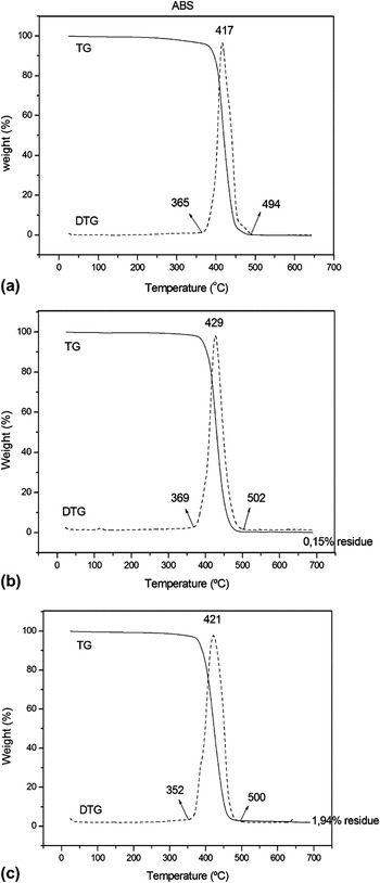
Figures 1(a)–1(c) present the TG/DTG of the samples ABS alone, coated ABS previously submitted to treatment with acid and containing the europium (III) complex [ABS (ac.)complex], and coated ABS previously treated with base containing the europium (III) complex [ABS (bc.) complex], respectively.
TG/DTG curves of (a) the organic polymer ABS before coating, (b) the ABS (ac.) complex, and (c) the ABS (bc.) complex.

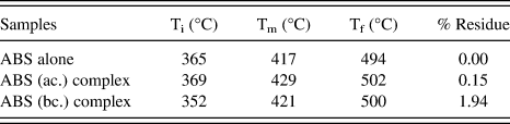
Thermal analyses were conducted for verifying the thermal properties of the ABS polymer before and after coating with the europium (III) complex. Decomposition of the noncoated ABS substrate occurred in a single stage, and the maximum decomposition temperature [Fig. 1(a)] appeared at 417 °C. In a previous work, we had observed that the decomposition temperature of the ABS polymer increased after coating with different compositions of sols.Reference Bandeira, De Campos, De Faria, Ciuffi, Calefi, Nassar, Silva, Oliveira and Maia23, Reference De Campos, Bandeira, Calefi, Ciuffi, Nassar, Silva, Oliveira and Maia24 Indeed, in the present work the decomposition temperature maximum changed from 417 °C for ABS alone to 429 and 421 °C for the ABS (ac.) complex and the ABS (bc.) complex, respectively, after treatment and coating of the ABS substrate. At a first glance, this increase appears to be very small. However, it should be borne in mind that the dip-coating technique usually affords thin films with 0.1–1 μm thickness, depending on the viscosity of the sol.Reference Bandeira, de Campos, Calefi, Ciuffi, Nassar, Silva, Oliveira and Maia25Table I lists the initial, maximum, and final decomposition temperatures as well as the percentage of residue remaining after TG analysis for the coated and noncoated ABS substrates prepared herein.
Initial (Ti), maximum (Tm), and final (Tf) decomposition temperatures and residue percentage of the as-prepared coated andnoncoated ABS substrates.
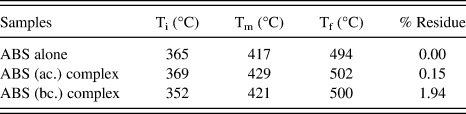
The decomposition temperatures obtained for the coated and noncoated ABS polymer are slightly different, which is attributed to the thickness of the coating. Observation of the temperature range over which ABS decomposition occurred (difference between the temperatures at which the decomposition started and finished) provides evidence that the ABS polymer was really coated with the europium (III) complex. These ranges were 129, 133, and 148 °C for ABS alone, the ABS (ac.) complex, and the ABS (bc.) complex, respectively. Interestingly, the sample that decomposed over the largest temperature range also yielded the highest residue percentage. The residual percentages of 0.15 and 1.94% are indicative of the presence of an inorganic compound in the ABS polymer substrate. Nevertheless, the amount of residue that remained after the thermal analysis was very low in all cases, suggesting that the coating was very thin. The residue consisted of sodium and europium oxide, and the fact that the ABS (bc.) complex gave a larger residue demonstrates that pretreatment of the ABS polymer with base (sodium hydroxide) was more efficient for europium (III) complex immobilization on the substrate as compared with acid (acetic acid) treatment.
B. XRD
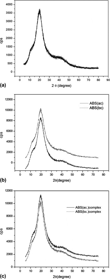
Figures 2(a), 2(b), and 2(c) correspond to the XRD patterns obtained for ABS alone, ABS treated with acid, ABS treated with base, the ABS (ac.) complex, and the ABS (bc.) complex.
XRD patterns for the (a) organic polymer ABS before coating (b) after treatment with acid or base, and (c) after treatment and coating with the europium (III) complex.

All the samples displayed broad peaks between 10 and 30°, characteristic of amorphous materials. The similarity between the patterns indicates that the acid and base treatment did not affect the structure of the ABS polymer. Coating with the europium (III) complex did not have any effect on the structure of ABS, either. The absence of a peak due to the presence of a crystalline phase in the case of the ABS (ac.) complex and the ABS (bc.) complex can be explained by the low thickness of the coating. Moreover, it should be borne in mind that the coating was prepared from silicon precursors, so an amorphous phase should be expected.
C. SEM
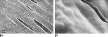
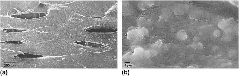
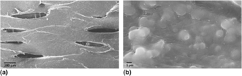
Figures 3–5 depict the SEM images obtained for ABS alone, the ABS (ac.) complex, and the ABS (bc.) complex, respectively.
Surface of the ABS polymer.

Surface of the ABS (ac.) complex.
Surface of the ABS (bc.) complex.

Figures 3(a) and 3(b) bring the SEM images for ABS at 75× and 5000× magnification, respectively. The surface of the ABS polymer is rough and presents micropores.
SEM clearly shows the existence of the coatings on the substrate surface (Figs. 4 and 5). The micropores present on the surface of ABS do not disappear after coating, which is explained by the small thickness of the layer. In another work, we had observed the disappearance of the pores because different alkoxide precursors had been used.Reference Bandeira, De Campos, De Faria, Ciuffi, Calefi, Nassar, Silva, Oliveira and Maia23 From Figs. 4(b) and 5(b) it can be noted that the coating was formed by synterization of the spherical particles with different sizes. There are no cracks in the coating, indicating that a homogeneous layer was obtained.
D. PL
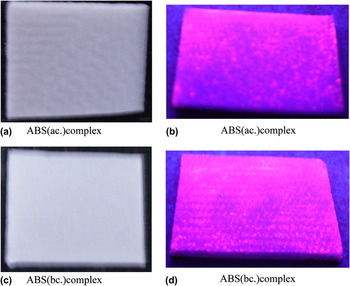
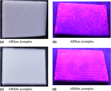
Figure 6 displays photographs of the ABS (ac.) complex and the ABS (bc.) complex under visible (a) and ultraviolet (b) light.
Photographs of the ABS (ac.) complex and the ABS (bc.) complex and under visible light (a and c) and ultraviolet light (b and d), respectively.

When ultraviolet light was used for excitation of the europium (III) complex, the strong red emission ascribed to the europium (III) ion was observed.
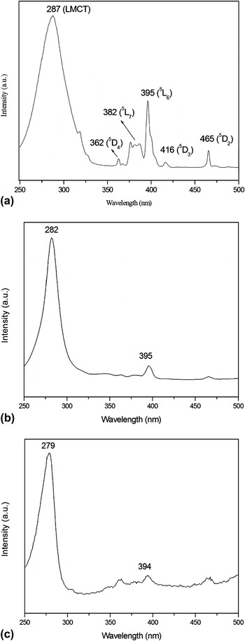
Figure 7 shows the excitation spectra of the europium (III) ion in the Eu3+:dpa complex [Fig. 8(a)] and of the europium (III) complex in the coating on the ABS polymer substrate [Fig. 7(b) and 7(c)], monitored at the 5D0 → 7F2 transition.
Excitation spectra for the europium (III) ion in the (a) Eu3+:dpa complex, (b) ABS (ac.) complex, and (c) ABS (bc.) complex. λem. = 615 nm.

The dpa molecule itself presents absorption maximum at 290 nm. The excitation spectrum displays a broad band in the 250–325 nm region, and the maximum for the europium (III) complex appears at 287 nm. This band is due to energy transfer from the dpa ligand to the europium (III) ion, the so-called ligand-to-metal charge transfer (LMCT). The f–f transitions appear as sharp lines in the 350–475 nm range and are ascribed to transitions from the 7F0 level to the 5L6, 5L7, and 5D2-4 levels. In Figs. 7(b) and 7(c), which correspond to the excitation spectra of the ABS (ac.) complex and the ABS (bc.) complex, respectively, there is a broad band between 250 and 300 nm, attributed to LMCT. The presence of the LMCT band indicates that the Eu3+:dpa complex is not affected by the sol-gel medium. Indeed, the energy of the LMCT band depends on the anion type, the binding strength of the valence band electrons, and the size of the site occupied by the europium (III) ion.Reference Oliveira, Cebim, da Silva and Davolos3 The slight decrease in the wave length of the LMCT band for the ABS (ac.) complex and the ABS (bc.) complex as compared with ABS suggests a small change in the symmetry of the europium (III) ion, due to possible distortion of the europium (III) complex D3 symmetry.Reference Binnemans, Van Herck and GiSrller-Walrand13
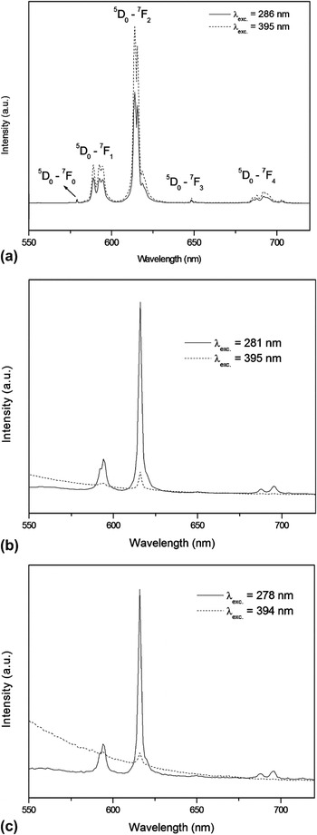
Figure 8 illustrates the emission spectra of the excitation spectra of the europium (III) ion in the Eu3+:dpa complex (Fig. 8a) and of the europium (III) complex in the coating on the ABS polymer substrate [Figs. 8(b) and 8(c)], when excited at the LMCT band and at the 5L6 Eu3+ level.
Emission spectra for the europium (III) ion in the (a) Eu3+:dpa complex, (b) ABS (ac.) complex, and (c) ABS (bc.) complex excited at the LMCT band and 5L6 Eu3+ level.

The emission spectrum of the Eu3+ ion in the complex displays emission lines corresponding to transitions from the excited state 5D0 to the fundamental level 7FJ (J = 0, 1, 2, 3 and 4), dominated by the 5D0 → 7F2 (∼614 nm) electric dipole transition that occurs in the red region of the electromagnetic spectrum. The emission spectra for the complexes in the samples ABS (ac.) complex and ABS (bc.) complex present the same transition when excited at the LMCT band, but the emission prompted by the excitation at the 5L6 level is very weak. This can be attributed to interaction between the Eu3+:dpa complex and the silica coating, which quenches the luminescence of the ion.
The emission spectrum of the Eu3+:dpa complex presents the 5D0 → 7F0 transition at 17,271 cm−1 [Fig. 8(a)], which can only be detected if the symmetry group of the complex is Cnv, Cn, or Cs.Reference Reinhard and Gudel12 The 5D0 → 7F0 transition disappears for the samples ABS (ac.) complex and ABS (bc.) complex, indicating that a change in the symmetry of europium (III) occurred.
The 5D0 → 7F1 transition appears as three peaks when the point group is orthorhombic (D2, C2v), monoclinic (C2, Cs), or triclinic (C1).Reference Kim, Yoon, Sohn and Kang14 Three peaks were observed for the Eu3+:dpa complex, showing that the europium (III) can occupy a D2, C2v, C2, Cs, or C1 symmetry site. The division of the 5D0 → 7F2 transition furnishes information about the site symmetry. Herein, three peaks were detected for this transition, evidencing that the point group is D2. The presence of the 5D0 → 7F0 transition is indicative of distortion or of a mixture of site symmetries in the Eu3+:dpa complex. Kim et al.Reference Kim, Yoon, Sohn and Kang14 have suggested a D3 site symmetry for europium (III) in Eu(dpa)33-.
In the present case the 5D0 → 7F0 transition is absent from the spectrum of the europium (III) complex incorporated into the silica sol coating the ABS polymer. The asymmetry of the band corresponding to 5Do → 7F1 transition suggests that the transition could be composed of three peaks, which is coherent with the D2 or D3 symmetry, according to the section rules.Reference Binnemans, Van Herck and GiSrller-Walrand13 Thus, incorporation of the europium (III) complex into the sol may have modified the site symmetry of the lanthanide ion.
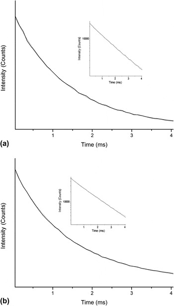
Figure 9 shows the lifetime of the 5D0 → 7F2 transition when the samples are excited at the LMCT band.
Lifetime curve for the europium (III) ion in the (a) ABS (ac.) complex, and (b) ABS (bc.) complex excited at the LMCT band.

The decay curve corresponds to a monoexponential decay; so only one lifetime is observed for the samples excited at the LMCT band. Excitation at this band hence demonstrates that energy transfer takes place in one europium (III) site only. The lifetime was the same for all the samples; i.e., 1.39 ms. This is one more evidence that the europium (III) complex is not destroyed upon incorporation into the silica sol.
IV. CONCLUSION
The luminescent properties of the europium (III) complex were preserved after its incorporation into the sol-gel silica matrix, later used as coating. Energy transfer is very important when increased emission of rare earth compounds is desirable, enabling application of the resulting materials in several fields. In this article, it has been demonstrated that the sol-gel process can be used for incorporation of organic compounds into inorganic matrices without compromising the integrity of the incorporated substance.
Many techniques have been used for the characterization of the as-prepared materials, but luminescence was the most effective property for detection of the complex in the coating and demonstration of the structural integrity of the incorporated compound. Thermal analysis together with photoluminescence showed that a small amount of the europium (III) complex is sufficient for application of the as-prepared coated ABS as luminescent material.