Introduction
Lymphatic filariasis (LF), commonly known as elephantiasis, is caused by infection with nematodes such as Wuchereria bancrofti (Wb), Brugia malayi (Bm) and Brugia timori (Bt). Over 120 million people are infected worldwide, with about 40 million incapacitated or disfigured with swollen limbs (lymphoedema) and genitals (hydrocele) (Michael & Bundy, Reference Michael and Bundy1997; WHO, 2010). More than 1 million people across 80 countries are at risk of infection. Infection is usually acquired in childhood, when the infective-stage larvae are transmitted to humans through mosquitoes. The visible manifestations of the disease, however, occur later in life, causing social disgrace. Hence detection of filarial antigens in endemic populations is essential.
The traditional thick blood examination is known to be inconvenient and insensitive (Abaru & Denham, Reference Abaru and Denham1976). Commercial assays like WbSXP-1 antibody detection (Baskar et al., Reference Baskar, Srikanth, Suba, Mody, Desai and Kaliraj2004), Og4C3 antigen detection assay (More & Copeman, Reference More and Copeman1990) and the immunochromatographic card test (ICT) (Weil et al., Reference Weil, Lammie and Weiss1997) are available for detecting W. bancrofti infection. Other techniques, such as membrane filtration (Desowitz & Hitchcock, Reference Desowitz and Hitchcock1974) and polymerase chain reaction (PCR)-based assays, are also available (Kluber et al., Reference Kluber, Supali, Williams, Liebau and Fischer2001; Rahmah et al., Reference Rahmah, Nurulhasanah, Norhayati, Zulkarnain and Norizan2010). However, all these methods have drawbacks, such as incompatibility for mass screening, lack of stability and specificity, high cost and time consumption. Earlier, a capture enzyme-linked immunosorbent assay (ELISA) was developed with rWbSXP-1 antibodies and was validated for the native microfilariae (mf) antigen (Pandey et al., Reference Pandey, Madhumathi, Karande and Kaliraj2011). In order to improve on the sensitivity of detection of parasite antigens, we explored the possibility of developing another antigen capture assay using the B. malayi vespid allergen homologue (BmVAH). This protein is expressed in the third larval (L3) stage as well as the mf stage, and it exhibits 91% identity with WbVAH (Anand et al., Reference Anand, Gnanasekar, Thangadurai, Prabhu, Kaliraj and Ramaswamy2007; Bennuru et al., Reference Bennuru, Semnani, Meng, Ribeiro, Veenstra and Nutman2009). The VAH/VAL-1 (venom allergen-like 1) family of proteins shares significant similarity with the hymenopteran vespid allergens (VA). VAH proteins are cysteine-rich secretory proteins (CRISP). Endemic normal (EN) and microfilaraemic (MF) populations harbour circulating antibodies to BmVAL-1, with high levels of immunoglobulin G4 subclass (IgG4) that may indicate stimulation by adult- or microfilaria-derived immunogens (Murray et al., Reference Murray, Gregory, Gomez-Escobar, Atmadja and Maizels2001).
In the present study, polyclonal and monoclonal antibodies (mAbs) against rBmVAH were developed and characterized. A capture ELISA was established with the aim to detect circulating filarial antigens (CFA) in asymptomatic and/or amicrofilaraemic individuals and was compared with the previously reported rWbSXP-1 ELISA (Pandey et al., Reference Pandey, Madhumathi, Karande and Kaliraj2011).
Materials and methods
Collection of blood samples
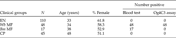
Based on clinical and parasitological examination using traditional methods, the study population was divided into microfilaraemic (MF), endemic normals (EN) and chronic patients (CP). The presence of circulating filarial parasite antigens was detected by the Og4C3 kit. The detection of microfilariae (mf) in night blood smears was considered as MF (n= 65) (brugian infection = 17, bancroftian infection = 48). Subjects with no mf were classified as EN (n= 110) and subjects with visible clinical symptoms of filariasis were grouped as CP (n= 45). Serum samples from healthy individuals residing in non-endemic areas NEN (n= 10) were kindly provided by Professor Murray Selkirk, Imperial College, London. There were no marked differences in the socio-economic status between the various groups and the details of patients selected in the present study are shown in table 1.
The occurrence in clinical populations of chronic pathology with visible symptoms of lymphoedema and asymptomatic bancroftian/brugian microfilaraemia; detection of microfilariae in blood samples at night and circulating antigens using the Og4C3 assay.

N, number of samples; EN, endemic normal; Wb MF, W. bancrofti microfilaraemic; Bm MF, B. malayi microfilaraemic; CP, chronic patients.
Preparation of filarial antigens
Infective larvae (L3) of B. malayi were obtained from the Mahatma Gandhi Institute of Medical Sciences, Sevagram, Maharashtra, India. Jirds (Meriones unguiculatus) were infected with 100 B. malayi infective larvae via the intra-peritoneal (i.p.) route and mf were obtained 3–4 months later by lavage of the peritoneal cavities of the jirds (McCall et al., Reference McCall, Malone, Hyong-Sun and Thompson1973). Wuchereria bancrofti mf were separated from the blood of infected individuals using a membrane filtration method (Chularerk & Desowitz, Reference Chularerk and Desowitz1970). The L3 and mf parasites were resuspended in 500 μl of 50 mm phosphate buffer, pH 7.2, containing 150 mm NaCl (phosphate-buffered saline, PBS) and sonicated (Sartorious AG, Goettingen, Germany) for 10 min at 10 cycles/min. The sonicated sample was centrifuged at 5000 × g, at 4°C for 10 min and the supernatant was collected. Protein concentration was determined by Bradford's method (Bradford, Reference Bradford1976) and the samples were stored at − 80°C.
Recombinant protein expression and purification
The recombinant filarial clones BmVAH (accession no.: AF334661 cloned in pRSETA) and WbSXP-1 (accession no.: AFO98861 cloned in pRSETB) were used in this study. Escherichia coliGJ1158 cells transformed with recombinant BmVAH (rVAH) or WbSXP-1 (rSXP-1) were induced with 300 mm NaCl for protein expression. Both the recombinant proteins are N-terminal histidine-tag fusion proteins. The proteins were purified, as described earlier (Anand et al., Reference Anand, Gnanasekar, Thangadurai, Prabhu, Kaliraj and Ramaswamy2007; Janardhan et al., Reference Janardhan, Pandiaraja, Thirugnanam, Balamurali, Fernando, Mody, Desai, Meenakshisundaram and Kaliraj2007), and the purity of the preparations was judged by sodium dodecyl sulphate–polycrylamide gel electrophoresis (SDS-PAGE) followed by staining with Coomassie Blue.
Generation of antibodies against rVAH
Monoclonal antibodies (mAbs)
BALB/c mice (6–8 weeks old, female, housed in a 12 h night–day cycle at a controlled temperature of 24°C and constant humidity, and food ad libitum) were immunized with recombinant VAH emulsified in Freund's complete adjuvant (Gangatirkar et al., Reference Gangatirkar, Gangadharan, Narendranath, Nagpal, Salunke and Karande2002). After an adequate increase in the titres of the circulating antibody, spleens were excised and splenocytes were fused with a myeloma cell line (Sp2/0-Ag14) using PEG 4000 (Merck, Rahway, New Jersey, USA). Specific antibody-secreting hybridoma clones were expanded and sub-cloned to monoclonality. High-affinity mAbs were selected by indirect ELISA using recombinant VAH antigen, native L3 parasite and Bm mf lysate. Monoclonality was confirmed by subclass isotyping.
Polyclonal antibodies
Laboratory-bred rabbits were immunized with purified rVAH as described previously (Harlow & Lane, Reference Harlow and Lane1988). Blood samples were collected 10–13 days after the final immunization dose and tested for immunoreactivity against rVAH by ELISA.
Western blot analysis
The recombinant protein (2 μg), lysate of L3 larvae (20 μg) and mf lysate antigen (25 μg) samples were electrophoresed on 12.5% polyacrylamide gel under reducing conditions and transferred to a nitrocellulose membrane. The blots were incubated with mAbs or polyclonal antibodies for 2 h, followed by incubation with the corresponding secondary antibodies conjugated to horseradish peroxidase (HRP) (Dako, Glostrup, Denmark) for 1 h. After extensive washes with PBS, the blots were developed using enhanced chemiluminescent HRP substrate (Millipore, Billerica, Massachusetts, USA) and images were acquired by Image Quant LAS 4000 (GE Healthcare, Little Chalfont, Bucks, UK).
ELISA
Indirect ELISA
The recombinant proteins (10 μg/ml) in PBS were coated in the wells of ELISA plates (Nunc Maxisorp, Nalgene Nunc International, Denmark) and unoccupied sites were blocked with 0.5% gelatin in PBS. The wells were incubated with monoclonal or polyclonal antibodies followed by incubation with the respective HRP-conjugated secondary antibodies (Dako). The bound peroxidase activity was detected using tetramethyl benzidine (TMB) at a concentration of 60 μg/ml in citrate phosphate buffer, pH 5.5, containing 0.03% H2O2. The reaction was stopped with 1 m H2SO4, and absorbance was read at 450 nm using a multi-scan ELISA reader (Molecular Devices, Inc., Sunnyvale, California, USA).
Sandwich ELISA
ELISA plates were coated separately with purified polyclonal or monoclonal antibodies (1 μg/well in PBS), followed by blocking with 0.5% gelatin in PBS. Human serum samples diluted 1:50 in PBS were added to the wells and incubated for 2 h, followed by incubation for 1 h with the biotinylated mAbs in PBS. After three washes with PBS, streptavidin–HRP conjugate (Banglore Genei, Bangalore, India) was added and incubated for 1 h. The plates were washed and peroxidase activity retained in the well was determined as described above. The results were expressed as mean absorbance at 450 nm for each group ± standard error. Samples were considered positive for the assay when the optical density was higher than the mean OD +3SD value of 10 NEN control sera.
Competitive ELISA
The appropriate concentration of Abs were incubated with varying concentrations of rVAH (50 μg/ml to 25 ng/ml) for 16 h at 25°C to attain equilibrium. The complexes were transferred to ELISA wells pre-coated with 1 μg of rVAH and incubated for 2 h. The remaining steps were followed as described above for indirect ELISA. The dissociation constant (K d) for each of the antibodies was calculated using the equation:
 $$\begin{eqnarray} \end{eqnarray}$$
derived from Scatchard and Klotz (Friguet et al., Reference Friguet, Chaffotte, Djavadi-Ohaniance and Goldberg1985), where A 0 and A are the absorbance measured for antibody in the absence and presence of antigen, respectively; K d is the dissociation constant and a 0 is the total antigen concentration.
Purification of mAbs and polyclonal antibodies
Antibodies were purified from hybridoma culture supernatants using a protein A-Sepharose column (GE Healthcare). Polyclonal antibodies were obtained from rabbit serum by precipitating with ammonium sulphate at 33% saturation. Antibodies were biotinylated using biotinamidohexanoic acid 3-sulpho-N-hydroxsuccinimide ester sodium salt (Sigma-Aldrich, St. Louis, Missouri, USA) as described previously (Harlow & Lane, Reference Harlow and Lane1988). For rWbSXP-1, previously generated 1F6H3 and 2E12E3 mAbs (Pandey et al., Reference Pandey, Madhumathi, Karande and Kaliraj2011) and anti-SXP-1 rabbit polyclonal antibodies were purified and biotinylated as described above.
Data analysis
All statistical analyses were performed using GraphPad Prism software version 5 (GraphPad Software, San Diego, California, USA). Comparisons between two individual data points were made using the Mann–Whitney test. For multiple comparisons, Bonferroni's post-test was used. P ≤ 0.05 was considered to be statistically significant.
Results
Selection of monoclones and Western blots
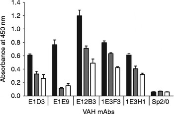
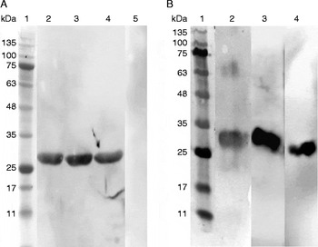
Of the several hybridoma clones established, five stable clones specific to rVAH were selected and tested for reactivity with L3 parasite and Bm mf. The VAH mAbs – E1D3, E12B3 and 1E3F3 – showed reactivity with the native antigens (fig. 1) and hence were selected. In Western blot analysis VAH mAbs reacted with the rVAH protein (fig. 2A). The pool of the three mAbs recognized native L3 parasite, Bm mf and Wb mf antigens at 28 kDa (fig. 2B). Similar results have been reported previously for native L3 and mf parasite antigens (Murray et al., Reference Murray, Gregory, Gomez-Escobar, Atmadja and Maizels2001). Therefore these mAbs were used for standardization of sandwich assays for the detection of CFA.
Screening of monoclones against rVAH and native parasite antigens. rVAH (0.5 μg) or L3 lysate (2 μg) or Bm mf lysate (2 μg) antigens were coated on ELISA plates and incubated with hybridoma culture supernatants with Sp2/0 as the negative control. Black bars, recombinant protein; grey bars, L3 larvae; white bars, B.malayi microfilariae.

Western blot analysis of rVAH and native parasite antigens. (A) Recombinant VAH protein was probed with mAbs and developed. Lanes: 1, molecular weight marker; 2, E1D3; 3, E12B3; 4, 1E3F3; 5, Sp2/0 cell supernatant. (B) Filarial parasites L3, Bm mf and Wb mf lysates were probed with the pool of all three VAH mAbs. Lanes: 1, molecular weight marker; 2, third larval stage (L3); 3, B. malayi microfilariae (Bm mf); 4, W. bancrofti microfilariae (Wb mf).

Characterization of VAH mAbs
The isotype of E12B3 mAb was found to be IgG2a and the remaining mAbs were identified to be of IgG1 isotype. The affinity (K d) values of mAbs E1D3, E12B3 and 1E3F3 were found to be 8.2 × 10⁻⁸, 1.63 × 10⁻⁷ and 4.1 × 10⁻⁷mol/l, respectively. The K d value of mAb E1D3 was one order of magnitude lower than that of the other antibodies.
Standardization of capture and detection Abs for VAH, SXP-1 and combination (VS) assays
Sandwich ELISA was standardized with various combinations of polyclonal and monoclonal Abs, as capture and detection Abs. The VAH assay, using a combination of rabbit anti-VAH polyclonals as capture Ab, and biotinylated E12B3 mAb as the detection Ab, could measure as little as15 ng of rVAH. The SXP-1 assay, using a combination of 1F6H3 SXP-1 mAb as capture Ab, and biotinylated SXP-1 polyclonal as detection Ab, exhibited the same sensitivity of detection as the VAH assay. The VS combination assay also detected as little as 15 ng of both rVAH and rSXP-1 antigens. However, the maximum binding (Bmax) obtained was significantly higher as compared to the individual VAH and SXP-1 assays (data not shown).
Detection of native parasite antigens in sandwich ELISA
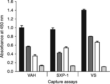
The combination (VS) assay detected the recombinant proteins as well as the native parasite antigens with higher binding values when compared to either of the individual, i.e. VAH and SXP-1, assays (fig. 3). However, the clinical serum samples were subjected to all the three immunoassays to detect CFA.
Capture ELISA to detect recombinant and native parasite antigens. Capture antibodies were coated in ELISA wells and incubated with rVAH or/and rSXP-1 (0.5 μg) or L3 lysate (2 μg) or Bm mf lysate (2 μg). Retained antigens were detected with biotinylated antibodies followed by streptavidin–peroxidase and developed. Black bars, recombinant protein; grey bars, L3 larvae; white bars, B. malayi microfilariae; dotted bars, no antigen.

Capture assays for detection of CFA in clinical samples
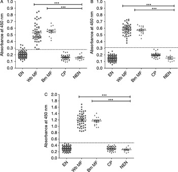
Sandwich ELISA was carried out for the detection of circulating antigens in the clinical serum samples of endemic areas for filariasis. VAH, SXP-1 and VS assays showed highest reactivity with MF and very low reactivity with EN, CP and NEN groups (table 2). The VS assay showed enhanced immunoreactivity with MF sera (fig. 4C) when compared with either VAH or SXP-1 assay (fig. 4A, B). When data were analysed using the t-test to compare the antigen levels in EN, MF and CP with the control NEN group, the MF group showed significantly higher antigen levels (P < 0.0001) compared to the other groups (fig. 4A, B, C). Data were analysed using two-way ANOVA to compare mean OD values of each group in all the assays. In the VS assay, Wb MF and Bm MF groups exhibited the highest optical density values (OD ± SD = 1.198 ± 0.21 and 1.167 ± 0.125, respectively; P < 0.001) when compared to other groups in all assays (table 2).
Detection of filarial antigens in clinical populations, expressed as mean values of optical density (OD) ± SD and positive reactivity (%) using VAH, SXP-1 and VS capture assays.

N, number of samples; EN, endemic normal; Wb MF, W. bancrofti microfilaraemics; Bm MF, B. malayi microfilaraemics; CP, chronic patients; NEN, non-endemic normal.
Mean values of optical density (OD) ± SEM plotted in triplicate to show circulating filarial antigens in clinical samples using sandwich ELISA. Capture antibodies were coated in ELISA wells followed by incubation with serum samples. Retained antigens were detected with biotinylated antibodies followed by streptavidin–peroxidase and developed. Mean OD +3SD of negative control non-endemic normal (NEN) samples are taken as the cut-off (dotted lines) with clinical groups categorized as endemic normal (EN), n= 110; B.malayi microfilaraemics (Bm MF), n= 17 and W. bancrofti microfilaraemics (Wb MF), n= 48; and chronic pathology (CP), n= 45. ***P< 0.0001, statistically significant when compared to NEN samples, n= 10.

Discussion
LF is the second most important vector-borne disease in endemic countries such as India, South-East Asia, Africa, the Western Pacific and parts of South America. The infection, which is both debilitating and disfiguring, may lead to long-term physical impairment. The disease is associated with a high degree of social stigma and the management of infection is an economic burden (Ramaiah et al., Reference Ramaiah, Das, Michael and Guyatt2000). Sensitive diagnostic methods to detect human LF in endemic populations of developing countries is essential for efficient surveillance, early detection and eradication of this disease, as emphasized by WHO (2010).
Until now, microscopy-based parasitological examination of blood smears has been the most commonly used test for the confirmation of mf circulation in highly populated endemic countries. This method has several drawbacks: difficulty in the collection of night blood samples, low sensitivity, variation due to disparity in blood volumes collected and the time frame for collection, which may not correspond to the window of appearance of mf in the circulation.
For some time now, ICT and Og4C3 ELISA have been available for the detection of circulating antigens of Wb in serum or whole blood. ICT was the first rapid-format antigen detection test to be introduced for detecting filariasis. This test shows positivity only for individuals who harbour a high number of circulating mf and the sensitivity is lower (71.9%) than that of the filtration technique (Pani et al., Reference Pani, Hoti, Elango, Yuvaraj, Lall and Ramaiah2000). Moreover, the kit needs to be stored at 2–8°C, which is not always possible in rural areas in India and, also, is not cost- effective (Pani et al., Reference Pani, Hoti, Elango, Yuvaraj, Lall and Ramaiah2000). The sensitivity of the Og4C3 test is lower than that of the ICT (Pani et al., Reference Pani, Hoti, Elango, Yuvaraj, Lall and Ramaiah2000) and neither of these tests detects Bm antigens. The more recently developed diagnostic methods available detect circulating antibodies to the filarial parasite antigens, namely Bm14, WbSXP-1 and BmR1, in patients. These assays are unable to distinguish current infection from past infections. Hence, the development of sensitive diagnostics to detect brugian and bancroftian CFA in blood, to identify active filarial infection in endemic countries, was necessary for appropriate surveillance after any intervention programmes. SXP-1 secretory antigen has already been exploited for detection of filarial infection (Rao et al., Reference Rao, Eswaran, Ravi, Gnanasekhar, Narayanan, Kaliraj, Jayaraman, Marson, Raghavan and Scott2000; Lalitha et al., Reference Lalitha, Eswaran, Gnanasekar, Rao, Narayanan, Scott, Nutman and Kaliraj2002). Another antigen identified as a diagnostic target molecule was VAH because it is a secretory protein of mf (Bennuru et al., Reference Bennuru, Semnani, Meng, Ribeiro, Veenstra and Nutman2009). We attempted to develop a capture assay for the detection of VAH in clinical samples with a view to improve the sensitivity of filarial antigen diagnostics.
Monoclonal and polyclonal antibodies were established against rBmVAH protein. Sandwich ELISA was standardized with the selected mAbs which were reactive with rBmVAH and, more importantly, also with the native mf antigens. In order to compare the VAH assay with an established detection method, two mAbs raised against rWbSXP-1 were also recruited (Pandey et al., Reference Pandey, Madhumathi, Karande and Kaliraj2011).
The individual and combination capture assays were standardized and evaluated with a large number of serum samples which were collected from different clinical groups, such as EN, asymptomatic MF, CP and a control NEN group. The capture assay using VAH antibodies detects Wb and Bm antigens in 97.91% (47/48) and 100% (17/17), respectively, of MF-infected individuals. The CP and EN samples showed minimum reactivity. The same clinical samples were also tested in the SXP-1 immunoassay. We found no difference in the detection levels between these two assays. Therefore, a combination capture assay was designed to detect filarial antigens by ELISA.
A capture assay using different combinations of VAH and SXP-1 antibodies was standardized. As expected, both Wb and Bm MF serum samples were positive in the assay and no reactivity was seen with EN and CP samples. However, the ELISA readings for the positive samples obtained in the VS capture assay was twice that of VAH or SXP-1 ELISA, thereby increasing the confidence levels.
In summary, monoclonal and polyclonal antibodies were raised against rBmVAH antigen and capture assays were standardized. Our results showed that the VAH capture assay was comparable to SXP-1 assay, and the combination (VS) assay detected positive samples with greater binding levels. Moreover, as the assay detects antigens, it can differentiate active infection from chronic infection. In endemic countries such as India, where there is co-prevalence of both bancroftian and brugian infections, it is advantageous to use a diagnostic tool that detects both these types simultaneously. Additional studies are needed to compare the cross-reactivity with Loa loa, Onchocerca and non-lymphatic filarial infected samples. The capture assay developed in this study gives preliminary assessment of the detection of filarial infections and adds value to the development of sensitive diagnostics for the Global Programme to Eliminate Lymphatic Filariasis (GPELF) in developing countries.
Acknowledgements
We sincerely thank Dr V. Gopalrathinam, Senior Entomologist, Zonal Entomological Research Laboratory, Vellore, Tamilnadu for his cooperation and support in sample collection. We thank to Dr M.V.R. Reddy, Professor and Head, Department of Biochemistry, Mahatma Gandhi Institute of Medical Sciences, Sevagram, Maharashtra, India, for providing third larval stage parasites. We also thank Dr Murray Selkirk, Professor of Biochemical Parasitology, Division of Cell and Molecular Biology, Imperial College, London, for providing the non-endemic sera samples for the study. R.R. would also like to thank Dr Sudarshan Gadadhar and Ms Jeya Prita for helping with data analysis.
Financial support
This work was supported by the Department of Biotechnology, Government of India, (grant no. BT/01/COE/06/13). The Senior Research Fellowship granted to R.R. by the Council of Scientific and Industrial Research, Government of India, is gratefully acknowledged.
Conflict of interest
None.
Ethical standards
The animal experiments have been approved by the ‘Institutional Animal Ethics Committee’, Indian Institute of Science, Bangalore, India. Blood samples were collected from volunteers after obtaining written consent and this was documented by the Department of Public Health and Preventive Medicine, Chennai, Tamil Nadu, India (Ref. No. 26433/VCII/SI//2012). The procedures followed were in accordance with the guidelines issued by the same department. The Institutional Review Board at the Centre for Biotechnology, Anna University, India has also approved the protocols.