INTRODUCTION
The ookinete is a motile stage of malaria parasites that forms in the midgut lumen of the mosquito vector within the infective bloodmeal bolus. It invades the intestinal epithelium and upon arriving at the basal lamina it transforms into an oocyst in which sporozoites, the parasite stage infective for vertebrates develop (Sinden, Reference Sinden1999). Sporozoites and ookinetes move by gliding, a locomotion form characteristic of the invasive stages of apicomplexan parasites (Kappe et al. Reference Kappe, Bruderer, Gantt, Fujioka, Nussenzweig and Ménard1999; Ménard, Reference Ménard2001; Fowler et al. Reference Fowler, Margos and Mitchell2004).
Gliding is a substrate-dependent movement driven by an actin-myosin motor that translocates surface thrombospondin-related adhesive protein (TRAP)-like molecules from the apical to the rear extreme of the parasite. As the ookinete moves, the TRAP-like CTRP and Pbs25 (Lecona et al. Reference Lecona, Rodriguez, Argotte-Ramos, Alvarado-Delgado and Rodriguez2010) are shed on the substrate. Aldolase acts as a metabolic enzyme as well as a bridge between the actomyosin system and the transmembrane adhesins (Daher and Soldati, Reference Daher and Soldati2009; Baum et al. Reference Baum, Richard, Healer, Rug, Krnajski, Gilberger, Green, Holder and Cowman2006; Soldati-Favre, Reference Soldati-Favre2008).
Several proteins have been described in Plasmodium ookinetes: P25, P28, CTRP, WARP, SOAP, MAOP, chitinase, myosin A, CDPK3, PbGCbeta, IMC1b, etc. (Huber et al. Reference Huber, Cabib and Miller1991; Tsuboi et al. Reference Tsuboi, Cao, Kaslow, Shiwaku and Torii1997; Naitza et al. Reference Naitza, Spano, Robson and Crisanti1998; Dessens et al. Reference Dessens, Beetsma, Dimopoulus, Wengelnick, Crisanti, Kafatos and Sinden1999; Yuda et al. Reference Yuda, Sawai and Chinzei1999; Margos et al. Reference Margos, Sidén-Kiamos, Fowler, Gillman, Spaccapelo, Lycett, Vlachou, Papagiannakis, Eling, Mitchell and Louis2000; Siden-Kiamos et al. Reference Siden-Kiamos, Vlachou, Margos, Beetsma, Waters, Sinden and Louis2000; Yuda et al. Reference Yuda, Yano, Tsuboi, Torii and Chinzei2001; Tomley and Soldati, Reference Tomley and Soldati2001; Dessens et al. Reference Dessens, Sidén-Kiamos, Mendoza, Mahairaki, Khater, Vlachou, Xu, Kafatos, Louis, Dimopoulos and Sinden2003; Kadota et al. Reference Kadota, Ishino, Matsuyama, Chinzei and Yuda2004; Siden-Kiamos et al. Reference Siden-Kiamos, Ecker, Nyback, Louis, Sinden and Billker2006; Hirai et al. Reference Hirai, Arai, Kawai and Matsuoka2006; Tremp et al. Reference Tremp, Khater and Dessens2008). These molecules participate in motility, invasion (Yuda et al. Reference Yuda, Sawai and Chinzei1999; Templeton et al. Reference Templeton, Kaslow and Fidock2000; Limviroj et al. Reference Limviroj, Yano, Yuda, Ando and Chinzei2002; Dessens et al. Reference Dessens, Sidén-Kiamos, Mendoza, Mahairaki, Khater, Vlachou, Xu, Kafatos, Louis, Dimopoulos and Sinden2003; Hirai et al. Reference Hirai, Arai, Kawai and Matsuoka2006; Tremp et al. Reference Tremp, Khater and Dessens2008), peritrophic matrix degradation (Huber et al. Reference Huber, Cabib and Miller1991), basal lamina recognition (Adini and Warburg Reference Adini and Warburg1999; Vlachou et al. Reference Vlachou, Lycett, Sidén-Kiamos, Blass, Sinden and Louis2001) and defence against proteolysis. However, the description of ookinete surface proteins and their participation in the complex process of mosquito midgut invasion is still incomplete.
In this work, we used a phage display strategy to identify a consensus sequence (PWWP) in peptides that bind to the surface of Plasmodium berghei ookinetes. Assuming that these peptides display the structural features of ookinete ligands in the vector mosquito midgut, we used them to search for new molecules on the ookinete surface. Two proteins were identified, actin and enolase.
MATERIALS AND METHODS
Parasites
Ookinetes of P. berghei strain Antwerpen Katanga (ANKA) clone 2.34 (kindly provided by R. E. Sinden, Imperial College, UK) were obtained as previously described (Rodríguez et al. Reference Rodriguez, Gerold, Dessens, Kurtenbach, Schwartz, Sinden and Margos2000). Briefly, 6-week-old BALB/c male mice were inoculated with 200 μl of phenylhydrazine (PH) (6 mg/ml) in PBS, by the intraperitoneal route (IP). Three days after PH treatment, mice were inoculated IP with 1×10⁸ P. berghei parasites. Three days post-infection, the presence of gametocytes was determined in thin blood films stained with Giemsa. The blood containing gametocytes was diluted 1:5 in RPMI containing 20% heat-inactivated bovine fetal serum (Hyclone, Thermo Scientific, Utah, USA). The suspension was incubated for 24 h at 20°C to allow ookinete development. Red blood cells were lysed by adding NH4Cl (0·17 m). Red blood cell debris was removed and ookinetes purified by centrifugation using a Nycodenz™ gradient.
Selection of phages that bind to P. berghei ookinete surfaces
The interaction of P. berghei ookinete surface molecules with a collection of peptides with random sequences was carried out using a phage display commercial kit (Phage Display Peptide Library Kit Ph.D. – 12™, New England Biolabs, #cat. E8110S) following the manufacturer's instructions. This phage collection displays linear peptides of 12 amino acids in length. Using freshly prepared ookinetes, 5 selection cycles were carried out. Before and after each selection cycle, the phage titres in the working solution and the eluted solution were determined. For each selection cycle, 4×10¹¹ plaque-forming units (pfu) were used diluted in 1 ml of Tris-Buffered Saline (TBS, Tris-base 0·5 m, NaCl 9%, pH 7·6). Phages were incubated with ookinetes for 1 h at room temperature (RT) with gentle shaking. The ookinete-phage suspension was centrifuged for 3 min at 1500 g and washed 10 times with TBS to remove the unbound phages. Ookinete-bound phages were eluted using glycine 0·2 m HCl, pH 2·2 in 1 mg/ml of bovine serum albumin (BSA). The sample was centrifuged for 3 min at 1500 g, and the supernatant recovered and neutralized with 1 m Tris-HCl, pH 9·1. The eluted phages were amplified, purified and titrated. In the second and third selection cycle, 0·01% Tween-20 (v/v) in TBS was used as washing solution. For the fourth and fifth selection cycles, 0·05% Tween-20 in TBS was used. After the fifth cycle, eluted phages were titrated and individual phage clones were amplified. The corresponding DNA from each clone was obtained and sequenced at the DNA Sequencing Unit of the National Institute of Public Health in Cuernavaca, Morelos, Mexico.
Ookinete binding pattern of selected phages
The binding patterns of 2 randomly selected phage clones (5 and 14) to P. berghei ookinetes were determined by immunofluorescence assays. Ookinetes and selected clones were mixed in a 1:12 500 ratio, incubated for 20 min at RT, washed 4 times with 1x phosphate buffered saline (PBS), twice with PBS-0·05% Tween-20, and a final wash with PBS. The samples were centrifuged for 3 min at 1500 g and the pellets containing the ookinetes and phages were suspended in 100 μl of PBS. An aliquot of each sample was placed on a slide and allowed to air dry. Ookinetes incubated with non-selected phage clones and/or secondary antibody only were included as control. The samples were incubated for 1 h with a murine monoclonal antibody anti-M13 (which recognizes the gp8 protein of M13) (Amersham, 27-9420-01) diluted 1:250 in PBS-2% BSA. After incubation, the unbound antibody was removed by washing 3 times with PBS-0·05%, Tween-20 and once with PBS, and incubated 1 h in the dark with an anti-mouse IgG coupled to FITC (American Qualex, A106FN) diluted 1:100 in PBS-2% BSA. Unbound secondary antibody was removed by washing 3 times with PBS-0·05% Tween-20 and once with PBS. Each sample was incubated with propidium iodide (0·5 μg/ml) and ribonuclease (to avoid staining of RNA) (500 μg/ml) in PBS for 5 min, washed twice with PBS, and covered with a drop of SlowFade Light Antifadant (Molecular Probes, S-7461, Invitrogen, UK) to preserve fluorescence. The preparations were analysed under a UV fluorescence microscope (Nikon, E600 Eclipse).
Detection of phage ligands by overlay assays
Overlay assays were used to detect interactions between the selected phages and ookinete proteins (Crawford et al. Reference Crawford, Michelsen and Beckerle1992). Ookinete proteins were separated by one-dimensional (1D) and two-dimensional (2D) SDS-PAGE (Laemmli, Reference Laemmli1970; O'Farrell, Reference O'Farrell1975). For 1D electrophoresis, proteins were extracted from 50×106 ookinetes using 600 μl of lysis buffer (Tris-HCl 50 mm, NaCl 150 mm, EDTA 15 mm, Tween-20 0·1%, glycerol 10%) containing protease inhibitors (AEBSF 104 mm, aprotinin 0·08 mm, leupeptin 2 mm, bestatin 4 mm, pepstatin 1·5 mm, E-64 1·4 mm) (Sigma P8340). The sample was centrifuged for 15 min at 10 000 g, the supernatant recovered and the protein concentration determined by Bradford methodology (Sigma, B6916). Then 50 μg of ookinete proteins were separated using 10% polyacrylamide preparative running gels, and transferred to nitrocellulose membranes (Towbin et al. Reference Towbin, Staehelin and Gordin1979).
For 2D electrophoresis, 60×106 ookinetes were treated with lysis buffer (urea 9·5 m, NP-40 2%, β-mercaptoethanol 5%, and ampholytes 5–7/3–10; GE Healthcare, Denmark) for 15 min at RT. The sample was centrifuged for 5 min at 13 000 g and the supernatant proteins were separated in an isoelectric focusing gel for 15 h at 400 V and 2·5 h at 800 V. After pI separation, proteins were resolved by molecular mobility using SDS-PAGE and transferred onto nitrocellulose membranes at 120 V for 1·5 h.
The nitrocellulose membrane obtained from the 1D gel was cut into 3 mm strips for overlay assays. The membrane obtained from the bi-dimensional gel was used intact in one overlay assay. Nitrocellulose membranes were blocked with 5% skimmed-milk in PBS for 1 h, then incubated at RT for 1 h with selected phage clones at a concentration of 1×10⁹ pfu/ml diluted in 2% skimmed-milk in PBS and 1% Tween-20. After incubation, membranes were washed for 4 h with Tween-20 1% in PBS and for 1 h with Tween-20 0·5% in PBS, with changes of washing solution every 30 min, and then given 2 final washings of 5 min each with PBS. The strips and the membrane were incubated for 1 h at RT with anti-M13 (Amersham, UK, 27-9420-01) at 1:5000 dilution, followed by 3 washings with 0·05% Tween-20 in PBS and 1 with PBS. The membranes were incubated with an anti-mouse IgG coupled to horseradish peroxidase (Amersham, UK, NIF 825) in a 1: 20 000 dilution of 2% skimmed milk in PBS for 1 h at RT. The unbound secondary antibody was removed by 3 washings with 0·05% Tween-20 with PBS and 3 washings with PBS. The reactions were visualized by chemiluminescence (ECL, Amersham, UK, RPN2209), after which membranes were stained with Coomassie blue to visualize protein spots.
Identification by mass spectrometry of ookinete proteins recognized by selected phages
Ookinete proteins were separated by 2D electrophoresis as described above. The gel was stained with Coomassie blue and the spots corresponding to proteins recognized by the selected phages were identified and excised from the gel. All steps of MALDI-sample were performed manually, including preparation, picking of gel spots, destaining of the gel pieces, in-gel trypsin digestion, and sample loading onto MALDI plates. The gel pieces were dehydrated by washing 3 times (∼10 min) with 25 mm NH4HCO3/50% acetonitrile (ACN), once with ACN, then dried in a Concentrator Plus (Eppendorf). Dried gel plugs were treated with freshly prepared 10 mm DTT in 50 mm NH4HCO3 for 45 min at 56°C. After incubation, the DTT was replaced by the same volume of freshly prepared 55 mm iodoacetamide in 50 mm NH4HCO3 for 30 min and dehydrated with 100% ACN. The dried gel pieces were incubated for 12 h at 37°C with 25 mm NH4HCO3 containing 0·02 μg/μl of mass spectrometry grade trypsin (Promega, Madison, WI, USA). Extraction of tryptic peptides was performed by the addition of 5% formic acid (FA)/50% ACN 3 times. The extracts were pooled and dried completely by centrifugal lyophilization. Tryptic peptides were suspended in 20 μl of 0·1% TFA, desalted and concentrated on C-18 ZipTips (Millipore, Billerica, MA, USA) following the manufacturer's protocol. ZipTips were eluted with 10 μl of 50% ACN/0·1% TFA solution and were mixed (1:1) with a matrix consisting of a saturated solution of alpha-cyano-4-hydroxycinnamic acid prepared in 50% ACN/0·1% TFA. Aliquots (0·7 μl) were spotted onto the MALDI sample target plate. Peptide mass spectrometry analyses were obtained on a MALDI-TOF-MS Voyager DE Pro (Applied Biosystems, Foster City, CA, USA). Peptide mass spectra were acquired in positive reflection mode, averaging 1500 laser shots with a resolution of 10 000. Spectra were processed and analysed by the MASCOT software against NCBInr 20080616 database and PlasmoDB release 5.4 (Matrix Science, London, UK) for peptide mass fingerprints. Taxonomy was restricted to Alveolata. NCBInr 20080616 database was used for all searches under Alveolata (148527 sequences) and PlasmoDB release 5.4. Database search parameters were as follows: carbamidomethylation as a fixed modification, oxidation of methionine as a variable modification and 1 missed tryptic cleavage. The peptide mass tolerance was 100 ppm. A confident identification had a statistically significant (P<0·05) protein score.
Co-culture of insect cells with P. berghei ookinetes pre-incubated with selected phages
To evaluate the effect of selected phages on mosquito cell invasion by the parasites, interaction assays (Varma and Pudney, Reference Varma and Pudney1969) were carried out using ookinetes and Mos20 cells (kindly provided by I. Sidén-Kiamos, IMBB, Greece). The Mos20 cells originated from Aedes aegypti and can be invaded by P. berghei ookinetes (Sidén-Kiamos et al. Reference Siden-Kiamos, Vlachou, Margos, Beetsma, Waters, Sinden and Louis2000). Parasites were pre-incubated with the selected phage clones (clones 5 and 14) at 1:5×10⁵ ookinete:phage ratio, washed once to eliminate excess phages and co-cultivated with Mos20 cells at 1:1 ookinetes:cells ratio. Ookinetes and cells were placed in a 24-well plate containing circular cover slips, allowed to interact for 3 h, and fixed with 1% formaldehyde in PBS for 1·5 h. The formaldehyde excess was blocked with a solution of PBS-2% BSA. To visualize phages, the preparations were incubated for 1 h with mouse anti-M13 antibody diluted 1:250 in PBS-2% BSA, washed 3 times with PBS, incubated for 1 h at RT with anti-mouse Alexafluor 594-conjugated antibody, diluted 1:300 in PBS-2% BSA, and washed 3 times with PBS.
As a control for surface protein (Rodríguez et al. Reference Rodriguez, Gerold, Dessens, Kurtenbach, Schwartz, Sinden and Margos2000), the samples were incubated for 1 h with a mouse anti-Pbs25 monoclonal antibody (Rodríguez et al. unpublished). Samples were washed 3 times with PBS, and incubated in the dark with anti-mouse FITC-conjugated antibody (Zymed, 816511) (1:250 dilution in PBS-2% BSA) for 1 h at RT, and finally washed 3 times with PBS. The preparations were covered with a drop of SlowFade Light Antifadant (Molecular Probes) and observed as above. In these preparations, phages were identified by red signal and Pbs25 on the ookinete surface was identified by green signal.
To identify the intracellular parasites that had invaded Mos20 cells, a permeabilization step after incubation with the primary antibody was performed as follows. After 3 h, co-cultures were fixed with paraformaldehyde 1% in PBS and incubated with the anti-Pbs25 monoclonal antibody for 1 h, unbound antibody was removed by 3 washes with PBS, and the co-cultures were then incubated for 1 h with a goat anti-mouse FITC-conjugated IgG diluted 1:250 in PBS-2% BSA and washed 3 times with PBS. Cells were permeabilized by adding 250 μl of cold (4°C) alcohol:acetone mix (1:1) for 2 min. Samples were allowed to air dry and were then washed with PBS. After permeabilization, the samples were incubated again with the anti-Pbs25 monoclonal antibody for 1 h, washed 3 times with PBS, incubated 1 h with an anti-mouse Alexafluor 594-conjugated IgG antibody, diluted 1:300 in PBS-2% BSA, and finally washed 3 times with PBS. Preparations were covered with a drop of SlowFade Light Antifadant (Molecular Probes) and assessed as above. In these preparations, intracellular parasites showed red fluorescence, while extracellular parasites, visualized by green and red fluorophores, were detected as yellow.
Western blot of P. berghei ookinetes with anti-actin antibodies
Plasmodium berghei ookinetes were produced as previously described (Rodríguez et al. Reference Rodríguez, Margos, Compton, Ku, Lanz, Rodríguez and Sinden2002) and used for Western blot analysis using the anti-actin antibody (Actin I-19:sc-1616-R, Santa Cruz Biotechnology, CA, USA), diluted 1:1000. Conditions used were as described previously (Rodríguez et al. Reference Rodriguez, Gerold, Dessens, Kurtenbach, Schwartz, Sinden and Margos2000). Incubation with a mouse irrelevant IgG was included as negative control.
Immunofluorescence assays of live-fresh, NH4Cl purified and permeabilized ookinetes probed with anti-actin antibody
To investigate the presence of actin on the surface of P. berghei ookinetes, immunofluorescence assays (IFI) were carried out using fresh-live ookinetes and those purified with NH4Cl. Ookinetes were obtained as previously described (Rodríguez et al. Reference Rodríguez, Margos, Compton, Ku, Lanz, Rodríguez and Sinden2002). Briefly, 5×10⁶ parasites were obtained and centrifuged for 10 min at 300 g and 4°C, washed twice with cold PBS and incubated in 400 μl of PBS containing anti-actin antibody (1:100) for 1 h at 4°C with gentle shaking.
For NH4Cl purification, ookinete cultures were centrifuged for 10 min at 300 g and 4°C, the cell pellet was diluted 1:20 with cold 0·17 m NH4Cl for 20 min at 4°C and constant shaking to eliminate red blood cells and P. berghei-infected red blood cells (Winger et al. Reference Winger, Tirawanchai, Nicholas, Carter, Smith and Sinden1988). Purified ookinetes were washed 3 times with cold PBS and incubated with the anti-actin antibody as above. Samples were incubated in the dark at 4°C with anti-FITC. propidium iodide (PI) (0·5 μg/ml in PBS), diluted 1:200 was added 10 min before the end of the 1 h incubation period. Samples were washed 3 times with PBS and centrifuged for 5 min at 300 g and 4°C. Samples were fixed with 2·5% formaldehyde in PBS for 15 min at 4°C, washed as above, placed on a glass slide, allowed to air dry and covered with a drop of SlowFade Light Antifadant (Molecular Probes) and examined under a fluorescence microscope (Nikon E600 Eclipse). Assays were carried out in triplicate and the number of ookinetes showing fluorescence with anti-actin antibody was recorded. A rabbit anti- β-tubulin antibody (Santa Cruz, Biotechnology) was included as control for intracellular labelling. Incubation of ookinetes with an irrelevant rabbit IgG was included as negative control.
To permeabilize ookinetes, NH4Cl purified parasites were fixed for 15 min with 2·5% formaldehyde and treated with 0·1% Triton X-100 in PBS for 15 min. After washing they were blocked with 1% BSA-50 mm NH4Cl and incubated with anti-actin and or anti-tubulin antibody as described above.
Analysis of P. berghei ookinetes treated with anti-actin antibody, by transmission electron microscopy
Purified ookinetes (8×10⁶) were mixed with 1 ml of RPMI, pH 8·3, and samples were placed on nickel grids covered with formvar-carbon and incubated for 15 min at 21°C in a wet chamber. Excess liquid was absorbed with filter paper and the samples were fixed with 2·5% paraformaldehyde in PBS 1X for 15 min. Slides were washed with distilled water and blocked overnight with 1% BSA in 50 mm PBS-NH4Cl at 4 °C in a wet chamber. Samples were probed with rabbit anti-actin antibody (Actin I-19:sc-1616-R, Santa Cruz Biotechnology) diluted 1:20 in PBS containing 50 mm NH4Cl, and then incubated for 1 h at RT in a wet chamber. After incubation, samples were washed once with PBS-0·05% Tween 20 and 3 times with PBS containing 50 mm NH4Cl, 5 min each wash. Samples were incubated with a goat anti-rabbit antibody coupled to 10 nm colloidal gold particles (EMS, cat. 25109) for 1 h at RT in a wet chamber. The same wash was carried out, with an additional PBS wash. To stabilize the secondary antibody binding, samples were fixed with 2·5% glutaraldehyde in PBS for 10 min, washed once with water and allowed to air dry. Uranyl acetate (2·5% aqueous solution) was used as a contrast solution for 3 min, after which samples were rinsed abundantly with H2O and allowed to dry. Ookinetes treated with an irrelevant rabbit IgG were included as control. All samples were examined under an electron microscope (JEOL 1011).
In silico analysis and hydrophobicity profiles of phage peptides
The BioEdit software (Hall, Reference Hall1999) was used to compare hydrophobicity profiles between phage displayed peptides and Anopheles gambiae proteins. The prediction of transmembrane domains was made using the TMpred (Hofmann and Stoffel, Reference Hofmann and Stoffel1993) and TMHMM (Sonnhammer, Reference Sonnhammer, von Heijne, Krogh, Glasgow, Littlejohn, Major, Lathrop, Sankoff and Sensen1998) programmes.
RESULTS
The selected phage clones share similar peptide sequences
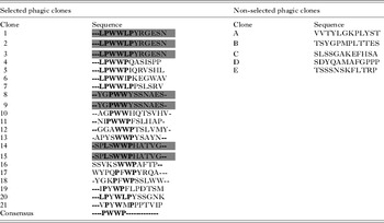
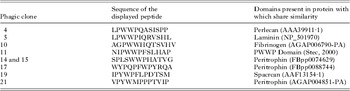
After 5 selection cycles, 21 phage clones positive for binding to the surface of P. berghei ookinetes were sequenced and their DNA sequences translated in silico into amino acid sequences. The 21 selected phage peptides had adjacent residues of tryptophan (W) and proline (P) within their sequence (Table 1), with proline residues flanking tryptophan. Prolines (8/21) were frequently associated with leucine (L) residues. Three groups of complete 12-amino acid peptides were repeatedly observed: LPWWLPYRGESN in clones 1, 2 and 3; YGPWWYSSNAES in clones 8 and 9; and SPLSWWPHATVG in clones 14 and 15 with a consensus sequence PWWP. The latter motif was not present in the 5 sequenced clones from the initial phage pool (Table 1), indicating that this sequence feature was positively selected.
Peptide sequences displayed by selected phages

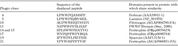
The selected phage peptide sequences showed identity with extracellular proteins and or the peritrophic matrix in the An. gambiae genome (Table 2), and with domains present in a wide variety of proteins that participate in protein-protein interactions (Stec et al. Reference Stec, Nagl, van Ommen and den Dunnen2000).
Peptide sequences displayed by selected phages show homology with proteins with domains typical of mosquito midgut components (intestinal lumen and basal lamina)
Selected phages bind to the ookinete surface
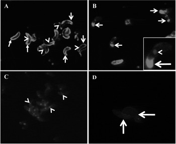
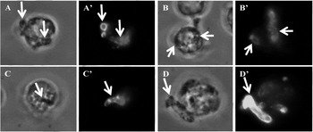
Specific peripheral immunofluorescence was uniformly observed on ookinetes incubated with selected phages (clones 5 and 14, Fig. 1A and B, arrows). In some cases the signal was observed only at one end of the parasite (Fig. 1B, inset and arrow). Ookinetes incubated with non-selected phage clone (C) and/or incubated with secondary antibody only showed no signal (D).
Immunofluorescence detection of selected phages bound to the surface of Plasmodium berghei ookinetes. (A) Phage clone 5 stained with anti-M13 FITC (green, big arrows) at the periphery of the ookinete; nuclei were stained with propidium iodide (red, small arrows). (B) Phage clone 14 binds preferentially to one end of the ookinete as shown by arrows. (C) Negative control, ookinetes incubated with non-selected phage clones. (D) Background fluorescence.

Phage distribution during ookinete invasion of Mos20 cells
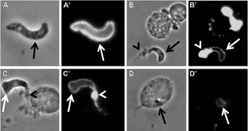
In experiments with ookinetes pre-incubated with phage clones 5 and 14 added to Mos20 cells, samples stained before and after cell permeabilization confirmed that ookinetes pre-incubated with selected phages invaded Mos20 cells (Fig. 3). The parasites interacting with Mos20 cells were devoid of phages, as evidenced by the anti-M13 staining (Fig. 2B', C'and D', arrows). A double staining using an anti-Pbs25, and anti-M13 was performed to track the location of bound phages during Mos20 invasion by ookinetes (Fig. 2). The Pbs25 protein and phages co-localized at one end of these parasites (Fig. 2A', arrow).
Immunofluorescence detection of selected phages (clones 5 and 14) bound to Plasmodium berghei ookinetes after co-culture of Mos20. The phase-contrast images (capitals without apostrophe) have their corresponding fluorescence images (capitals with apostrophe). (A) An ookinete covered with phage clone 5 detected with anti-M13 and anti-Pbs25 (arrow). In (B′) and (C′) ookinetes are shown devoid of phages but positive for Pbs25 (arrows). An intense co-location of both phage clones and Pbs25 can be observed at one pole of the ookinete (arrow heads). In (D) a Mos cell is invaded by an ookinete (arrow).

Ookinetes pre-incubated with selected phages invade Mos20 cells. The phase-contrast images (capitals without apostrophe) have their corresponding fluorescence images (capitals with apostrophe). Ookinetes were pre-incubated with selected phage clone 14 and afterwards with Mos20 cells. A double staining was applied to identify intracellular and extracellular parasites, both probed with anti-Pbs25 antibody. (A) Transverse view of an extracellular ookinete (left arrow) next to a longitudinal view of an intracellular ookinete (right arrow). (B) Two intracellular ookinetes invading the same cell (left and right arrows). (C) An ookinete in the process of invading a Mos cell (arrow). (D) A Mos-20 cell with an extracellular ookinete (arrow).

The selected phages recognized four ookinete proteins
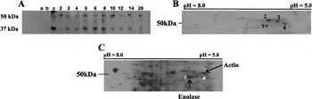
Overlay assays using ookinete proteins separated by 1D gel electrophoresis showed that different selected phage clones (clones 2–6, 8, 10, 12, 14 and 20) recognized the same 2 main bands between ∼37 and ∼50 kDa (Fig. 4A). Using 2D electrophoresis, clone 14 recognized at least 4 proteins in the range of ∼50 kDa (Fig. 4B, 1–4). Only 2 of these proteins were visualized in the 2D gel (1 and 4) (Fig. 4C, arrows). The analysis of these spots by mass fingerprint identified proteins gi|68070065 enolase from Plasmodium berghei and PFL2215w actin I from Plasmodium falciparum 3D7. The orthologue in Plasmo DB version 7.1 is the protein PBANKA_103010 actin II, putative from Plasmodium berghei str. ANKA. The observed masses that matched with actin I were 1961·07, 1193·60, 1177·70, 1123·54, 905·48, 767·44, and 644·07. The masses used for enolase identification were 840·43, 1180·60, 1199·57, 1619·85, 1754·94, 1801·87, 1833·9498 and 1896·94.
Western blot of phage clones bound to ookinete proteins using anti-M13 antibody. (A) SDS–PAGE electrophoresis, selected phages recognized 2 protein bands of 37 and ∼50 kDa. Lanes: (a) no phage; (b) native phage (without peptide); (c) pool of random-selected phages. Numbered lanes correspond to clone number. (B) In 2D electrophoresis, phage clone 14 bound to 4 proteins around 50 kDa. (C) Coomassie-stained 2D gel of ookinete proteins. The arrows indicate 2 proteins recognized by phage clone 14 that were identified as actin and enolase by mass spectrometry.

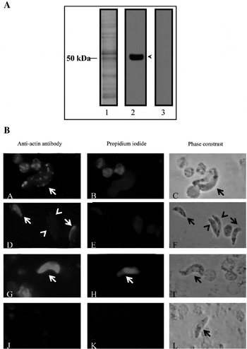
In Western blot analysis of ookinete protein extracts, the anti-actin antibody recognized a band of approximately 47 kDa, corresponding to actin (Fig. 5A, lane 2, arrow head), no signal was observed when ookinete protein extracts were incubated with a mouse irrelevant IgG (Fig. 5A, lane 3). We further characterized actin in P. berghei purified ookinetes, permeabilized and unpermeabilized, and by electron microscopy (EM).
(A) Western blot of ookinetes and anti-actin antibody. Lane 1: ookinete extract stained with Coomassie. Lane 2: ookinete extract probed with anti-actin antibody (1:1000) recognizing a band of approx. 50 kDa (arrow). Lane 3: ookinete extract probed with an anti-rabbit IgG only. Molecular weights are expressed in kilodaltons (kDa). Blot was developed with a chemiluminescence kit. (B) Immunofluorescence analysis of Plasmodium berghei ookinetes with anti-actin antibody. (Ba) Live-fresh ookinetes show patchy actin on their surface (arrows) and do not stain with propidium iodide (Bb). Ookinetes exposed to NH4Cl show patchy actin (Bd, arrow heads), uniform actin (Bd, arrows), and do not stain with PI (Be). Permeabilized ookinetes stained with anti-actin (Bg, arrow) and PI (Bh, arrow). Ookinetes are negative to an irrelevant rabbit IgG (Bj), to incubation with an anti-rabbit-FITC only (data not shown) and to PI (Bk). Images on Bc, f, i and l are the phase-contrast images of Ba, d, g and j, respectively.

Actin is detected on the surface of live, fresh P. berghei ookinetes
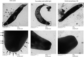
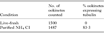
In immunofluorescence assays of live-fresh (intact) ookinetes treated with the anti-actin antibody, 99·37% of parasites depicted a patchy pattern signal (Table 3 and Fig. 5B, panel A, arrow) but were negative to the anti-tubulin antibody. While only 68·37% of purified (NH4Cl-exposed) ookinetes incubated with the same antibody presented this patchy anti- actin signal (Table 3 and Fig. 5B, panel D, arrow heads), 27·71% presented a uniform surface pattern (Fig. 5B, panel D, arrows). Of the NH4Cl exposed ookinetes, 83·3% were stained with the anti-tubulin antibody (see Supplementary Fig. S1, online version only, and Table 4). Live-fresh and purified ookinetes were not stained with propidium iodide (Fig. 5B, panels B and E, respectively). Permeabilized ookinetes stained completely with the anti-actin antibody and propidium iodide (Fig. 5B, panels G and H, respectively), and with the anti-tubulin antibody (Supplementary Fig. S2, online version only). No fluorescence was detected on ookinetes incubated only with an irrelevant rabbit IgG (Fig. 5B, panel K) or an anti-rabbit IgG-FITC (S2). A positive signal (anti-actin gold-labelled antibody) was detected on the surface of ookinetes (Fig. 6A and D, square and arrows), but no signals were detected on ookinetes probed with secondary anti-rabbit IgG (Fig. 6, B and E, respectively) or with an irrelevant primary rabbit IgG (Fig. 6, C and F).
Plasmodium berghei ookinetes probed with anti-actin antibody and evaluated by electron microscopy analysis. (A) P. berghei ookinetes probed with anti-actin. (B) P. berghei ookinetes probed with an anti-rabbit IgG conjugated to gold particles (incubation with a primary antibody omitted). (C) P. berghei ookinetes probed with an irrelevant rabbit IgG. Images in D, E and F are magnifications of A, B and C, respectively.

Percentage of live-fresh and purified ookinetes expressing actin

Percentage of live-fresh and purified ookinetes expressing tubulin

Selected peptides share features with An. gambiae midgut proteins
Several peptides displayed by selected clones, despite not having identical sequences, shared a hydropathicity profile (values based on Kyte and Doolittle, 1981) characterized by 2 maximums separated by a valley of hydropathicity (data not shown). When a homology analysis was conducted in the An. gambiae gene database, searching for proteins with similarity with the selected peptides, a protein with Peritrophin-A domains (AGAP004851-PA) was found (Table 2). This protein (AGAP004851-PA) had a 274 amino acid sequence and a region (positions 213–224) homologous to the peptide displayed by clone 21. The similarity between both profiles was significant (Pearson correlation=0·926, P=0·00004) in a window of 11 amino acids.
DISCUSSION
Using a phage display we identified a PWWP peptide consensus sequence, which bound to the P. berghei ookinete surface. Given that selection was carried out using live ookinetes with presumed unaltered membrane permeability, it was expected that the selected phages bind to the ookinete surface. All selected phage clones that bound to ookinetes share structural features that could reflect some of the structural requirements of native mosquito ligand molecules. The peptide sequences displayed by these phages have sequences and hydropathicity profiles similar to proteins with domains typical of insect peritrophic membrane and basal lamina, such as peritrophin-A (AGAP004851-PA). Therefore, these peptide sequences seem to indicate structural similarities of the phages to the mosquito digestive tract molecules, which interact with ookinete surfaces during their migration. The peritrophin-A domain is found in chitin-binding proteins, particularly in midgut peritrophic matrix proteins of mosquitoes and other insects (Shen and Jacobs-Lorena, Reference Shen and Jacobs-Lorena1998). This structure represents a potential barrier with which the ookinete needs to interact and through which it needs to pass during its journey to the basal lamina.
The displayed peptides have tryptophan and proline residues with relative positions that might be functionally important. Several protein-protein interactions involve sequences less than 10 residues long (Geysen et al. Reference Geysen, Barteling and Meloen1985) and within these the proline residues are essential for the interaction (Kay et al. Reference Kay, Williamson and Sudol2000).
Selected phages bound to ookinetes presented various patterns. In some cases phages were bound only to one end of parasites, indicating that phages interact with components from the apical end of live ookinetes. During ookinete interaction with Mos20 insect cells, tagged phages were displaced from the anterior to the posterior parasite end and shed along with Pbs25. This indicates that the parasite molecules recognized by the phages may be involved in parasite gliding. However, as interacting and intracellular parasites were devoid of phages, it is also possible that the parasite-mosquito cell interaction does not require the molecules recognized by phages.
Two parasite proteins were identified by mass spectrometry assays of the ookinete protein spots: enolase and actin I. Because these are typically intracellular proteins, this finding was unexpected. However, identification has been made in several organisms of surface enolases with what are most likely non-enzymatic functions (Pancholi and Fischetti Reference Pancholi and Fischetti1998; Bergmann et al. Reference Bergmann, Rohde, Chhatwal and Hammerschmidt2001; Pancholi, Reference Pancholi2001; Pitarch et al. Reference Pitarch, Jimenez, Nombela and Gil2006), including apicomplexan parasites (Labbé et al. Reference Labbé, Péroval, Bourdieu, Girard-Misguich and Péry2006; Pal-Bhowmick et al. Reference Pal-Bhowmick, Vora and Jarori2007b). Enolase has been documented in asexual and sexual stages (merozoites, schizonts, gametocytes and sporozoites) of P. falciparum and P. yoelli (Pal-Bhowmick et al. Reference Pal-Bhowmick, Mehta, Coppens, Sharma and Jarori2007a), and our findings indicate that it is also present on the surface of P. berghei ookinetes. Interestingly, this molecule was localized on the merozoite surface, and anti-enolase antibodies blocked erythrocyte infection, indicating that it participates in invasion (Pal-Bhowick et al. Reference Pal-Bhowmick, Vora and Jarori2007b). The participation of enolase in invasion has been documented in Eimeria tenella, where the molecule was localized on the sporozoite apex and appears to be secreted into the medium (Labbé et al. Reference Labbé, Péroval, Bourdieu, Girard-Misguich and Péry2006). The ability of enolase to bind human plasminogen opens the possibility of tethering this molecule (Labbé et al. Reference Labbé, Péroval, Bourdieu, Girard-Misguich and Péry2006), and suggests that it is able to activate matrix metalloproteases (Gong et al. Reference Gong, Hart, Shchurin and Hoover-Plow2008) on the merozoite surface, enabling cell invasion. Similarly, the possibility that enolase recruits vertebrate plasminogen (from the bloodmeal bolus) to the ookinete surface and that it participates in the mosquito midgut invasion warrants investigation.
The interaction of actin with myosin A and myosin light chain (MLC 1) to form a molecular motor for gliding has been well documented in Apicomplexa (Keeley and Soldati, Reference Keeley and Soldati2004). In the current gliding model, the actin-myosin complex (located under the parasite plasma membrane) is anchored through GAP45 (glideosome associated protein 45) to the inner membrane complex. Aldolase functions as a bridge between the submembranous actin and transmembrane adhesins (Jewett and Sibley, Reference Jewett and Sibley2003). These adhesins in Plasmodium belong to the thrombospondin-related Anonymous Protein family (Buscaglia et al. Reference Buscaglia, Coppens, Hol and Nussenzweig2003), represented in ookinetes by CTRP (Lecona et al. Reference Lecona, Rodriguez, Argotte-Ramos, Alvarado-Delgado and Rodriguez2010). The polymerization of actin filaments drives the motile process (Wetzel et al. Reference Wetzel, Håkansson, Hu, Roos and Sibley2003; Ganter et al. Reference Ganter, Schüler and Matuschewski2009).
Besides the submembranous actin, our results (phage overlay, IFI, immuno-electron microscopy) documented the presence of this molecule on the ookinete surface. To rule out the possibility that this atypical location resulted from parasite plasma membrane damage, anti-actin fluorescence was compared on fresh live, NH4Cl treated, and detergent treated parasites. The integrity of fresh parasites was tested by their negative staining with an anti-tubulin antibody and propidium iodide. Fresh parasites presented a patchy pattern of anti-actin staining, while treatment with NH4Cl and permeabilization increased the proportion of parasites with a uniform signal.
Actin has been observed on the surface of different cells, such as lymphocytes, monocytes and endothelial cells (reviewed by Smalheiser, Reference Smalheiser1996), and various functions have been attributed to the surface location of this protein in different systems (Moroianu et al. 1993; Liu et al. Reference Liu, Clarke and Baker2005). The interaction of cells with extracellular matrix components could determine cellular phenotype induction and maintenance by cytoskeleton proteins and integrins (Cody and Wicha, Reference Cody and Wicha1986; Kikkawa et al. Reference Kikkawa, Sanzen, Fujiwara, Sonnenberg and Sekiguchi2000). Accordingly, it has been proposed that the interaction of ookinetes with components of the midgut basal lamina provides possible cues for their transformation into oocysts (reviewed by Sinden, Reference Sinden1999). The fact that Plasmodium ookinetes interact with laminin, a major component of the mosquito basal lamina (Arrighi et al. Reference Arrighi, Ebikeme, Jiang, Ranford-Cartwright, Barrett, Langel and Faye2008), and integrin, probably from midgut epithelial cells, has been documented (Mahairaki et al. Reference Mahairaki, Lycett, Sidén-Kiamos, Sinden and Louis2005; Nacer et al. Reference Nacer, Underhill and Hurd2008). It is possible that the integrin that coats invading ookinetes does so by binding to laminin (Sonnenberg Reference Sonnenberg1993). The ookinete surface proteins P25 (Vlachou et al. Reference Vlachou, Lycett, Sidén-Kiamos, Blass, Sinden and Louis2001), SOAP (Dessens et al. Reference Dessens, Sidén-Kiamos, Mendoza, Mahairaki, Khater, Vlachou, Xu, Kafatos, Louis, Dimopoulos and Sinden2003) and CTRP (Mahairaki et al. Reference Mahairaki, Voyatzi, Siden-Kiamos and Louis2001) with affinity for laminin have been proposed as the parasite counterparts for these interactions. However, SOAP and CTRP are not essential for ookinete transformation into oocysts (Nacer et al. Reference Nacer, Underhill and Hurd2008), and P25 is shed off as the parasite moves, indicating that they are unlikely to function as mediators for the cues leading to oocyst transformation. On the other hand, we could not detect actin on the tracks left by P. berghei ookinetes on a solid substrate during gliding, suggesting that surface actin is not shed off. We propose that actin on the parasite surface could anchor the parasite to the basal lamina and transmit signals for parasite transformation, as could possible interactions of the parasite with transmembrane proteins. Further investigation is needed regarding these possible interactions, as well as the specific routes followed by enolase and actin to the parasite surface.
The identification of molecules involved in Plasmodium-Anopheles interactions is crucial to the development of new strategies that could interrupt malaria transmission by disrupting the establishment of the parasite in the vector. Identification of Plasmodium proteins with functions related to the motility and invasion of the ookinete could enable the development of new transmission blocking strategies capable of controlling malaria.
ACKNOWLEDGEMENTS
The authors thank Robert E. Sinden for critically reviewing the manuscript and for useful suggestions. Thanks to Fidel de la Cruz Hernández-Hernández and Adriana Mendoza Rodriguez for technical help. This work was supported by a CONACYT grant (g-37186M) to M.H.R. and a CONACYT grant (CB200801107006) to M.C.R.