Introduction
Worldwide close to 50 million people are living with dementia with the numbers projected to double almost every 20 years (Prince et al., Reference Prince, Guerchet, Ali, Wu and Prina2015). Alzheimer’s disease (AD) is the predominant cause of dementia and cognitive deficits including deficits in working memory are core features of the illness (Baddeley et al., Reference Baddeley, Bressi, Dellasala, Logie and Spinnler1991). Current treatment of cognitive deficits in mild-to-moderate AD relies primarily on acetylcholinesterase inhibitors (AChEIs) which provide modest symptomatic benefits while causing several adverse effects (Birks, Reference Birks2006). Search for novel treatments has not been successful to date with a failure rate of 99.6% for pharmacological trials (Cummings et al., Reference Cummings, Morstorf and Zhong2014). Studies have explored the effects of noninvasive brain stimulation on cognition in AD using transcranial magnetic stimulation (TMS) (Lee et al., 2016; Sabbagh et al., Reference Sabbagh2019). Still, these studies did not assess mechanisms underlying the improvement in cognition. Thus, there is an urgent need to advance our understanding of the physiological mechanisms underlying cognitive deficits in AD and explore novel interventions for cognitive enhancement.
Synaptic plasticity refers to the use and time-dependent alteration of synapses and is a key mechanism underlying learning and memory (Draganski et al., Reference Draganski, Gaser, Busch, Schuierer, Bogdahn and May2004; Kim and Linden, Reference Kim and Linden2007). Cortical plasticity is critically important for sustaining complex cognitive functions of higher cortical regions such as the dorsolateral prefrontal cortex (DLPFC) (Fuster et al., Reference Fuster, Bodner and Kroger2000). DLPFC is important for the maintenance of executive function that includes the abilities to select, maintain, and manipulate information online, collectively referred to as working memory (Fuster et al., Reference Fuster, Bodner and Kroger2000; Pasupathy and Miller, Reference Pasupathy and Miller2005; Baddeley, Reference Baddeley1996). AD pathology involves the DLPFC early on in the course of illness, and is associated with several neurophysiological changes that cause neurodegeneration and impairment in plasticity (Kaufman et al., Reference Kaufman, Pratt, Levine and Black2012; Rowan et al., Reference Rowan, Klyubin, Cullen and Anwyl2003; Crary et al., Reference Crary, Shao, Mirra, Hernandez and Sacktor2006). DLPFC plasticity is not only important for sustaining executive tasks, but also compensates for neuropathology and dysfunction in other regions secondary to AD pathology (Kaufman et al., Reference Kaufman, Pratt, Levine and Black2012; Voytek et al., Reference Voytek, Davis, Yago, Barcelo, Vogel and Knight2010; Grady et al., Reference Grady, McIntosh, Beig, Keightley, Burian and Black2003). Thus, DLPFC plasticity could be an appropriate potential target and an intermediate marker for interventions aimed at enhancing working memory in AD.
Long-term potentiation (LTP) is considered a prototype of synaptic plasticity and it can be used to assess neuroplasticity in vitro (Malenka and Bear, Reference Malenka and Bear2004; Malenka and Nicoll, Reference Malenka and Nicoll1999). Paired associative stimulation (PAS) is a TMS paradigm that can induce LTP-like activity in the human brain by simulating spike-timing-dependent plasticity protocols (Ziemann et al., Reference Ziemann2008; Vallence and Ridding, Reference Vallence and Ridding2014). PAS-induced LTP-like activity is accomplished by combining electrical stimulation of a peripheral nerve with magnetic stimulation of the contralateral cortex (Ziemann et al., Reference Ziemann2008; Vallence and Ridding, Reference Vallence and Ridding2014). PAS-induced LTP-like activity meets key criteria that define LTP, i.e. input specificity, associativity, cooperativity, and persistence (Stefan et al., Reference Stefan, Kunesch, Cohen, Benecke and Classen2000). While several molecular markers of plasticity are proposed, to our knowledge, there is no evidence of change in synaptic or other molecular markers of plasticity in response to PAS in humans (Geddes et al., Reference Geddes, Wilson, Miller and Cotman1990). However, there may be differential effects of brain-derived neurotrophic factor (BDNF) single-nucleotide polymorphisms on PAS-induced plasticity, with BDNF “Met” allele associated with reduced response to PAS (Cheeran et al., Reference Cheeran2008). Plasticity impairments have been shown in the motor cortex of patients with AD using single session PAS (Battaglia et al., Reference Battaglia2007; Terranova et al., Reference Terranova2013). Further, PAS combined with electroencephalography (EEG) can be used to detect plasticity in the DLPFC (Rajji et al., Reference Rajji2013).
We designed and conducted a randomized controlled trial (RCT) in which a 2-week course of daily repetitive PAS (rPAS) was delivered to patients with early AD and compared to control rPAS for its effect on DLPFC plasticity and working memory performance at post-intervention days 1, 7, and 14 (https://clinicaltrials.gov/ct2/show/NCT01847586). The first aim of this trial was to compare DLPFC plasticity in AD and healthy individuals at baseline and we reported the results of this comparison elsewhere (Kumar et al., Reference Kumar2017). Here, we report the results of the RCT phase of this study. Our primary hypothesis was that active rPAS will result in better DLPFC plasticity post-intervention compared to control rPAS. Our secondary hypothesis was that active rPAS will result in better working memory performance post-intervention compared to control rPAS.
In addition, we used this RCT as a platform to study other neurophysiological mechanisms relevant to plasticity and working memory. Working memory in the DLPFC is supported by local re-entrant neuronal circuits within the DLPFC and re-entrant circuits connecting it to more posterior regions (Buzsaki, Reference Buzsaki2002; Pignatelli et al., Reference Pignatelli, Beyeler and Leinekugel2012). Function of these circuits has been associated with theta and gamma oscillations as measured using EEG (Gevins et al., Reference Gevins, Smith, McEvoy and Yu1997; Howard et al., Reference Howard2003). More specifically, modulation of gamma amplitude by theta phase (“theta–gamma coupling”) has been associated with working memory performance in animal and human studies (Lisman and Idiart, Reference Lisman and Idiart1995; Rajji et al., Reference Rajji, Zomorrodi, Barr, Blumberger, Mulsant and Daskalakis2017). Impaired theta–gamma coupling was associated with impaired cognition in a mouse model of AD (Stoiljkovic et al., Reference Stoiljkovic, Kelley, Horvath and Hajos2018). In humans, theta–gamma coupling predicted impairments in working memory in patients with mild cognitive impairment (Goodman et al., Reference Goodman2018). Thus, we also report on the impact of rPAS on theta–gamma coupling during working memory performance.
Materials and methods
This study was conducted at the Centre for Addiction and Mental Health (CAMH), a teaching hospital at the University of Toronto. CAMH Research Ethics Board approved the study in accordance with the declaration of Helsinki. All participants provided their informed written consent. The trial was registered at Clinicaltrials.gov # NCT01847586.
Participants and clinical and cognitive assessments
Participants were recruited from CAMH and other collaborating hospitals in Toronto from May 2013 to October 2016. To be eligible, they had to meet the criteria for probable AD following the National Institute of Neurological and Communicative Disorders and Stroke, and the Alzheimer’s Disease and Related Disorders Association (NINCDS-ADRDA) criteria (Dubois et al., Reference Dubois2007); score 17 or above on the Mini-Mental State Examination (MMSE) (Folstein et al., Reference Folstein, Folstein and McHugh1975); either not be taking an AChEI or be on a stable dose for at least 3 months; and not have any contraindication for TMS. Participants were assessed using the NINCDS-ADRDA criteria and the Structured Clinical Interview for DSM-IV (First, Reference First2002) to verify the diagnosis and rule out exclusionary psychiatric illnesses. They also underwent a thorough clinical assessment by a study psychiatrist. All participants underwent assessment of cognition using MMSE, Repeatable Battery for the Assessment of Neuropsychological Status (RBANS) (Randolph et al., Reference Randolph, Tierney, Mohr and Chase1998), and Executive Interview (EXIT) (Royall et al., Reference Royall, Mahurin and Gray1992) at baseline. Mood was assessed at baseline using the Cornell Scale for Depression in Dementia (CSDD) (Alexopoulos et al., Reference Alexopoulos, Abrams, Young and Shamoian1988).
Sample size calculation
In a previous study, we observed an effect size, Cohen’s d = 1.4, between active and sham PAS conditions (Rajji et al., Reference Rajji2013). We estimated that a sample of 32 (16 in each arm) will provide us with 80% power to detect a time × condition interaction for acute PAS effects at post-rPAS day 1 for an effect size of Cohen’ d = 1.02 at alpha = 0.05.
Plasticity, working memory, and theta–gamma coupling assessments
Baseline DLPFC plasticity was assessed using PAS-EEG as previously described (Kumar et al., Reference Kumar2017). EEG recordings were done using the TMS-EEG protocol through a 64-channel Synamps 2 Neuroscan EEG system. Electrodes were placed as per 10–20 International System using an EEG cap, and the impedance at each electrode was set at ≤5 kΩ. EEG signals were recorded using DC and a low-pass filter of 100 Hz at a 20 kHz sampling rate as per protocol in our published TMS-EEG experiments (Rajji et al., Reference Rajji2013; Daskalakis et al., Reference Daskalakis, Farzan, Barr, Maller, Chen and Fitzgerald2008). Working memory was assessed using N-back task (Kumar et al., Reference Kumar2017). Only 1- and 2-back tasks were used because AD participants were not able to perform the 3-back or more difficult conditions. All participants were offered both tasks. A prime (A′) was used as the outcome measure for N-back task as it takes into account both the hit rate and false alarm rate (Kumar et al., Reference Kumar2017). To assess baseline, theta–gamma coupling EEG was also recorded while participants were performing the N-back task (Goodman et al., Reference Goodman2018). Assessments of DLPFC plasticity using PAS-EEG, of working memory using N-back task, and of theta–gamma coupling using EEG during the N-back task were repeated at days 1, 7, and 14 after the rPAS intervention.
Randomization
Participants were randomized 1:1 in a double-blinded manner using a balanced random assignment to 10 sessions (daily, 5 days per week) of active or control rPAS (further details below). Randomization sequence was generated on the computer by assigned staff who was not involved in any other study procedures. Intervention setup was done by a staff person not involved in the study to keep the interventionists blinded. Participants, interventionists, assessors, and investigators remained blinded to the treatment allocation.
Intervention
Active rPAS consisted of a repetitive pairing of electrical stimulation of the median nerve at the right wrist (180 pulses at 0.1 Hz) with TMS of the contralateral left DLPFC with an interstimulus interval of 25 ms. An identical procedure was followed for control rPAS except that the interstimulus interval was 100 ms, which has been shown not to induce LTP-like activity (Rajji et al., Reference Rajji2013). Site of stimulation at the DLPFC was localized using the MRIcro/reg software and the MINIBIRD system (Ascension Technologies, USA) as previously described (Daskalakis et al., Reference Daskalakis, Farzan, Barr, Maller, Chen and Fitzgerald2008). TMS pulses were delivered at stimulus strength sufficient to induce a peak-to-peak 1 millivolt motor-evoked potential using a 7 cm figure-of-eight coil and a Bistim module (Magstim Company Ltd., UK). Median nerve stimulation was delivered at electrical stimulus strength equivalent to 300% of the participants’ sensory threshold. Participants were asked to keep a count of the sensory stimuli and were randomly asked about the count to focus their attention on the stimulus which has been shown to be important for LTP induction using PAS (Rajji et al., Reference Rajji2013).
Data processing
EEG data was processed offline using MATLAB (The MathWorks Inc., USA) and the EEGLAB toolbox using published methods (Kumar et al., Reference Kumar2017; Rajji et al., Reference Rajji, Zomorrodi, Barr, Blumberger, Mulsant and Daskalakis2017). For TMS-EEG analyses, the data were downsampled to 1000 Hz and segmented from −1000 to 2000 ms relative to the onset of TMS pulse, baseline corrected, re-segmented, and then digitally filtered using the second order, Butterworth, zero-phase shift 1–55 Hz band-pass filter. EEG signals from pre-PAS, and 0, 17, and 34 min post-PAS were concatenated, and then cleaned using a combination of manual and automated techniques (Kumar et al., Reference Kumar2017). EEG data was then re-referenced to the average electrode for further analysis. DLPFC plasticity was calculated as potentiation of cortical-evoked activity (hereafter referred to as PAS-LTP), defined as the maximum ratio of post/pre-PAS cortical-evoked activity between 50 and 275 ms post-TMS pulse (Kumar et al., Reference Kumar2017).
For N-back EEG analyses, data were filtered and segmented from −1400 to +3100 ms relative to the stimulus onset, and then cleaned using a combination of automated and manual methods as described above (Rajji et al., Reference Rajji, Zomorrodi, Barr, Blumberger, Mulsant and Daskalakis2017; Goodman et al., Reference Goodman2018). Then, we filtered the raw EEG data for theta (4–7 Hz) and gamma (30–50 Hz) frequencies with second-order zero-phase shift filter and created the time series for gamma amplitude and theta phase using the Hilbert transform. Subsequently, we created a concatenated signal of 5000 ± 150 ms separately for different N-back trial types (target correct, target not correct, nontarget correct, and nontarget not correct) and conditions (1- and 2-back) at each electrode. All included epochs had to include the time interval from the stimulus onset to the time of response. We used modulation index (MI) as the measure of theta–gamma coupling (Rajji et al., Reference Rajji, Zomorrodi, Barr, Blumberger, Mulsant and Daskalakis2017; Goodman et al., Reference Goodman2018; Tort et al., Reference Tort, Komorowski, Eichenbaum and Kopell2010). To calculate MI, each phase of theta was binned into 18 intervals of 20 ° each. The average amplitude of gamma at each theta bin was calculated and normalized, resulting in phase–amplitude distribution function. We then calculated the MI for each electrode by measuring the divergence of the observed amplitude distribution from a uniform distribution (Rajji et al., Reference Rajji, Zomorrodi, Barr, Blumberger, Mulsant and Daskalakis2017; Goodman et al., Reference Goodman2018; Tort et al., Reference Tort, Komorowski, Eichenbaum and Kopell2010). Finally, MI during target trials was calculated as a weighted average based on the number of correct and incorrect responses for all target trials.
Statistical analyses
All data were analyzed using the Statistical Program for Social Sciences (SPSS) version 24.0 (SPSS Inc., Chicago, IL, USA). Data distribution was examined using box plots. Data were transformed to Ln distribution to achieve normality as needed. χ2 and independent samples t-tests were employed to evaluate differences between the active and control rPAS groups on demographic variables. Further analyses were conducted using repeated-measures analysis of variance (ANOVA) to compare DLPFC plasticity, working memory, and theta–gamma coupling across post-days 1, 7, and 14 after the intervention. For DLPFC plasticity, repeated-measure ANOVA was conducted with PAS-LTP as dependent variable, intervention group (active and control rPAS) as between-subject variable and time points (baseline, post-intervention days 1, 7, and 14) as within-subject independent variables. For working memory, separate repeated-measures ANOVAs were conducted for 1- and 2-back conditions with A′ as dependent variable, intervention group (active and control rPAS) as between-subject variable and time points (baseline, post-intervention days 1, 7, and 14) as within-subject independent variables. Following the same procedure, separate repeated-measures ANOVAs were carried out with MI during 1- and 2-back tasks as dependent variables to compare theta–gamma coupling between the groups across time. Additionally, to generate hypotheses for future research, we calculated simple main effects of time in both groups and conducted post hoc within-group analyses using independent sample and paired sample t-tests to compare DLPFC plasticity (PAS-LTP), working memory (A′), and theta–gamma coupling (MI) between baseline and post-intervention days 1, 7, and 14 across and within the groups. Effect sizes were calculated using Cohen’s d using G * Power and confidence intervals were calculated using established methods (Faul et al., Reference Faul, Erdfelder, Lang and Buchner2007; Smithson, Reference Smithson2003). Pooled pre–post-standard deviation and paired group correlations were used to calculate effect sizes for paired t-tests. Further, we conducted exploratory subgroup analyses after selecting participants based on the degree of cognitive impairment (MMSE ≤ 24), executive function impairment (EXIT interview score ≥15), and use of AChEIs, and compared the plasticity, working memory performance (2-back), and theta–gamma coupling during working memory performance between the active and control groups at baseline and post-day 1 using independent sample t-tests. Finally, we examined the relationship between DLPFC plasticity, working memory, and theta–gamma coupling using Pearson’s correlation. For all analyses, the level of statistical significance was set at α = 0.05.
Results
Demographic and baseline characteristics
Thirty-two AD participants were included out of which 16 (females = 9, mean (SD) age = 76.5 (6.8)) were randomized to active rPAS and 16 (females = 7, mean (SD) age = 76.4 (6.0)) to control rPAS (Figure 1). There were no significant differences between the two groups at baseline in age, gender, education, or cognition as assessed by MMSE, RBANS, and EXIT, or mood symptoms as assessed by CSDD. The two groups did not differ in baseline resting motor threshold, baseline pre-PAS cortical-evoked activity, DLPFC plasticity, attention during PAS (assessed by the difference between participant’s count of sensory stimuli during PAS and the actual number of sensory stimuli), 1- and 2-back tasks, or theta–gamma coupling during N-back. Only the sensory threshold at the wrist was lower in the active rPAS group than in the control rPAS group at baseline (Table 1). All 16 participants in each group performed 1-back task at baseline and follow-up points. In the active group, 15 participants performed the 2-back task at baseline, 14 at post-day 1, 15 at post-day 7, and 15 at post-day 14. In the control group, 13 participants performed the 2-back task at baseline, 14 at post-day 1, 13 at post-day 7, and 13 at post-day 14.
Consort chart showing recruitment and flow of participants in the study.

Baseline participant characteristics including demographics, cognitive status, and neurophysiological characteristics

Abbreviations: PAS, Paired Associative Stimulation with interstimulus interval = 25 ms (active condition); C-PAS, Control rPAS condition with interstimulus interval = 100 ms; M, Male; F, Female; MMSE, Mini-Mental State Examination (score out of 30, a higher score indicates better performance); CSDD: Cornell Scale for Depression in Dementia (score ranges from 0 to 38, a higher score indicates worse depression); EXIT, Executive Interview (score ranges from 0 to 50, a higher score indicates worse performance); RBANS, Repeatable Battery for the Assessment of Neuropsychological Status (score ranges from 40 to 160, a higher score means better performance); PAS-LTP, Ratio of post-PAS to pre-PAS cortical-evoked activity, a measure of long-term potentiation-like activity.
* Natural log of modulation index, the measure of theta–gamma coupling.
** Motor threshold is expressed in terms of the percentage of maximum TMS machine output.
*** Statistically significant, for all statistical tests, the level of significance was set for α = 0.05.
DLPFC plasticity
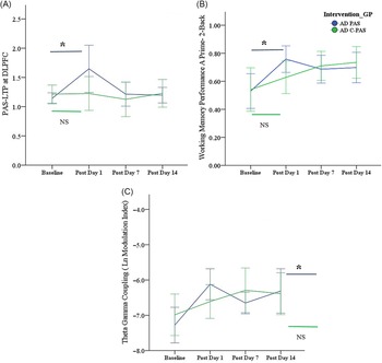
On the primary analysis, there was no significant group × time interaction for DLPFC plasticity (PAS-LTP) (F 3,90 = 1.9, p = 0.14). There was a significant simple main effect of time on DLPFC plasticity only for the active rPAS group (F 3,45 = 3.65, p = .019, partial η 2 = 0.20). However, on post-rPAS day 1, there was no significant difference in PAS-LTP between the active (mean (SD) = 1.65 (0.81)) and control group (mean (SD) = 1.22 (0.58), t = 1.7, df = 30, p = 0.1, Cohen’s d = 0.6, 95% CI [–0.11, 1.30]). Further, post hoc within-group comparisons showed that only the active rPAS group experienced a significant increase in DLPFC plasticity from baseline to post-rPAS day 1 (t = 2.27, df = 15, p = 0.038, Cohen’s d = 0.7), while there was no such change in DLPFC plasticity in the control rPAS group (t = 0.06, df = 15, p = 0.954, Cohen’s d = 0.02). There were no within-group differences between DLPFC plasticity at baseline versus any other time points (post-intervention days 7 and 14) in either group (Figure 2A).
Changes in DLPFC plasticity, working memory, and theta–gamma coupling in active and control rPAS groups. A. Line diagram showing DLPFC plasticity as assessed at the site of stimulation (DLPFC) using PAS, across baseline and post-intervention days 1, 7, and 14. PAS-LTP is defined as the ratio of post-PAS to pre-PAS cortical-evoked activity. Active rPAS group received a 2-week course of rPAS with ISI = 25 ms, control rPAS group received a 2-week course of rPAS with ISI = 100 ms. B. Line diagram showing working memory performance (2-back task) in active and control rPAS groups at baseline and at post-intervention days 1, 7, and 14. Y-axis represents A prime (A′) which takes into account the hit rate and false alarm rate and is corrected for extreme values. C. Line diagram showing theta–gamma coupling assessed from electroencephalography (EEG) recorded during the working memory task (2-back task) and represented as natural log of modulation index in active and control rPAS groups at baseline and at post-intervention days 1, 7, and 14. Y-axis represents modulation index. Abbreviations: DLPFC, Dorsolateral Prefrontal cortex; PAS, Paired Associative Stimulation; LTP, Long-Term Potentiation; ISI, Interstimulus Interval (between median nerve stimulation and DLPFC stimulation); NS – not statistically significant. Error bars represent +/− 2 standard error. * – Statistically significant (p < 0.05).

Working memory
There was no significant group × time interaction for working memory performance on 2-back (F 3,54 = 0.9, p = 0.444) or 1-back (F 3,66 = 0.3, p = 0.824) tasks with A′ as dependent variable and the intervention groups and follow-up time points as independent variables. There was a significant simple main effect of time on 2-back performance only for the active rPAS group (F 3,27 = 3.74, p = .023, partial η 2 = 0.29). Again, on post-rPAS day 1, there was no significant difference between working memory (2-back task) performance between the active rPAS (mean (SD) A′ = 0.75 (0.16)) and control rPAS group (mean (SD) A′ = 0.63 (0.20), t = 1.7, df = 21, p = 0.102, Cohen’s d = 0.7, 95% CI [-0.14, 1.55]). However, on post hoc within-group analyses for performance on the 2-back task, only the active rPAS group showed improvement at post-rPAS day1 as compared to baseline (t = 2.3, df = 10, p = 0.043, Cohen’s d = 0.7), while there was no such change in the control rPAS group (t = 0.5, df = 10, p = 0.7, Cohen’s d = 0.2). The control rPAS group showed improvement in 2-back performance only at post-day 14 as compared to baseline (t = 2.4, df = 11, p = 0.033) (Figure 2B). There was no change in 1-back performance across time in either group.
Theta–Gamma coupling
There were no group × time interactions for theta–gamma coupling (MI) during either 2-back (F 3,48 = 1.6, p = 0.21) or 1-back, F 3,78 = 1.4, p = 0.3) tasks. Similar to what we observed for DLPFC plasticity and working memory performance, there was a significant simple main effect of time on theta–gamma coupling during 2-back performance only for the active rPAS group (F 3,27 = 4.88, p = 0.008, partial η 2 = 0.35). There was no significant difference between theta–gamma coupling (2-back task) between the active rPAS (mean (SD) Ln MI = −6.1(0.8)) and control rPAS group (mean (SD) Ln MI = −6.6 (0.8), t = 1.5, df = 23, p = 0.2, Cohen’s d = 0.6, 95% CI [–0.21, 1.40]) (Figure 2C). Again, on post hoc tests, there was an enhancement of theta–gamma coupling during the 2-back task in the active rPAS group at post-day 1 as compared to baseline (t = 2.9, df = 11, p = 0.02, Cohen’s d = 0.9), and not in the control group (t = 0.9, df = 8, p = 0.4, Cohen’s d = 0.3). Theta–gamma coupling enhancement during the 2-back task was also noted at post-day 14 in both active (t = 2.3, df = 10, p = 0.043) and control groups (t = 3.2, df =8, p = 0.013).
There was no change in theta–gamma coupling across time during the 1-back task in either of the groups, which is similar to the fact that the two groups did not also experience any change in performance on the 1-back task.
Exploratory subgroup analyses
Twenty-five participants had MMSE ≤ 24, out of which 12 were randomized to active rPAS. There were no differences between the active and control groups at baseline, but on post-day 1, active rPAS group had better 2-back performance (mean (SD) A′ = 0.82 (0.12)) as compared to the control group (mean (SD) A′= 0.59 (0.19), t = 3.05, df = 16, p = 0.008). There were no differences between the groups on DLPFC plasticity or theta–gamma coupling. Further, among participants with MMSE > 24 or those selected based on EXIT interview scores, there were no differences between the active and control groups on plasticity, working memory or theta–gamma coupling. Seventeen participants were taking AChEIs, out of which nine were randomized to active rPAS. There were no differences between the groups on DLPFC plasticity or working memory performance. For theta–gamma coupling, there were no differences between the active and control groups at baseline, however, on post-day 1, the active rPAS group had higher theta–gamma coupling (mean (SD) Ln MI = −5.9 (0.48)) as compared to the control group (mean (SD) Ln MI = −6.7 (0.65), t = 2.43, df = 10, p = 0.04). Among participants not taking AChEIs, there were no differences between the active and control groups on plasticity, working memory, or theta–gamma coupling.
Relationships among DLPFC plasticity, working memory, and theta–gamma coupling
There was a significant positive correlation between working memory performance (A′) and theta–gamma coupling (MI) during the working memory task, with both groups analyzed together. Pearson’s correlation analyses showed a significant positive correlation between 1-back A′ and Ln MI during 1-back at baseline (r = 0.6, n = 31, p < 0.001), post-day 1 (r = 0.5, n = 28, p = 0.006), post-day 7 (r = 0.8, n = 29, p < 0.001), and post-day 14 (r = 0.7, n = 26, p < 0.001).
Similarly, there was a positive correlation between 2-back A′ and Ln MI during 2-back at baseline (r = 0.5, n = 25, p = 0.003), post-day 7 (r = 0.4, n = 23, p = 0.03), and post-day 14 (r = 0.5, n = 21, p = 0.009) (Figure 3). There was no significant correlation between 2-back A′ and Ln MI at post-day 1 (r = 0.3, n = 22, p = 0.09).
Scatterplot showing the correlation between working memory performance (Y-axis – as assessed by N-back tasks 2-back condition, and represented as A′) and theta–gamma coupling (X--axis – assessed from EEG recorded during the working memory task, and represented as natural log of modulation index – Ln MI). Pearson’s correlation, r = 0.5, n = 25, p = 0.003.

There were no significant correlations between working memory performances and DLPFC plasticity or between theta–gamma coupling and DLPFC plasticity.
Adverse effects
There were no serious adverse events in either arm and there were no early dropouts in either of the two groups. There were 11 adverse events in the active rPAS group and 7 in the control rPAS group. Two participants experienced sleep problems in the active rPAS group and none in the control group. There was one instance of transient blurry vision and one instance of transient muscle weakness in the active group with no such events reported in the control group. These events did not happen during or immediately following the rPAS sessions and were determined to be not related to PAS. Please see Table 2 for details of adverse events in both groups.
Details of adverse events experience by participants in active and control rPAS groups

Discussion
This was a pilot randomized double-blind-controlled study to investigate the effects of rPAS delivered to the DLPFC on DLPFC plasticity and working memory in patients with AD. The successful completion of rPAS course by all randomized participants and lack of any serious adverse events shows that the intervention was well tolerated. The study was negative on primary outcome measures in terms of detecting differences between active and control rPAS groups. However, within-group analyses show promising results, mainly that right after the intervention (i.e. post-day 1), active rPAS and not control rPAS, results in enhanced DLPFC plasticity, working memory performance on 2-back, and theta–gamma coupling during 2-back performance. After post-day 1, and without any booster rPAS sessions, the improvement in DLPFC plasticity does not persist while the improvement in working memory and theta–gamma coupling becomes more variable. Our post hoc analyses also showed that changes in working memory performances parallel changes in theta–gamma coupling at all time points for both groups (except for one time point for the active group) and that these two measures are strongly correlated, providing further support to the role of theta–gamma coupling in working memory.
To our knowledge, this is the first study to investigate the effects of DLPFC rPAS on DLPFC plasticity and working memory in patients with AD. One small study in nine healthy volunteers showed that a modified rPAS protocol targeted at the motor cortex can result in motor cortex reorganization (McKay et al., Reference McKay, Ridding, Thompson and Miles2002). Several small studies have reported beneficial effects of repetitive TMS (rTMS) applied to DLPFC and other brain regions on cognitive function in AD; however, these studies did not assess DLPFC plasticity (Lee et al., 2016; Sabbagh et al., Reference Sabbagh2019; Bentwich et al., Reference Bentwich2011; Rabey et al., Reference Rabey, Dobronevsky, Aichenbaum, Gonen, Marton and Khaigrekht2013; Liao et al., Reference Liao2015; Dong et al., Reference Dong2018). Notwithstanding the possibility that rPAS is not effective in enhancing plasticity or working memory, several factors could have contributed to not finding a significant effect. First, this was designed as a small pilot study with no prior pilot data in AD to adequately estimate the sample size needed to detect the effect. The study was powered to detect a large effect size (Cohen’s d = 1.02), whereas the observed between-group effect sizes were moderate and nonsignificant (for plasticity, Cohen’s d = 0.6, 95% CI [–0.11, 1.30] and for working memory, Cohen’s d = 0.7, 95% CI [–0.14, 1.55]). Second, the primary outcome of this pilot study was to determine whether rPAS could enhance DLPFC plasticity. Thus, rPAS was delivered unilaterally to the left DLPFC. One could argue that to enhance working memory, and possibly plasticity, bilateral rPAS should have been delivered. Third, the variable results after post-day 1 could be due to the lack of ongoing or at least booster rPAS sessions. The goal of having these assessments was in fact to assess the durability of any effect without any booster sessions.
Implications of the preliminary analyses for future research
Our preliminary finding of within-group improvement in DLPFC plasticity and working memory with active rPAS may have important implications for future research. It has been shown that environmental enrichment can promote neurogenesis and LTP in the hippocampus of AD mice (Hu et al., Reference Hu, Xu, Pigino, Brady, Larson and Lazarov2010). Further, it has been proposed that exercise can have positive effects on brain plasticity based on the measurement of indirect markers of plasticity such as BDNF and neurotrophic gene expression (Rolland et al., Reference Rolland, Abellan van Kan and Vellas2008). It has also been shown postmortem that the brains of AD patients may be capable of mounting an adaptive plastic response (Geddes et al., Reference Geddes, Monaghan, Cotman, Lott, Kim and Chui1985). Some recent studies have shown the importance of frontal brain regions for apathy and other behavioral symptoms in AD (Padala et al., Reference Padala, Padala, Samant and James2020; Nowrangi et al., Reference Nowrangi2020). Our findings of within-group plasticity and working memory enhancement in the active rPAS group support future use of brain stimulation interventions aimed at frontal brain regions to enhance plasticity, cognition, and behavior in patients with AD. Our results also support the investigation of rPAS with alternative sites (such as bilateral stimulation) or parameters (potentially longer duration) or additional booster sessions and testing these paradigms in larger samples stratified by their cognitive status and other clinical variables such as behavioral symptoms and use of AChEIs.
Our exploratory findings of enhanced theta–gamma coupling in the active rPAS group along with enhanced working memory and of robust correlations between theta–gamma coupling and working memory performance may have important implications for a mechanistic understanding of working memory and cognition in AD. Diagnosis and treatment of dementia remains challenging and may lead to fewer people seeking help for dementia (Poole et al., Reference Poole, Wilcock, Rait, Brodaty and Robinson2020; Parker et al., Reference Parker, Barlow, Hoe and Aitken2020). A better understanding of biomarkers underlying cognition is the key to enhance diagnostic accuracy and develop novel treatment interventions. Hippocampal theta–gamma coupling has been associated with memory performance in animal models and humans with surgically implanted electrodes (Tort et al., Reference Tort, Scheffer-Teixeira, Souza, Draguhn and Brankack2013; Lega et al., Reference Lega, Burke, Jacobs and Kahana2016). Impaired theta–gamma coupling and its association with cognition has been shown in a mouse model of AD linking it with AD pathology (Stoiljkovic et al., Reference Stoiljkovic, Kelley, Horvath and Hajos2018). A recent study in older healthy adults showed that enhancing theta–gamma coupling using transcranial Alternating Current Stimulation resulted in enhanced working memory and there was an association between changes in theta–gamma coupling and changes in working memory (Reinhart and Nguyen, Reference Reinhart and Nguyen2019). Thus, our findings suggest that theta–gamma coupling may be used as an intermediate biomarker of working memory performance and as a potential target for cognitive-enhancing interventions in AD.
Limitations
The following are additional limitations of this study. First, we screened 109 patients to successfully recruit 32 participants (Figure 1). The top reason for failing screen was the travel and time commitment to the study, which raises the importance of adapting noninvasive brain stimulation to less mobile populations such as AD. In contrast, the retention rate was excellent in our study, demonstrating the high tolerability of noninvasive brain stimulation in AD. Second, 17/32 participants in our study were taking cognitive enhancer medications (Table 1). While this could have contributed to variability overall, it is unlikely to have confounded the working memory or DLPFC plasticity results between groups as the distribution was similar between two groups. Third, we relied on the clinical diagnosis of AD and did not include pathologic markers of AD. Fourth, we did not correct for coil-to-cortex distance to factor in cortical atrophy to determine the intensity of DLPFC stimulation using TMS. However, the intensity of stimulation was individualized by assessing the TMS intensity required to produce a 1 millivolt motor-evoked potential. Finally, this study did not include participants with mild cognitive impairment who could be more amenable to the enhancement of DLPFC plasticity and working memory owing to the earlier stages of illness.
Further directions and potential for translation to a treatment approach
Preliminary findings of our study highlight the need for further research into the effects of rPAS in AD before its translation into clinical care. Still, our findings suggest that using rPAS to enhance cognition in a group of patients with an objectively defined cognitive impairment could result in enhanced cognition. Future well-powered studies targeting such a population are needed, and if successful, additional studies should assess the effects of rPAS on other biological markers and behavioral symptoms of AD and as well as focus on overcoming barriers to implementation of rPAS for clinical use. Future studies should also examine potential relationships between working memory, DLPFC plasticity, and theta–gamma coupling.
Conclusions
This study was negative on the primary outcome and did not show significant differences between active and control rPAS groups with respect to DLPFC plasticity or working memory performance at post-intervention days 1, 7, or 14. Exploratory within-group analyses across time, conducted to generate hypotheses for future research, detected a moderate-to-large effect size for improved DLPFC plasticity, working memory performance, and theta–gamma coupling acutely post-intervention only in the active rPAS group. There was also a robust positive correlation between working memory performance and theta–gamma coupling. Finally, the rPAS intervention was well tolerated without any serious adverse effects. These results indicate the need for future studies to investigate the effect of rPAS in AD with more intensive protocols in larger samples and to also include populations at earlier stages of the illness before the onset of dementia.
Conflict of interest
The authors have no conflicts of interest to report related to this work.
Source of funding
This work was supported by the Weston Brain Institute (RR120070 to T.K.R.), in part by the Canada Research Chairs program (950-230,879 to T.K.R.), research support from Canada Foundation for Innovation (CAM-14-002 to T.K.R.), Centre for Addiction and Mental Health (CAMH) (2014 Fellowship Award to S.K.), University of Toronto (2018 Academic Scholars Award to S.K.), and in-kind support from the Temerty Centre for Therapeutic Brain Intervention at CAMH.
Description of authors’ roles
Sanjeev Kumar – Resources, Data curation, Formal analysis, Investigation, Methodology, Project administration, Validation Writing – original draft, Writing – review and editing.
Reza Zomorrodi – Data curation, Investigation, Methodology, Formal analysis, Writing – review and editing.
Zaid Ghazala – Data curation, Investigation, Methodology, Project administration, Writing – review and editing.
Michelle S. Goodman – Data curation, Investigation, Methodology, Formal analysis, Writing – review and editing.
Daniel M. Blumberger – Conceptualization, Project administration, Supervision, Writing – review and editing.
Zafiris J. Daskalakis – Conceptualization, Project administration, Supervision, Writing – review and editing.
Corinne E. Fischer – Supervision, Resources, Writing – review and editing.
Benoit H. Mulsant – Conceptualization, Project administration, Supervision, Writing – review and editing.
Bruce G. Pollock – Conceptualization, Project administration, Supervision, Writing – review and editing
Tarek K. Rajji – Conceptualization, Funding acquisition, Resources, Data curation, Formal analysis, Methodology, Project administration, Supervision, Validation, Writing – original draft, Writing – review and editing.
Acknowledgments
Dr. Blumberger has received research support from the Canadian Institutes of Health Research (CIHR), National Institute of Health (NIH), Brain Canada, and the Temerty Family through the CAMH Foundation and the Campbell Research Institute. He received research support and in-kind equipment support for an investigator-initiated study from Brainsway Ltd., and he is the principal site investigator for three sponsor-initiated studies for Brainsway Ltd. He received in-kind equipment support from Magventure for investigator-initiated research. He received medication supplies for an investigator-initiated trial from Indivior. He has participated in an advisory board for Janssen. In the last 5 years, Dr. Daskalakis has received research and equipment in-kind support for an investigator-initiated study through Brainsway Inc. and Magventure Inc. His work was supported by the Ontario Mental Health Foundation (OMHF), the CIHR, the NIH, and the Temerty Family and Grant Family and through the CAMH Foundation and the Campbell Family Institute. Corinne Fischer received grant funding from Roche Pharmaceuticals and is the North American PI for a device trial sponsored by Vielight Inc. Dr. Kumar has received grant support from Brain Canada, NIH, Brain and Behavior Foundation (NARSAD), Weston Brain Institute, and Canadian Centre for Ageing and Brain Health Innovation and in-kind equipment support from Soterix Medical Inc. During the past 5 years, Dr. Mulsant has received research funding from Brain Canada, the CAMH Foundation, the CIHR, and the NIH; research support from Bristol-Myers Squibb (medications for an NIH-funded clinical trial), Eli-Lilly (medications for an NIH-funded clinical trial), Pfizer (medications for an NIH-funded clinical trial), Capital Solution Design LLC (software used in a study funded by CAMH Foundation), and HAPPYneuron (software used in a study funded by Brain Canada). He directly owns the stocks of General Electric (less than $5,000). Dr. Rajji has received research support from Brain Canada, Brain and Behavior Research Foundation, BrightFocus Foundation, Canada Foundation for Innovation, Canada Research Chair, CIHR, Centre for Aging and Brain Health Innovation, NIH, OMHF, Ontario Ministry of Research and Innovation, and the Weston Brain Institute. Dr. Rajji also received in-kind equipment support for an investigator-initiated study from Magstim, and in-kind research accounts from Scientific Brain Training Pro. The remaining authors have no potential conflicts of interest to declare.