Acquisition of or colonization with pathogens during hospitalization increases the risk of later infection.Reference Huang, Diekema and Warren 1 The microbial environment in patient care settings plays an important role and may be a reservoir leading to the transmission of pathogens.Reference Chemaly, Simmons and Dale 2 Environmental interventions have demonstrated improvement in the bacterial contamination levelReference Stibich, Stachowiak and Tanner 3 as well as reduction in hospital acquired infections (HAIs).Reference Levin, Riley, Parrish, English and Ahn 4 – Reference Haas, Menz, Dusza and Montecalvo 6 New tools are needed to better understand the interplay of the environment, environmental interventions, and risk of nosocomial transmission and/or infection.
We examined the occurrence of genetically indistinguishable strains of vancomycin-resistant enterococci (VRE) from patients who were negative on admission but subsequently tested positive during weekly screenings on the leukemia and hematopoietic stem cell transplant (HCT) units at the University of Texas MD Anderson Cancer Center in Houston, Texas. An analysis of temporal and spatial patterns of strain repetition provided an improved understanding of the patterns of transmission and an improved means of evaluating interventions.
METHODS
Sampling
We targeted the leukemia unit (39 beds) and HCT unit (52 beds) which each spanned 2 floors between January 1, 2013, and December 31, 2013, with a combined 65,811 patient days. All patients admitted into these units were screened for VRE on admission with a rectal swab and weekly thereafter until discharge, according to our routine infection control practice. If the culture was positive for VRE within 48 hours of admission, the patient was considered to have community-acquired VRE; if the culture became VRE positive after 48 hours following admission, the patient was considered to have acquired VRE. After receiving approval from the institution’s Quality Improvement Assessment Board, the recovered isolates were saved and analyzed by polymerase chain reaction (PCR).
PCR
Repetitive sequence-based polymerase chain reaction (REP-PCR) strain typing has been used in other studies to discriminate isolates as genetically identical.Reference Svec, Vancanneyt and Seman 7 – Reference Higgins, Dammhayn, Hackel and Seifert 9 To perform REP-PCR using a UltraClean Microbial DNA Isolation Kit (MO-BIO Laboratories, Solana Beach, CA), genomic DNA was extracted from 1 µL of organism. A working concentration of 25–50 ng/mL DNA was verified using a NanoDropTM ND-1000 spectrophotometer. Using the Diversilab Enterococcus kit, REP-PCR was performed to produce the repetitive elements of the genome for each VRE isolate. The DNA fingerprints were automatically downloaded onto the Diversilab VRE Web site, and the VRE isolates were analyzed using Kullback-Leibler distance comparison software. This software also used an unweighted pair-group method to create dendrograms, scatter plots, and electropherograms for data interpretation. Each individual electropherogram was examined by a microbiologist with extensive experience (Thomas Huber, Central Texas Veterans Healthcare System) by comparing its overlay pattern to the Diversilab VRE library (DL) and a unique facility-specific library created for Diversilab unmatched strains.
Validation of the similarity scores given from the Diversilab process was necessary because a high percentage of agreement (≥95% similarity) between isolates does not guarantee that strains are identical. Furthemore, a few small peaks of difference do not impact the percent agreement score enough to substantially reduce the similarity score, but these differences may indicate that the strains are not identical. Hence, each pair of isolates with >90% similarity was overlaid. The tracings of the overlay were examined closely to determine whether peaks were in the same position and had the same height. If 2 peaks were different, the isolates were classified as “not identical,” and if the isolates were indistinguishable, they were considered to be of the same clone and one of the isolates was arbitrarily selected as an MD Anderson type strain.
Analysis
For each isolate, date, location, and service unit (leukemia or HCT) were recorded. When isolates were determined to be indistinguishable clones, the number of days, the distance (same room, same pod, same floor, and different floor), and the service unit were compared between the immediately previous occurrence and the occurrence being analyzed.
RESULTS
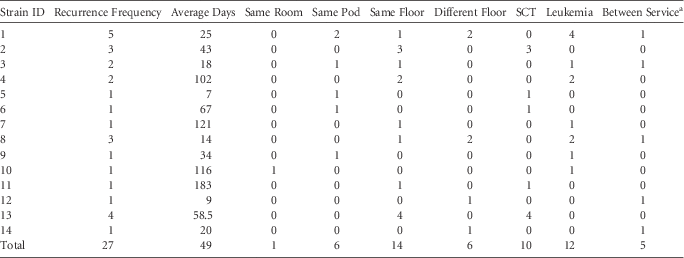
In 2013, a total of 82 nosocomial acquisitions of VRE were detected on these units. Of 82 VRE patient isolates tested, there were 55 genetically unique strains, 14 of which repeated at least once, making 27 recurrences of a strain. The average time between recurrences of a strain was 49 days (median, 36; range, 0–183 days) (Table 1). One strain recurred in the same room as the previous occurrence of the strain; 6 strains recurred on the same pod; 14 strains recurred on the same floor; and 6 strains recurred on a different floor (Table 1). Among the recurrences, 12 recurrences were among patients on the leukemia floors, 10 were among patients on the HCT floors, and 5 were in rooms on different services.
Descriptive Statistics for VRE Strain Repetition (N=27) of 14 Recurring Strains

NOTE. VRE, vancomycin-resistant Enterococci; SCT, stem-cell transfer unit.
aIndicates a repetition that occurred on SCT and then appeared on leukemia units of vice versa.
DISCUSSION
The methodology of REP sequencing for determining clonality is important because similarity output is inadequate. For example, 4 pairs with 95%+ similarity were determined to be “different” after variance in smaller peaks was observed during the overlay process, including 1 pair with 99% similarity.
The temporal data shows that VRE strains persist in some reservoir for an average of 49 days with a maximum of 183 days. This number exceeds the reported survival time of VRE in the environment of approximately 14 days.Reference Wendt, Wiesenthal, Dietz and Ruden 10 The majority of the recurrences (77%) were on the same floor as the previous occurrence of the strain, indicating a location-linked source of transmission. The within-service recurrence rate was high (81%).
Geographical data show recurrences not, as expected, in the same room but instead primarily on the same floor. This finding indicates that interventions focused on disinfecting only the rooms of VRE-positive patients may be suboptimal. Evidence of transmission to other rooms on the same floor or rooms on different floors indicates transfer of the VRE strains either by healthcare workers or via equipment. Other possible mechanisms for transfer of the pathogen include travel of patients for tests such as computerized tomography or magnetic resonance imaging, bronchoscopy or endoscopy or other procedures that require patients to be taken out of their rooms and transported through the unit.
The limitations of this study include the lack of data prior to and after the study period. This lack of data means that additional clonal repetitions could have taken place but would not have been detected. Another weakness is the inability to determine whether the “unique” strains are generally present in the community, although the lack of appearance in the Diversilab database and the striking dissimilarity of most of the isolates indicates uniqueness. Additionally, data other potential reservoirs such as environment, equipment, healthcare workers, visitors, etc. are lacking. Furthermore, we elected not to type the community-acquired strains of VRE because of cost and because we were interested in establishing a practical method for infection control. Adding the community-acquired VRE strains would have greatly increased the laboratory cost and burden and potentially made this method impractical for routine use in infection control.
This analysis demonstrated more VRE clonal repetition than anticipated and shows a clear nosocomial transmission risk associated with the units investigated. Additionally, this method seems feasible for routine use to investigate nosocomial transmission in healthcare settings. More research is needed to pair this method with interventions to prove the benefit of a particular intervention and potentially reach conclusions about its effectiveness.
ACKNOWLEDGMENTS
Dr. Huber and Dr. Chemaly report personal fees from Xenex Disinfection Services, LLC, during the conduct of the study. Drs. Ghantoji, Raad, Jinadatha have nothing to disclose. Dr. Stibich is a shareholder and employee of Xenex Disinfection Services, LLC.