BRIEF HISTORY OF THE MEDICAL USES OF CLAYS
Biblical and Quranic narratives mention that man was created from clay (Wikipedia (https://en.wikipedia.org/wiki/Biblical_and_Quranic_narratives, Reference Kelly, Deasy, Ziaka and Claffey2018)), an indication of the importance of these minerals to human life on our planet. In fact, since prehistoric times, humans have taken advantage of clays for diverse basic applications in everyday life with emphasis on health care as reported extensively in the past decade (Aguzzi et al. Reference Aguzzi, Cerezo, Viseras and Caramella2007; Carretero and Pozo Reference Carretero and Pozo2009, Reference Carretero and Pozo2010; Rautureau Reference Rautureau, Figueiredo Gomes, Liewig and Katouzian-Safadi2010, Liewig et al. Reference Lisuzzo2012; Rautureau et al. Reference Rebitski, Aranda, Darder, Carraro and Ruiz-Hitzky2017). Raw clays present in soils and mud have been used in medicine for centuries against infections and diseases, and in antidotes to diverse toxins and poisons (Allègre Reference Allègre2012). In many cases clays were applied topically for dermatologic treatments, as in the case of malignant ulcers (Williams and Haydel Reference Yao, Tan, Fang and Yu2010; Williams Reference Williams and Haydel2018) and at other times by geophagy (Allègre Reference Allègre2012). This latter application, which could be denoted more precisely as ‘clayphagy’, refers to the natural tendency to consume “earthy matter,” generally observed among all types of animals (Allègre Reference Allègre2012), including invertebrates, reptiles, birds, and mammals, including non-human primates such as the Japanese macaques (Wakibara et al. Reference Wang, Mynar, Yoshida, Lee, Lee, Okuro, Kinbara and Aida2001), Ugandan chimpanzees (Mahaney et al. Reference Merino, Ollier, Lanfranconi and Alvarez2005), and Sumatran orangutans from Indonesia (Allègre Reference Allègre2012; Stambolic-Robb Reference Sun and Qing1997). Humans from around the world practice ‘clayphagy’ to cure diverse diseases, particularly gastrointestinal disorders including, for instance, Asian cholera (Aufreiter et al. Reference Aufreiter, Hancock, Mahaney, Stambolic-Robb and Sanmugadas1997) or to eliminate parasites (Knezevich Reference Liewig, Rautureau and Gomes1999).
A very interesting property of clays, which is of great interest in terms of improving health, is their detoxification ability, which is based mainly on their adsorption and ion-exchange properties. Clays can thus act positively in the entrapment and extraction of diverse types of toxins and poisons, such as bacterial toxins (including the cholera toxin) and mycotoxins, as well as toxic molecular compounds (alkaloids, strychnine, etc.) and even radionuclides such as 137Cs. Likewise, ammonia and other nitrogen compounds harmful to the human organism can be adsorbed and eliminated by smectites and other clay minerals. The excellent contribution on this topic by Allègre (Allègre Reference Allègre2012) shows clearly the benefits of administering to humans as well as to animals a diverse range of clay minerals (kaolinites, smectites, and fibrous clays) with few or no contraindications, even at high doses. In fact, clays are particularly effective for a large variety of pathologies although the mechanisms explaining their mode of action are complex and in many cases remain unclear.
A potential problem for medical applications, however, may be the pollution of the clay by man, introducing contamination to soils and clay-mineral deposits in the form of polychlorinated biphenyl substances, pesticides, industrial and urban wastes containing heavy metals and pharmaceutical drugs, among others. Pharmacology and medicinal uses of clays require careful selection, control, and purification of the starting raw materials.
CURRENT RESEARCH ON CLAY MINERALS FOR HUMAN HEALTH
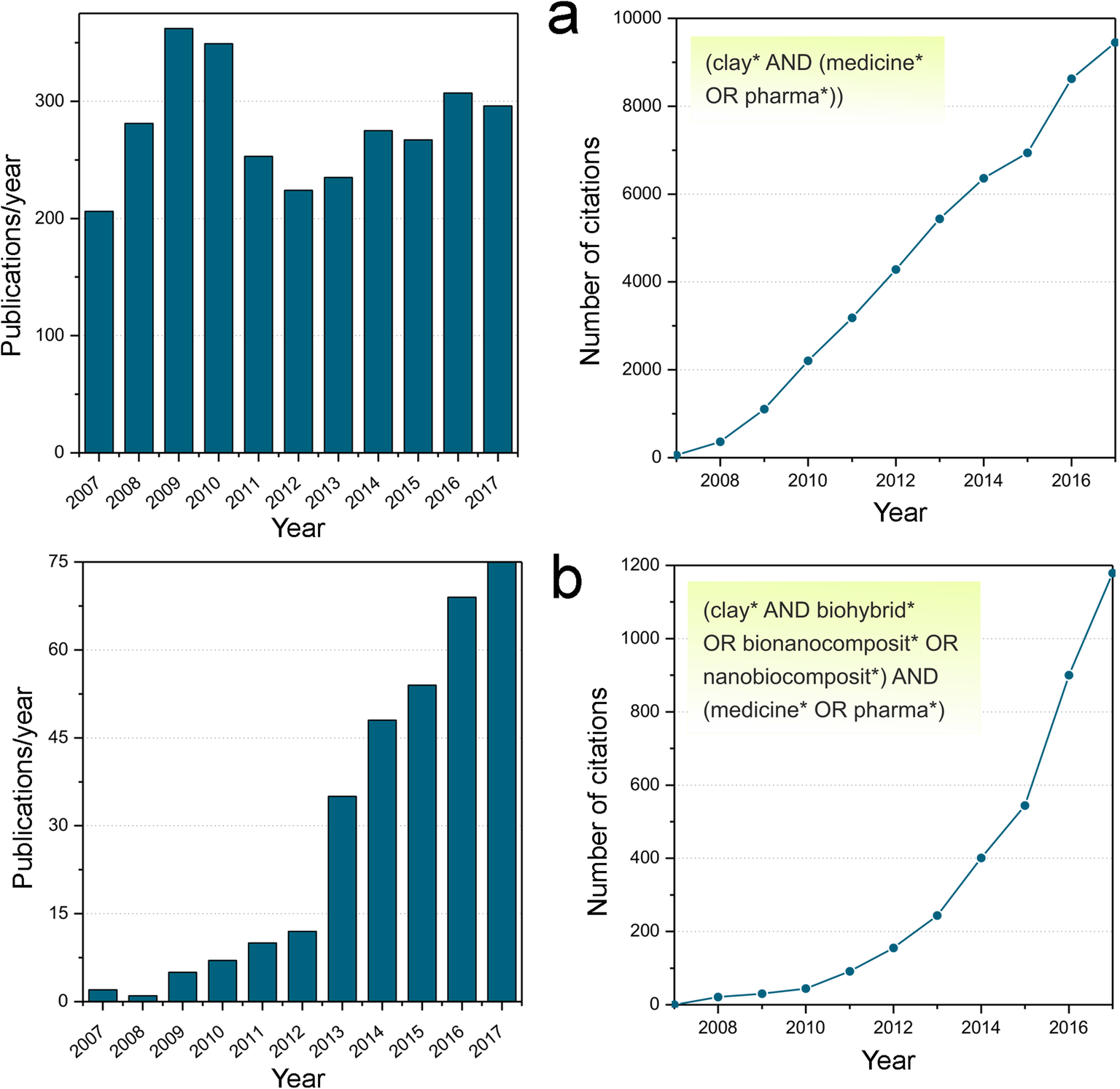
In recent times the ‘clay/medicine’ topic has generated ~200–300 publications per year with a total of ~10,000 citations in the past decade according to the Clarivate Web of Science database (Fig. 1a). During this period, the three most cited articles, which could be considered as representative ‘trending papers’ in the field, include the following topics: (1) the development of new hydrogels derived from clays as promising moldable materials for tissue engineering, a paper which has received ~800 citations thus far (Wang et al. Reference Wicklein, Darder, Aranda and Ruiz-Hitzky2010); (2) a review paper on bionanocomposites, which refers mainly to biohybrid materials derived from layered and fibrous clay minerals assembled in diverse compounds of biological origin, with ~350 citations (Darder et al. Reference Darder, Aranda and Ruiz-Hitzky2007); and (3) a paper on the ability of layered double hydroxides (LDH) to form hybrids by association with nucleic acids and the successful participation in the process of gene transfection (i.e. deliberate introduction of genetic material), with ~360 citations (Choy et al. Reference Choy, Choi, Oh and Park2007).
Publications and citations from ISI-WOS database, accessed in November 2018, searched for the 2007-2017 period, involving: (a) (clay* AND (medicine* OR pharma*)) and (b) (clay* AND biohybrid* OR bionanocomposit* OR nanobiocomposit*) AND (medicine* OR pharma*), as topics, respectively.
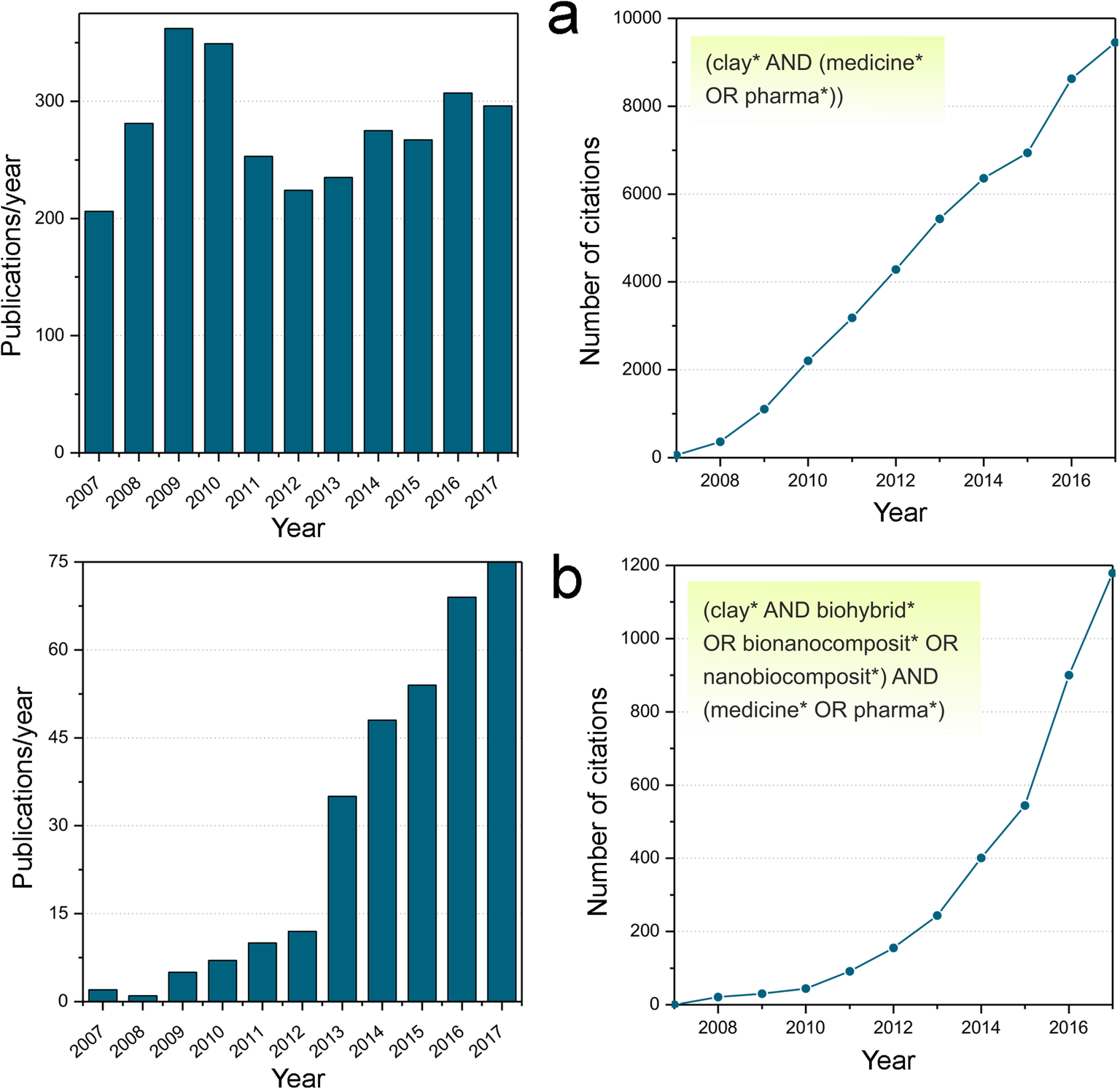
The three aforementioned contributions make reference to clay-based hybrid materials for biomedical and pharmaceutical uses. Within these materials, the number of papers about biohybrids and bionanocomposites, in relation to medicine and pharmacology, has increased exponentially during the past 10 years (Fig. 1b). The three most cited papers were: (1) the review paper of bionanocomposites emphasizing clay-based biohybrids ((Darder et al. Reference Darder, Aranda and Ruiz-Hitzky2007), mentioned above); (2) an article on bionanocomposites synthesized from poly(lactic acid) and acetylated microfibrillated cellulose showing the tailored properties of these materials as a function on their composition (Tingaut et al. Reference Viseras, Cerezo, Sanchez, Salcedo and Aguzzi2010); and (3) a feature article on clay-based hybrid materials for environmental and biomedical applications that discusses potential applications for drug delivery-systems (DDS), scaffolds, and regenerative medicine, and new substrates to immobilize biological species from enzymes to viruses among other topics (Ruiz-Hitzky et al. Reference Ruiz-Hitzky, Aranda, Darder and Ogawa2010).
BIOHYBRID–CLAY MATERIALS
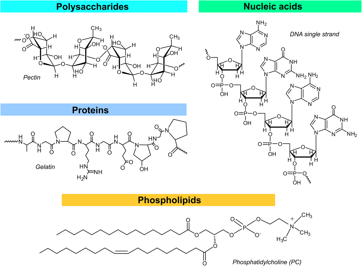
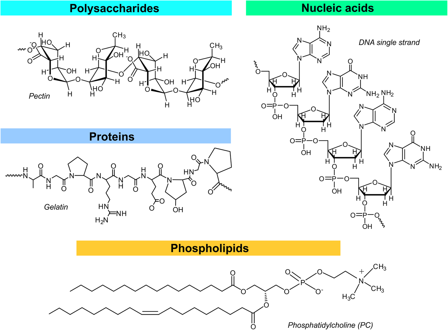
As indicated above, within the field of organic-inorganic hybrid materials, biohybrids involving species of biological origin are attracting increasing interest aimed, in particular, at applications related to biomedicine. These materials are produced by means of bottom-up processes, resulting from the assembly of naturally occurring molecular or polymeric components with inorganic solids at the nanometer scale (Darder et al. Reference Darder, Aranda and Ruiz-Hitzky2007; Ruiz-Hitzky et al. Reference Rytwo, Mendelovits, Eliyahu, Pitcovski and Aizenshtein2011). Among them, the so-called bionanocomposites involving biopolymers such as polysaccharides, proteins, lipids, or nucleic acids (Fig. 2) constitute a remarkable group of biohybrid materials. In certain cases, the development of bionanocomposites is inspired by natural materials produced in a template process referred to as ‘biomineralization’. For instance, the well organized stacked structure of the calcium carbonate polymorph, aragonite, with proteins in nacre has served as inspiration to develop bionanocomposites based on diverse biopolymers and layered clay minerals, trying to mimic the outstanding mechanical properties of this natural material (Yao et al. Reference Yao, Huang, Tang, Xu, Buntkowsky, Berglund and Zhou2010, Reference Yoshida and Sato2017). Typically, the development of clay-based bionanocomposites follows a bottom-up approach referred to as a ‘blocks assembly approach,’ where the natural clay minerals are used as nano-building units to assemble the nanocomposite material (Ruiz-Hitzky et al. Reference Rytwo, Mendelovits, Eliyahu, Pitcovski and Aizenshtein2011; Aranda and Ruiz-Hitzky Reference Aranda and Ruiz-Hitzky2018). In contrast, the synthesis of bionanocomposites involving LDH is carried out following an alternative bottom-up approach. In this case, the inorganic solid is commonly synthesized in the presence of biopolymer chains by precipitation of diverse salts at basic pH values by controlled addition of NaOH (Darder et al. Reference Darder, López-Blanco, Aranda, Leroux and Ruiz-Hitzky2005). In both cases, the processing of bionanocomposite materials follows water-based approaches, given the high hydrophilicity of both components, i.e. the clay minerals and most of the biopolymers.
Biopolymers involved in the preparation of bionanocomposite materials, showing an illustrative example of each type
One of the main advantages of bionanocomposites is their eco-friendly character, representing an ecological alternative to conventional polymer nanocomposites. The biopolymers used in this type of material are obtained from renewable and very abundant natural resources, and these are of significant interest in terms of replacing petroleum-based polymers in the production of nanocomposites. The biodegradability of these new materials is an important feature as they undergo quick biodegradation under composting conditions in contrast to petroleum-derived plastics; this helps to reduce environmental pollution due to plastic waste.
Biocompatibility is also a desirable noteworthy characteristic of bionanocomposites, particularly when considering their applications for diverse biomedical purposes (Mousa et al. Reference Nagy, Bíró, Berkesi, Benczédi, Ouali and Dékány2016; Ruiz-Hitzky et al. Reference Ruiz-Hitzky, Darder, Aranda, Cao and Brinker2008, Reference Ruiz-Hitzky, Darder, Aranda, Martin del Burgo and del Real2009a, Reference Ruiz-Hitzky, Aranda, Darder and Rytwob). An essential feature in regenerative medicine, e.g. for bone or cartilage repair, requires biocompatible scaffolds with suitable macroporosity as well as mechanical stability. Diverse bionanocomposites involving clay minerals assembled with polysaccharides, such as chitosan (Ambre et al. Reference Ambre, Katti and Katti2015), and structural proteins, such as gelatin (Zheng et al. Reference Zhuang, Zheng, Gao and De Yao2007) or collagen (Olmo et al. Reference Pacwa-Plociniczak, Plaza, Piotrowska-Seget and Cameotra1987), have proven to be suitable scaffold materials for this type of application. The clay particles play an important role in these scaffolds as they act as physical cross-linkers enhancing the mechanical stability and contributing to the reduction of the biodegradation rate in body fluids (Zhuang et al. Reference Zhuang, Zheng, Gao and De Yao2007), while recent studies have also reported a remarkable performance of clay minerals in the proliferation and differentiation of mesenchymal stem cells to bone cells (Ambre et al. Reference Ambre, Katti and Katti2015; Ghadiri et al. Reference Ghadiri, Chrzanowski and Rohanizadeh2015).
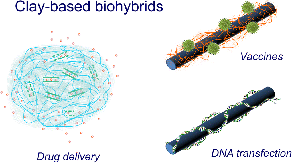
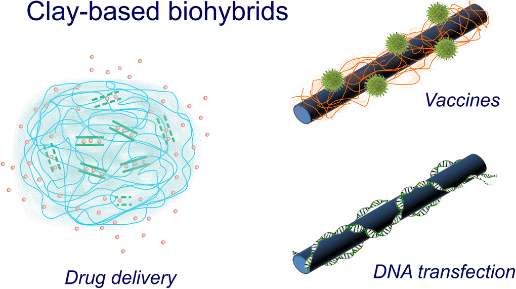
Bionanocomposites also offer many advantages for drug-delivery applications. While the layered or tubular clays usually act as nanocontainers, where the drug molecules are entrapped in order to provide sustained release (Aguzzi et al. Reference Aguzzi, Cerezo, Viseras and Caramella2007; Lvov et al. Reference Mahaney, Milner, Aufreiter, Hancock, Wrangham and Campbell2015; Rives et al. Reference Ruiz-Hitzky, Van Meerbeek, Bergaya, Theng and Lagaly2014), the biopolymers are used to provide the drug-loaded clays with a protective matrix that affords additional control during release of the drug. In this way, the entrapped drugs can be liberated gradually avoiding overdosing or underdosing episodes during treatment (Viseras et al. Reference Wakibara, Huffman, Wink, Reich, Aufreiter, Hancock, Sodhi, Mahaney and Russel2010). The biopolymers have another important role in drug-delivery systems, as they can help to reduce or eventually avoid the tendency of non-modified clay minerals to flocculate when dispersed in physiological media, enhancing the stability of dispersions of drug-delivery systems (Luckham and; Rossi Reference Lvov and Abdullayev1999). Besides controlled release of pharmaceuticals, clay minerals have also been evaluated in other advanced biomedical applications, including their use as nanocarriers of virus particles in the preparation of novel vaccines and of DNA in non-viral transfection. In the first case, the clay acts as an adjuvant (i.e. an additive to increase the efficacy of a vaccine) and the presence of a biomolecule as a clay modifier is a key point in creating a biomimetic environment which preserves the viral activity (Ruiz-Hitzky et al. Reference Ruiz-Hitzky, Aranda, Darder and Rytwo2009b; Wicklein et al. Reference Wicklein, Aranda, Ruiz-Hitzky and Darder2012). In non-viral gene therapy, the DNA chains are the biopolymers assembled with clay minerals such as sepiolite (Castro-Smirnov et al. Reference Castro-Smirnov, Piétrement, Aranda, Bertrand, Ayache, Le Cam, Ruiz-Hitzky and Lopez2016) or with layered double hydroxides (Choy et al. Reference Choy, Kwak, Jeong and Park2000). Illustrative examples of the application of biopolymer-clay nanocomposites as drug-delivery systems, vaccines, and DNA transfection (Fig. 3) will be discussed below, with special emphasis on biohybrid materials developed by the authors’ research group for these purposes.
Schematic representation of biohybrid beads used as drug-delivery systems, biopolymer-modified sepiolite fibers used as nanocarriers of virus particles in vaccines, and sepiolite fibers used as nanocarriers of DNA in non-viral gene therapy
BIONANOCOMPOSITES FOR DRUG-DELIVERY APPLICATIONS
One of the most useful applications of bionanocomposites in biomedicine is their deployment in the design of new, efficient DDS (Ruiz-Hitzky et al. Reference Ruiz-Hitzky, Aranda, Darder and Ogawa2010). Clays of diverse structure, including layered silicates such as smectites, tubular ones such as halloysite nanotubes (HNT), or fibrous ones such as sepiolite and palygorskite, as well as LDH have been explored for applications as DDS per se, i.e. without modification (Viseras et al., Reference Wakibara, Huffman, Wink, Reich, Aufreiter, Hancock, Sodhi, Mahaney and Russel2010). Biopolymers, and polysaccharides in particular, are commonly employed in the preparation of diverse systems for biomedical applications including drug delivery and other nano-therapeutic usages (Mizrahy and Peer Reference Mousa, Dong and Davies2012; Nitta and Numata Reference Oh, Park, Choi and Choy2013). The association of clays and biopolymers in the preparation of hybrid systems for drug-delivery applications seeks to combine the advantages offered by each of the two components. For instance, the incorporation of pharmaceutical drugs in the interlayer space of layered clays and LDH or within the lumen of HNT offers advantages in terms of protection from the atmosphere, light exposure, or changes in pH; this is a common tool used in the design of pharmacological formulations for cutaneous and oral administration. The combination of these hybrids with biopolymers can be used to produce bionanocomposite systems where controlled drug release is possible, as reported for tetracycline loaded within the lumen of HNT nanotubes and further coated with chitosan tested in vivo for treatment of periodontitis in dogs (Kelly et al. Reference Khan, Lei, Norquist and O'Hare2004). Below, various examples of the main contributions made by the authors’ research group regarding the use of bionanocomposites for drug-delivery applications will be introduced.
Layered double hydroxides, mainly Mg-Al LDH, have been explored as host matrices for intercalation of diverse drugs in the anionic form (Khan et al. Reference Knezevich2001; Rives et al. Reference Ruiz-Hitzky, Van Meerbeek, Bergaya, Theng and Lagaly2014). The particular sensitivity of the LDH matrices to pH can be used to provoke the release of the intercalated active species in a controlled manner. In this context, ibuprofen-intercalated LDH has been used widely as a model drug system for evaluation of drug-delivery methods, related mainly to new systems proposed for oral delivery in which the LDH-drug hybrid is, for instance, coated with a polymeric matrix. One of the approaches developed by the present authors deals with the preparation of a magnetic, bionanocomposite DDS based on the association of a Mg-Al LDH incorporating ibuprofen with alginate and magnetic graphite nanoparticles (Ribeiro et al. Reference Ribeiro, Alcântara, Darder, Aranda, Araújo-Moreira and Ruiz-Hitzky2014a). The bionanocomposites were prepared by dispersing the three components in water and then adding a 15% CaCl2 solution to produce the cross-linking of the biopolymer, which enabled easy processing of the suspension as beads or films. The presence of the magnetic graphite nanoparticles contributes to the incorporation of magnetic properties and also to improve the mechanical properties of the bionanocomposites. The graphite also improves the stability of the system in water solutions, crucial in terms of their potential application as a DDS. In vitro release tests of ibuprofen in a phosphate buffer (pH 7.4, 37 °C) confirm that the release rate was clearly improved when the drug was incorporated in the LDH matrix instead of directly within the biopolymer matrix. These experiments also showed how the hydrophobic character of graphite nanoparticles provided a protective barrier that reduces the rate of release in comparison to bionanocomposites prepared without it. Interestingly, the release of ibuprofen can be stimulated by an external magnetic field, proving that the DDS developed could take advantage of the presence of the magnetic component to produce a modulation in the dose of drug that can be released from the system (Ribeiro et al. Reference Ribeiro, Alcântara, Darder, Aranda, Araújo-Moreira and Ruiz-Hitzky2014a). The mechanism that affords this behavior is not entirely clear but is probably related to changes in the alignment of magnetic moments with the external magnetic field. This may lead to alterations in the organization of the polymer chains within the bionanocomposite, facilitating the release of the entrapped drug, similarly to that reported for other hybrid magnetic hydrogels (Giani et al. Reference Giani, Fedi and Barbucci2012). As indicated above, preliminary tests of this new magnetic DDS have been carried out, using ibuprofen, and processed as beads and films; other drugs and active species could also be incorporated and they could also be processed as foams. Interesting applications are, thus, suggested in wound dressings and as scaffolds in tissue engineering.
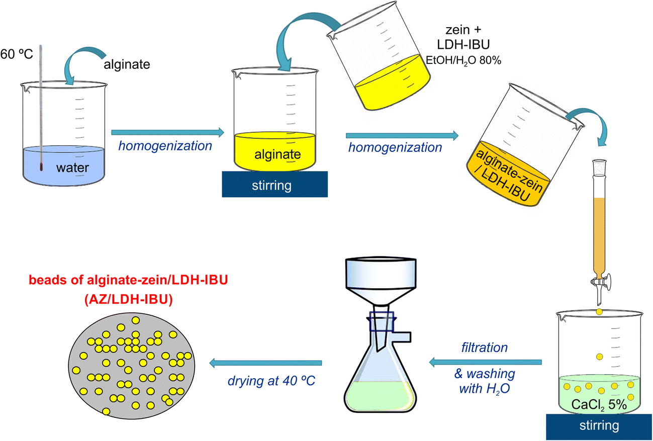
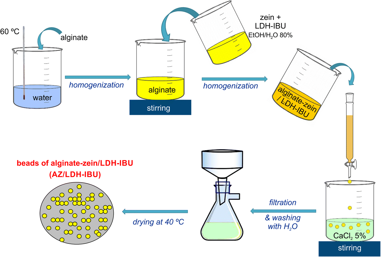
The control of drug released from DDS designed for oral administration must consider the different environments the drug will find along its passage through the gastrointestinal tract. The changes in pH occurring are of particular relevance considering the stability of the DDS at certain pH values and the necessity to stimulate and control the release of the drug in a specific region of the tract. First of all, the strong acidic pH in the stomach determines that the drug intercalated in LDH will be released readily if the intercalation compound is used directly as a DDS. Thus, most of the systems involve an association with other components to produce convenient and more effective formulations (Costantino et al. Reference Costantino, Nocchetti, Tammaro and Vittoria2012; Oh et al. Reference Olmo, Lizarbe and Gavilanes2012). Alginate is one of the polymers most commonly employed in the preparation of these formulations, possibly due to the facility for cross-linking with Ca2+ and other polyvalent cations. The quick dissolution of these cations in basic media such as the intestinal fluid (pH 6.8–7.4) may lead to uncontrolled release of the drug. For this reason, the possibility of developing a DDS which introduces a second biopolymer of hydrophobic character to control the drug release was investigated. The chosen polymer was zein, the major storage protein present in corn, which is insoluble in water due to the presence of a large amount of non-polar amino acid residues (Alcantara et al. Reference Alcantara, Aranda, Darder and Ruiz-Hitzky2010). A schematic illustration of the protocol employed to prepare beads based on mixtures of alginate and zein in which ibuprofen (as a model drug) is incorporated though intercalation in Mg-Al LDH is given in Fig. 4. The design of this type of ternary bionanocomposite takes advantage of the protective effect afforded by the LDH matrix, the simplicity of alginate processing, e.g. as beads, and the hydrophobic character that zein may convey to the DDS, which can be useful in controlling the swelling and rapid disintegration typically observed in alginate-based DDS. The use of the ibuprofen-LDH hybrid instead of the neat drug facilitates the integration of alginate and zein, which produces very homogeneous systems in comparison to those prepared by combining both biopolymers in the presence of ibuprofen. Water uptake and swelling properties are influenced heavily by the relative composition of alginate and zein, the presence of larger amounts of the protein favors the stability of microspheres as it inhibits the incorporation of water molecules. These characteristics are also relevant in terms of controlling the release of drugs in conditions that simulate the passage through the gastrointestinal tract as the presence of zein also influences the uptake of calcium and other ions that may favor the rapid disintegration of the bead and, thus, the uncontrolled release of the drug. In this way, in vitro tests simulating the release of ibuprofen from bionanocomposite beads prepared with different alginate-zein ratios show that these DDS give practically complete protection during the passage through the stomach, and the rate at which the drug is delivered is reduced as the zein content increases (Alcantara et al. Reference Alcantara, Aranda, Darder and Ruiz-Hitzky2010). The ability to adjust the composition of the DDS is significant in terms of producing tunable systems with different kinetics in the release of the drug, which could be very interesting for application in other therapies also.
Schematic representation of the general process reported by Alcantara et al. (Reference Alcantara, Aranda, Darder and Ruiz-Hitzky2010) for the preparation of microspheres of alginate–zein bionanocomposite DDS incorporating ibuprofen intercalated into a Mg-Al LDH (LDH-IBU)
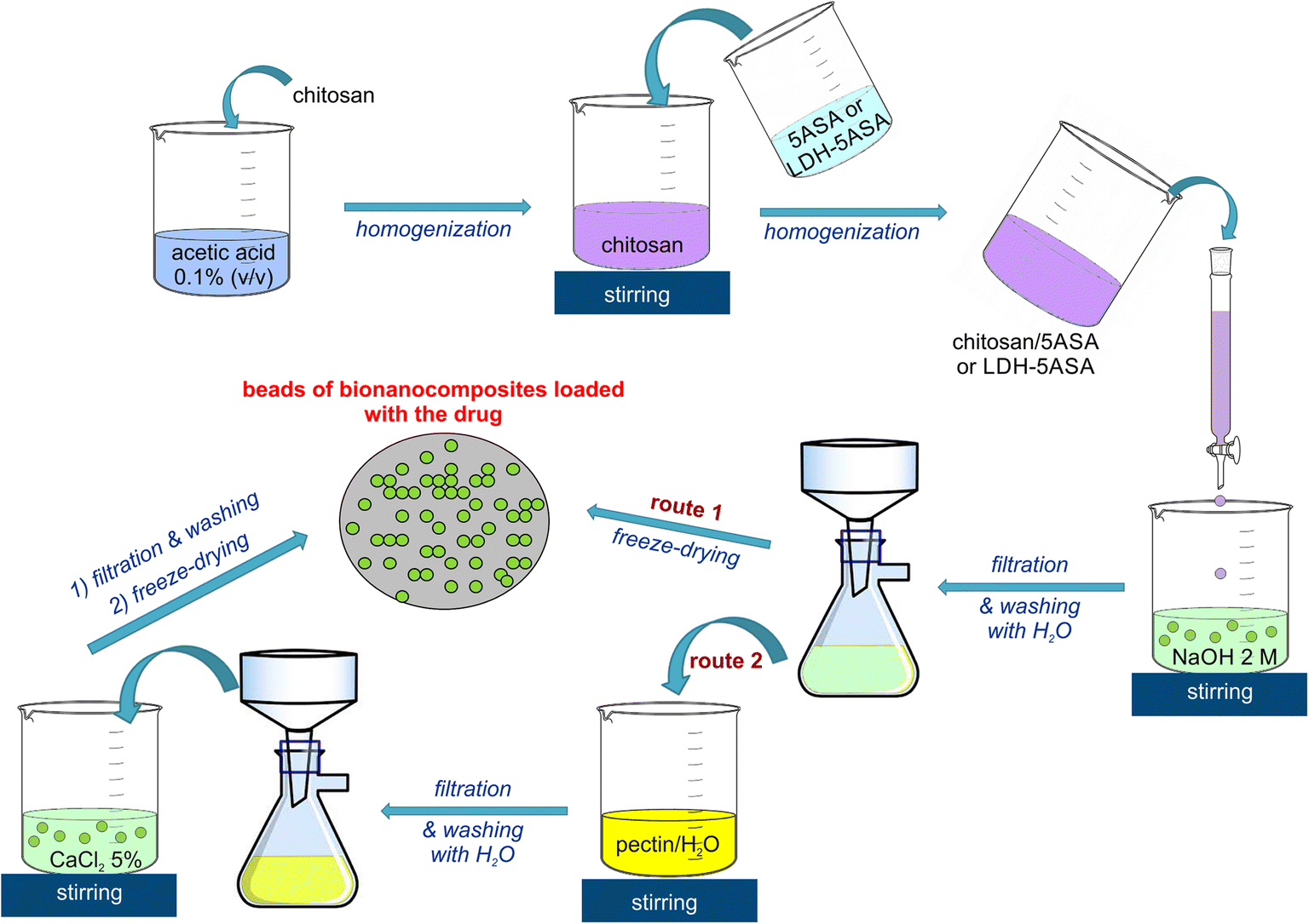
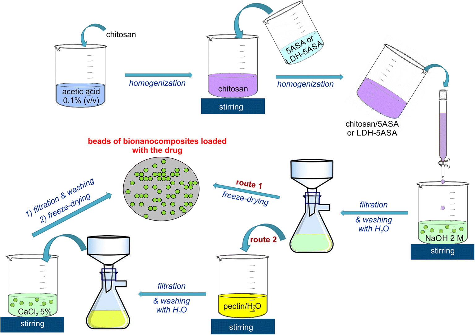
Certain biopolymers used widely in the preparation of DDS, e.g. chitosan, show interesting mucoadhesive properties which may be of interest in the development of systems for treatment at specific sites in the intestinal tract. This could be relevant in the treatment of colon diseases where controlled release of the drug at the precise location is most desirable and would help to reduce side effects. The development of a new bionanocomposite system for the selective delivery in the colon of 5-aminosalicylic acid (5ASA), the most widely used non-steroidal-anti-inflammatory drug employed in the treatment of ulcerative colitis and Crohn’s disease (Ribeiro et al. Reference Rives, del Arco and Martín2014b), was investigated, therefore. To prepare the DDS (Fig. 5), the 5ASA drug was first intercalated into a Mg-Al LDH and then associated with chitosan, producing beads of the bionanocomposite. The resulting microspheres show muco-adhesion (i.e. adhesion to mucus tissue) but they cannot be employed as oral DDS because of their low stability in acid media. The beads produced were further modified with a coating of the polysaccharide pectin, therefore, which was chosen because of its stability at low pH and its solubility at more basic pH as found in the intestine. Thus, the ternary drug delivery system designed will merge the following advantages afforded by each of the three components: (1) a protective coating on the outside of the bead afforded by pectin, which may ensure the preservation of the bead at low pH through its stage in the stomach; (2) the stability at high pH values and muco-adhesion properties of chitosan for specific adsorption in the intestinal tract once the pectin coating is removed; and (3) controlled release of the 5ASA drug achieved by its presence immobilized within the LDH host matrix (Ribeiro et al. Reference Rives, del Arco and Martín2014b). With this in mind, beads of diverse compositions were prepared to confirm in vitro tests which simulated changes in pH and time of residence that occur during the in vivo passage of the drug through the gastrointestinal tract; the pectin coating introduces stability in terms of water swelling and ensures a controlled release of the drug along its passage through the simulated gastrointestinal tract. Changes in the pectin coating thickness influence the kinetics of the release of 5ASA which is relevant when producing formulations where the application of this drug and others of interest in the treatment of intestinal diseases requires the action at specific sites. The option to include specific functionalities in chitosan to target specific locations for the release made the results obtained just the first stage in the design of more efficient formulations for treatment of colonic diseases.
Schematic representation of the general process reported by Ribeiro et al. (Reference Rives, del Arco and Martín2014b) for the preparation of microspheres of alginate–zein bionanocomposite DDS incorporating ibuprofen intercalated into a Mg-Al LDH (LDH-IBU)
Smectites and other clay minerals have also been employed as substrates for the immobilization of pharmaceutical drugs when designing DDS. In these cases, as the clay host is negatively charged, the drugs of choice need to be cationic species, e.g. certain antibiotics such as tetracyclines (Aguzzi et al. Reference Aguzzi, Cerezo, Sandri, Ferrari, Rossi, Bonferoni, Caramella and Viseras2014). Gentamicin is another antibiotic that is produced by fermentation of Micromonospura purpurea and commonly used to treat various types of bacterial infections, particularly those caused by Gram-negative organisms. From a chemical point of view, gentamicin is an aminoglycoside which contains three glycoside units and five amino groups which are able to protonate to various extents at pH <10. The intercalation of gentamicin into montmorillonite has been explored, followed by incorporation into hydroxyl-propylmethylcellulose (HPMC), to produce films with antimicrobial activity for burn-wound dressing applications. Gentamicin dissolved in water is intercalated into Na-montmorillonite (Cloisite®Na) by a cation-exchange mechanism, which enabled the incorporation of ~0.22 mmol/g of gentamicin in the clay, in accordance with the accommodation of gentamicin cations with a net charge of +5 (He Reference He2013). The antibiotic incorporated within the clay layers is also very stable against degradation by light. In vitro tests of gentamicin release carried out at pH 7 (phosphate buffer, 37 °C) show poor removal from the clay, even less than those reported recently when intercalated in Veegum®F pharmaceutical magnesium-aluminum silicate (Rapacz-Kmita et al. Reference Rautureau2017). The release results prove that the clay mineral may counteract a rapid release of the antibiotic upon water uptake, which was one of the disadvantages of antibiotic-loaded natural polymer; the hybrids were further incorporated in HPMC to produce bionanocomposites with various loadings in gentamicin-montmorillonite and its antibiotic activity checked. In vitro tests of the bionanocomposites processed as films showed antimicrobial activity towards Staphylococcus aureus with a maximum efficacy even for small amounts of gentamicin, proving the potential interest in applications for healing wound tissue (Aranda et al. Reference Aranda, He, Darder, Fernández-Saiz, Monte-Serrano, Charlet and Ruiz-Hitzky2014).
Recently, the present authors explored the use of montmorillonite in the development of a DDS that can be used for controlled delivery of metformin, the oral drug of choice for the treatment of type II diabetes. The chemical formula of metformin can be described as biguanide (3-(diaminomethylidene)-1,1-dimethylguanidine), a molecule which contains five nitrogen atoms and presents a complex tendency towards protonation with diverse species stable as a function of pH but which are mainly mono-protonated species between pH 1.5 and 10.5. Also reported recently was the strong affinity of metformin for being adsorbed in Na-montmorillonite (Cloisite®Na) from water solution of metformin hydrochloride (Rebitski et al. Reference Ribeiro, Alcântara, Darder, Aranda, Herrmann, Araújo-Moreira, García-Hernández and Ruiz-Hitzky2018). Some of the metformin adsorbed can be removed easily just by washing with water, leaving ~90 meq of drug per 100 g of clay, which coincides with the cation exchange capacity of the clay used. X-ray diffraction data confirm the intercalation of metformin, which is arranged in the interlayer space as a monolayer of monoprotonated molecules incorporated by a cation-exchange mechanism into the clay and stabilized by electrostatic interactions. Preliminary in vitro tests of metformin delivery from the intercalation compound, in conditions that simulate the liberation kinetics at pH values that vary, mimicking the gastrointestinal tract, show rapid release at low pH values (i.e. typical of the stomach), which indicates clearly the necessity to optimize the DDS. In this way, the use of bionanocomposite beads in which the intercalation compound is incorporated in a matrix of chitosan that is later covered with pectin as in the DDS developed for treatment of colon diseases (Ribeiro et al. Reference Rives, del Arco and Martín2014b) is being explored. Again, the mucoadhesive properties of chitosan may increase the permanence of the DDS in the intestinal tract with the clay acting as a reservoir, thereby reducing the applied doses of metformin required in standard treatments (where these have to be kept large due to the high solubility of metformin and its scarce interaction with substrates).
Halloysite nanotubes have been proposed and studied widely for use as a DDS alone or in combination with polymers (Lvov and Abdullayev Reference Lvov, Wang, Zhang and Fakhrullin2013). Recently, a methodology has been developed which allows the formation of homogeneous and stable aqueous suspensions of HNT in the presence of sepiolite and in which the incorporation of other hydrophilic nanoparticles, e.g. cellulose nanofibers (CNF), is possible (Aranda et al. Reference Aranda, Lo Dico, Lisuzzo, Wicklein, del Real, Lazzara and Ruiz-Hitzky2018). These suspensions can be processed easily as self-supported films forming hybrid nanopapers in which the presence of sepiolite and HNT clays allows further functionalizations (Lisuzzo Reference Lisuzzo, Aranda, Wicklein, Real, Lazzara and Ruiz-Hitzky2017; Lisuzzo et al. Reference Liu, Wu, Yu, Li, Gong, Zhu, Dang and Yang2018). In this way, the incorporation of various drugs such as ibuprofen, amoxicillin, and salicylic acid in the lumen of the HNT involved in the preparation of the hybrid nanopapers has been explored. Biohybrid films incorporating HNT loaded with salicylic acid thus proved to be effective in the inhibition of bacterial growth of gram-positive microbes such as Staphylococcus aureus. Antimicrobial tests were carried out at pH 5.5, typical of the human skin pH, proving the potential usefulness of the bionanocomposite materials developed for application as antiseptic dressings.
CLAY-LIPID HYBRIDS
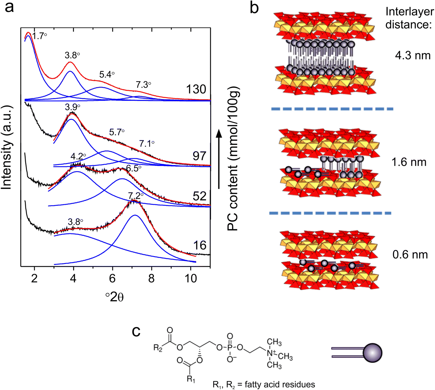
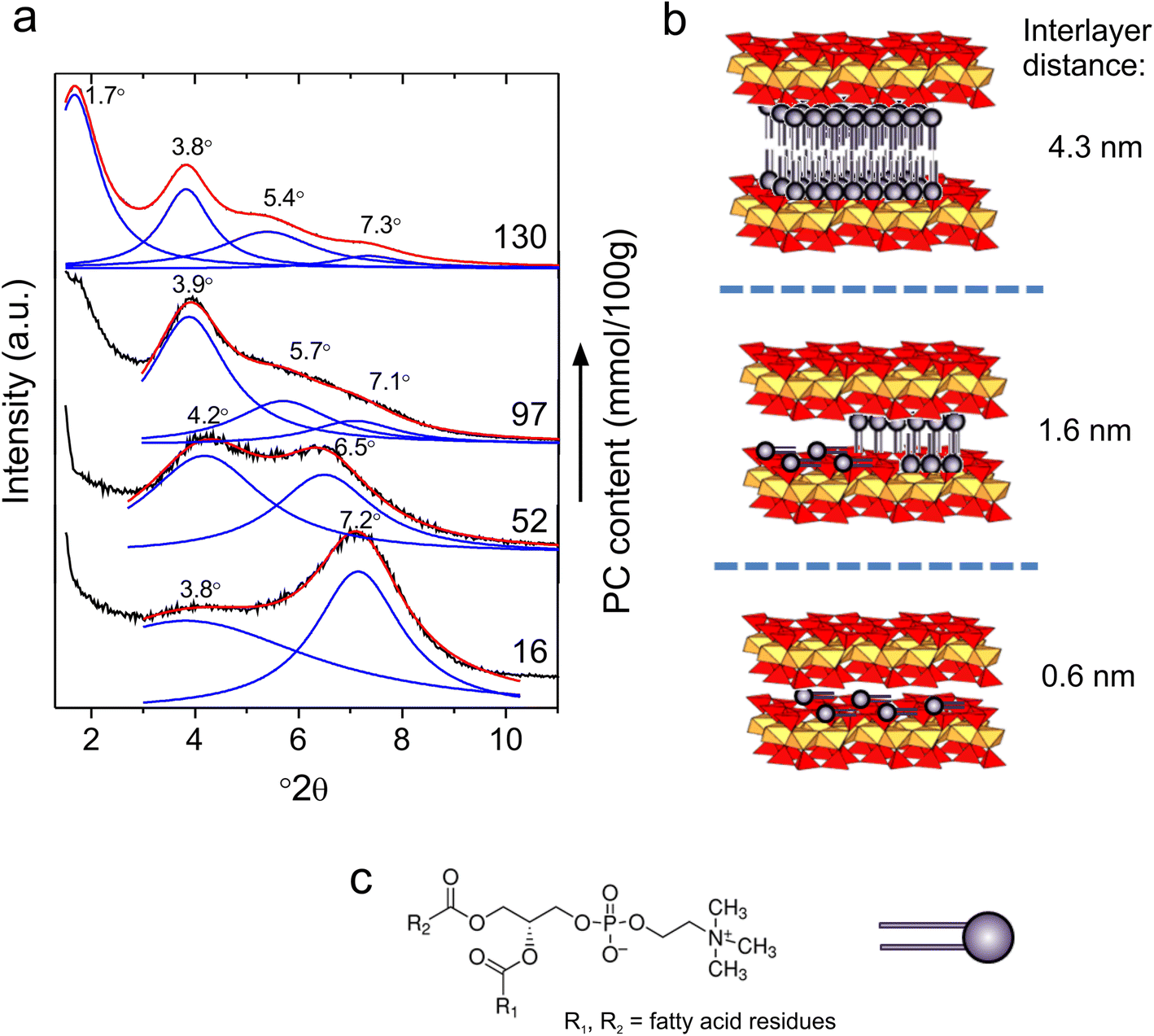
Naturally occurring surfactants such as fatty acids, phospholipids, lipopeptides, etc. (Pacwa-Plociniczak et al. Reference Park, Hwang, Oh, Yang and Choy2011) are environmentally benign and versatile alternatives to synthetic surfactants such as quaternary alkylammonium salts (Ruiz-Hitzky and Van Meerbeek Reference Ruiz-Hitzky, Aranda and Darder2006) used widely for the organophilization of clay minerals. These biosurfactants can form solid-supported bilayers with biomimetic features, which can be prepared via self-assembly routes (Ariga et al. Reference Ariga, Hill, Lee, Vinu, Charvet and Acharya2008) and find potential applications in biomedical or other biotechnological fields (Sun and Qing Reference Tingaut, Zimmermann and Lopez-Suevos2011). A fibrous clay–lipid hybrid was first reported by Wicklein et al. (Reference Wicklein, Darder, Aranda and Ruiz-Hitzky2010) who adsorbed phosphatidylcholine (PC) on sepiolite in a controlled manner. Lipid deposition was carried out either by liposome adsorption from the aqueous phase or by molecular adsorption from organic solvent (Wicklein et al. Reference Wicklein, Aranda, Ruiz-Hitzky and Darder2012). In both cases, the adsorption isotherms show the characteristic shape of mono- and bilayer formation as a function of the equilibrium PC concentration. This study also showed that liposomal deposition is more effective for the formation of a lipid membrane in this specific system. The reason is that, in aqueous media, PC adsorbs as aggregates (i.e. bilayered liposomes), while in ethanol, single PC molecules adsorb on the clay surface. The molecular interaction between the lipid head group and the clay surface is hydrogen bonding between the ester and phosphatidyl groups of the lipid and the silanol groups located on the sepiolite surface. The adsorption of phospholipids on sepiolite also has a significant influence on the surface wettability of the resultant material. The initially hydrophilic sepiolite becomes more hydrophobic as the first monolayer is completed, while with the subsequent deposition of lipid molecules the wetting increases gradually again as hydrophilic lipid head groups assemble at the external surface of the lipid bilayer. Similarly, phospholipids from egg yolk or soy lecithin can also be adsorbed onto layered smectite clays, e.g. montmorillonite (Wicklein et al. Reference Wicklein, Darder, Aranda and Ruiz-Hitzky2010; Merino et al. Reference Mizrahy and Peer2016) or vermiculite (Liu et al., Reference Luckham and Rossi2017), through cation exchange, which places intercalated lipid bilayers in the interlayer space of the clays (Fig. 6). In the latter case, the resulting bio-organo-clay was tested as an efficient adsorbent for the water remediation of the antibiotics oxytetracycline and ciprofloxacin due to the enhanced hydrophobic interactions between the antibiotics and the lipid layer (Liu et al. Reference Luckham and Rossi2017). Likewise, lecithin and lysolecithin in their deprotonated form were intercalated into LDH for the subsequent uptake of limonene (Nagy et al. Reference Nitta and Numata2013).
XRD pattern of phosphatidylcholine-montmorillonite hybrids showing the shift of the basal (001) reflection as a function of the lipid intercalation (a). Schematic illustration of the lipid intercalation in the clay interlayer space including the basal distance calculated from the XRD pattern (b). Molecular structure and drawing of phosphatidylcholine showing the zwitterionic headgroup (c)
Self-assembly of biosurfactants can be used to prepare mixed lipid membranes that offer diverse functionalities, which can be of interest in various technological applications (Plant Reference Rapacz-Kmita, Stodolak-Zych, Dudek, Gajek and Ziąbka1999). Hybrid layers consist of individual membrane leaflets of various molecules. Such a hybrid membrane was assembled on sepiolite where the inner leaflet comprised PC and the outer leaflet, the sugar-based surfactant n-octyl-β-d-galactoside (OGal) (Wicklein et al. Reference Wicklein, Martín del Burgo, Yuste, Darder, Escrig Llavata, Aranda, Ortín, del Real and Ruiz-Hitzky2011). The PC monolayer has been assumed to serve as a nucleation site for the growth of the OGal outer-membrane leaflet by hydrophobic interaction between the hydrocarbon chains. Non-ionic sugar-based surfactants such as OGal are a type of biosurfactant of interest for properties such as antifouling behavior or protein-stabilization ability.
The biomimetic character of the clay-supported lipid interface is also advantageous for the immobilization of certain proteins, especially membrane-bound enzymes. The catalytic activity of such enzymes is very sensitive to the immobilization site and can be compromised readily by protein-structure deformations. Cholesterol oxidase (COx) was shown by Wicklein et al. (Reference Wicklein, Martín del Burgo, Yuste, Darder, Escrig Llavata, Aranda, Ortín, del Real and Ruiz-Hitzky2011) to maintain its catalytic activity when supported on sepiolite modified with a bilayer lipid membrane. On the contrary, after immobilization on sepiolite hybrids displaying cetyltrimethylammonium or hybrid lipid–octyl-galactoside layers, the activity of COx was diminished significantly, underscoring the importance of the supported biomimetic lipid membrane. This offers possibilities for preparing selective biocatalysts (Wicklein et al. Reference Wicklein, Darder, Aranda, Martín del Burgo, del Real, Esteban and Ruiz-Hitzky2013) and sensitive biosensors (Wicklein et al. Reference Wicklein, Martín del Burgo, Yuste, Darder, Escrig Llavata, Aranda, Ortín, del Real and Ruiz-Hitzky2011) employing immobilized enzymes on sepiolite–lipid hybrids as active species.
Owing to their biocompatibility, clay–lipid hybrids are interesting candidates for in vivo applications such as enterosorbents for mycotoxins in cattle and poultry to ease infestations by fungi. In fact, bio-organo-clays such as sepiolite–lipid hybrids demonstrated more in vitro sequestration efficiencies for aflatoxin B1 compared to the pristine clay analogue (Wicklein et al. Reference Wicklein, Darder, Aranda and Ruiz-Hitzky2010) and, thus, could be a promising candidate material for in vivo applications.
CLAY BIOHYBRIDS AS VACCINE ADJUVANTS
Clays have been recognized as alternative immunogen supports in vaccine formulations due to their large specific surface area, vector behavior (i.e. targeting vehicle), and inherent biocompatibility (Ruiz-Hitzky et al. Reference Ruiz-Hitzky, Aranda, Darder and Rytwo2009b; Rytwo et al. Reference Stambolic-Robb2010). An important aspect of increasing the efficacy of vaccines is the stabilization of immunogens, i.e. inactivated or live-attenuated virus particles, viral vectors, proteins, or plasmids, on particulate carriers that can enable easy administration, protection from proteolysis, and improved thermal-storage performance (Heegaard et al. Reference Heegaard, Dedieu, Johnson, Le Potier, Mockey, Mutinelli, Vahlenkamp, Vascellari and Sørensen2011). In several studies, adsorption of antigens on the pristine clay surface was observed to alter their protein structure, possibly through strong electrostatic interactions with the highly charged clay surfaces (Rytwo et al. Reference Stambolic-Robb2010). This resulted in reduced immunogenicity of the antigen and, eventually, compromised the vaccination efficacy (Patil et al. Reference Piétrement, Castro-Smirnov, Le Cam, Aranda, Ruiz-Hitzky and Lopez2004). Other strategies have been developed by presenting a bio-organic surface layer on the clay particles to avoid these disadvantageous interactions.
Xanthan-sepiolite biohybrids were prepared as adjuvants in an influenza vaccine (Ruiz-Hitzky et al. Reference Ruiz-Hitzky, Aranda, Darder and Rytwo2009b). Here, the main idea was to provide a tissue-like environment for the influenza virus to be adsorbed, similar to the nasal mucous membrane, the natural location of influenza virus entry into a body. An inactivated influenza A virus was adsorbed on sepiolite-xanthan and immunization of mice and posterior challenge with infectious influenza A virus demonstrated the high level of seroprotection elicited by this vaccine. An interesting aspect of using microfibrous clays such as sepiolite is its needle-like morphology thought to enhance mucosal immune responses provoked by irritation of the nasal mucous through the fibers. This can help to improve the efficacy of vaccination and, thus, to reduce the dose necessary for immunization.
The possibility of using clays with a berberine-based bio-organic interface on montmorillonite in order to accommodate antigens was explored by Rytwo et al. (Reference Stambolic-Robb2010). In that study, the organic modification of the clay was shown to permit the co-adsorption of two antigenic proteins, heat-labile eteroxin and viral protein 2, without conformational protein alterations, as noted from evidence in the infrared spectra. The bioactivity retained by these supported proteins enabled efficacious binding to cellular GM1 receptors, an important step in the stimulation of an immune response to the infectious bursal disease virus.
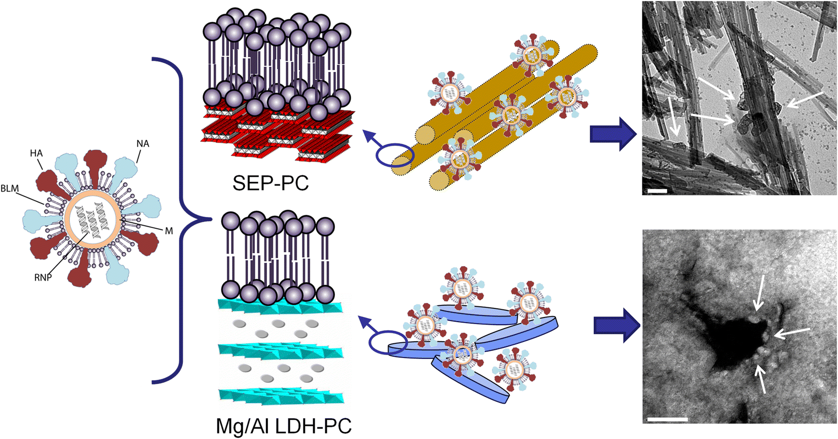
In another vaccine application, sepiolite-lipid and Mg-Al layered double hydroxide-lipid hybrids served as a nanocarrier for influenza A antigens (i.e. whole virus particles, hemagglutinin protein) (Wicklein et al. Reference Wicklein, Aranda, Ruiz-Hitzky and Darder2012) (Fig. 7).
Immobilization of influenza A viral particles on sepiolite-lipid and Mg-Al LDH-lipid biohybrids. The white arrows in the FE-SEM images indicate the immobilized viral particles. The scale bars = 100 nm (image reproduced with permission, from (Wicklein et al. Reference Wicklein, Aranda, Ruiz-Hitzky and Darder2012)). Copyright (2012) John Wiley & Sons, Inc.
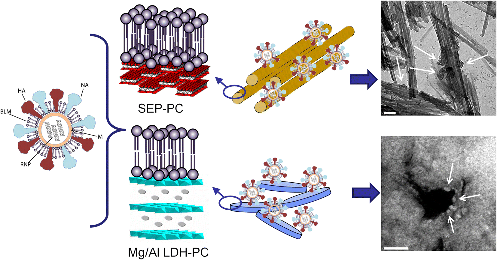
Functional studies in mice revealed that, in contrast to a standard aluminum hydroxide adjuvant, influenza vaccines based on sepiolite–lipid induced high titers of specific antibodies with a Type 1 T helper (Th1) cell profile that is often associated with efficient clearance of viral infections.
An important challenge in vaccination is the thermostability of vaccines, which becomes critical in stockpiling of prepandemic vaccines and in discontinuous cold-chains, e.g. in low-income countries. The stability against accidental freezing during transportation and storage can also compromise vaccine efficacy (~30% of all vaccines are freeze-sensitive) (Chen and Kristensen Reference Chen and Kristensen2009). Both scenarios represent economic and healthcare threats in the multimillion dollar range. The Ruiz-Hitzky group (Wicklein et al. Reference Wicklein, Aranda, Ruiz-Hitzky and Darder2012) explored sepiolite-lipid hybrids as adjuvants in thermostable influenza A vaccines. In vitro studies showed improved thermal stability at elevated temperatures up to 48 °C together with enhanced resistance against lyophilization-induced antigen denaturation as often seen for Alum-stabilized antigens (Clapp et al. Reference Clapp, Siebert, Chen and Jones Braun2011). This improvement in thermal stability was suggested to be related to the creation of a chemical microenvironment by the sepiolite-lipid biohybrid forming a kind of thermally protective scaffold for the influenza virus particles adsorbed.
The versatility of clay-lipid hybrids also allows for the immobilization of other biological molecules such as plasmid DNA vectors for genetic immunization. Layered double hydroxide-phosphatidylcholine (LDH-PC) hybrids were studied as nanocarriers of the plasmid pMAX-GFP (4.7 Kb), which expresses the Leishmania infantum LACK antigen (Wicklein et al. Reference Wilharm, Lepka, Faber, Hofmann, Kerrinnes and Skiebe2016). This antigen has been shown to elicit protection against the tropical disease leishmaniasis. The results suggested increased cytokine secretion of the DNA vaccine adjuvanted by the LDH-PC biohybrid as compared to the LDH-adjuvanted or non-adjuvanted DNA vaccine. LDH-PC also demonstrated high transfection efficacy of the pMAX-GFP plasmid into 293 T cells, which was superior to the commercial transfection agent Lipofectamine®. Combined, these results suggest the expediency of the LDH-PC biohybrid to improve the efficacy of DNA vaccines.
CLAY-DNA BIOHYBRIDS FOR GENE TRANSFER
Inorganic nanoparticles have been used as non-viral vectors for controlled gene delivery. Among these solids, LDH, HNT, layered clays (smectites), and fibrous clay minerals (sepiolite) have been used as nanocarriers of diverse types of nucleic acids (Choy et al. Reference Choy, Kwak, Jeong and Park2000, Reference Choy, Oh, Park, Sohn and Kim2004; Lvov and Abdullayev, Reference Lvov, Wang, Zhang and Fakhrullin2013; Park et al. Reference Patil, Venkatesh, Krishnappa and Srinivasa Gouda2013; Oh et al. Reference Olmo, Lizarbe and Gavilanes2012). Using sepiolite nanofibers, biohybrid materials have been prepared by adsorption of nucleic acids (DNA and RNA) and tested as part of promising therapeutic strategies based on gene transfer with potential impact on the treatment of genetic diseases, cardiovascular diseases, AIDS, Alzheimer’s disease, and several types of cancer (Castro-Smirnov Reference Castro-Smirnov2014). This approach represents the use of a new type of intracellular nano-platform for DNA transfection in mammalian cells.
Sepiolite was shown to facilitate the transfer of DNA into bacteria by a process known as the “Yoshida effect” (Yoshida and Sato Reference Zheng, Wang, Wang, Wang, Zhuang and Yao2009; Wilharm et al. Reference Williams2010), which was first applied using magnesium silicates of the asbestos type. The Yoshida effect is based on the perforation of the bacterial membrane by friction forces exerted by the fibers. In this case, it is not absolutely required that the DNA first binds to the fibers, but it can take advantage of the holes generated by the friction forces from the fibers to penetrate into the bacteria. The Yoshida effect, however, cannot work with mammalian cells because they would not survive such friction treatments. Consequently, alternative strategies based on natural internalization/externalization capacities of the mammalian cells are preferable. These strategies imply that DNA should first bind to sepiolite to form a sepiolite/DNA (Sep/DNA) biohybrid. Two important points must first be verified and characterized, therefore: (1) the capacity of sepiolite to bind directly to DNA prior to contact with the cells; and (2) the capacity of mammalian cells to uptake the Sep/DNA biohybrid without requiring friction forces, but still leading to the stable transfer of the DNA molecules into the cells (Piétrement et al. Reference Plant2018). As reported by Castro-Smirnov et al. (Reference Castro-Smirnov, Piétrement, Aranda, Bertrand, Ayache, Le Cam, Ruiz-Hitzky and Lopez2016), the length of the selected sepiolite nanofibers is of great importance to determine the suitability for its cellular uptake. After analyzing the aspect and size distribution of sepiolite fibers by transmission electron microscopy (TEM), an average width of 15 nm was found and ~80% of fibers were in the 200–400 nm length range (Castro-Smirnov et al. Reference Castro-Smirnov, Piétrement, Aranda, Bertrand, Ayache, Le Cam, Ruiz-Hitzky and Lopez2016). Working concentrations of sepiolite suspensions in the range of 1–10 ng/μL were non-toxic to different mammalian cell lines, such as V79 hamster cells and U2OS human cancerous cells. Importantly, sepiolite displays a high natural, stable fluorescence (green excitation at 488 nm and emission between 498 nm and 530 nm, red excitation at 532 nm and emission between 542 nm and 685 nm), which allows the uptake process of the sepiolite nanofibers by the cells without adding any further compounds as required for other gene-transfer vectors to be followed directly (Castro-Smirnov et al. Reference Castro-Smirnov, Ayache, Bertrand, Dardillac, Le Cam, Piétrement, Aranda, Ruiz-Hitzky and Lopez2017a). By taking advantage of this natural fluorescence, therefore, it is possible to: (1) follow easily the fate of sepiolite into cells; (2) achieve high contrast in TEM, which facilitates their intracellular localization; and (3) select cells containing sepiolite using conventional cell-sorting techniques, in order to increase the transfection efficiency (Piétrement et al. Reference Plant2018).
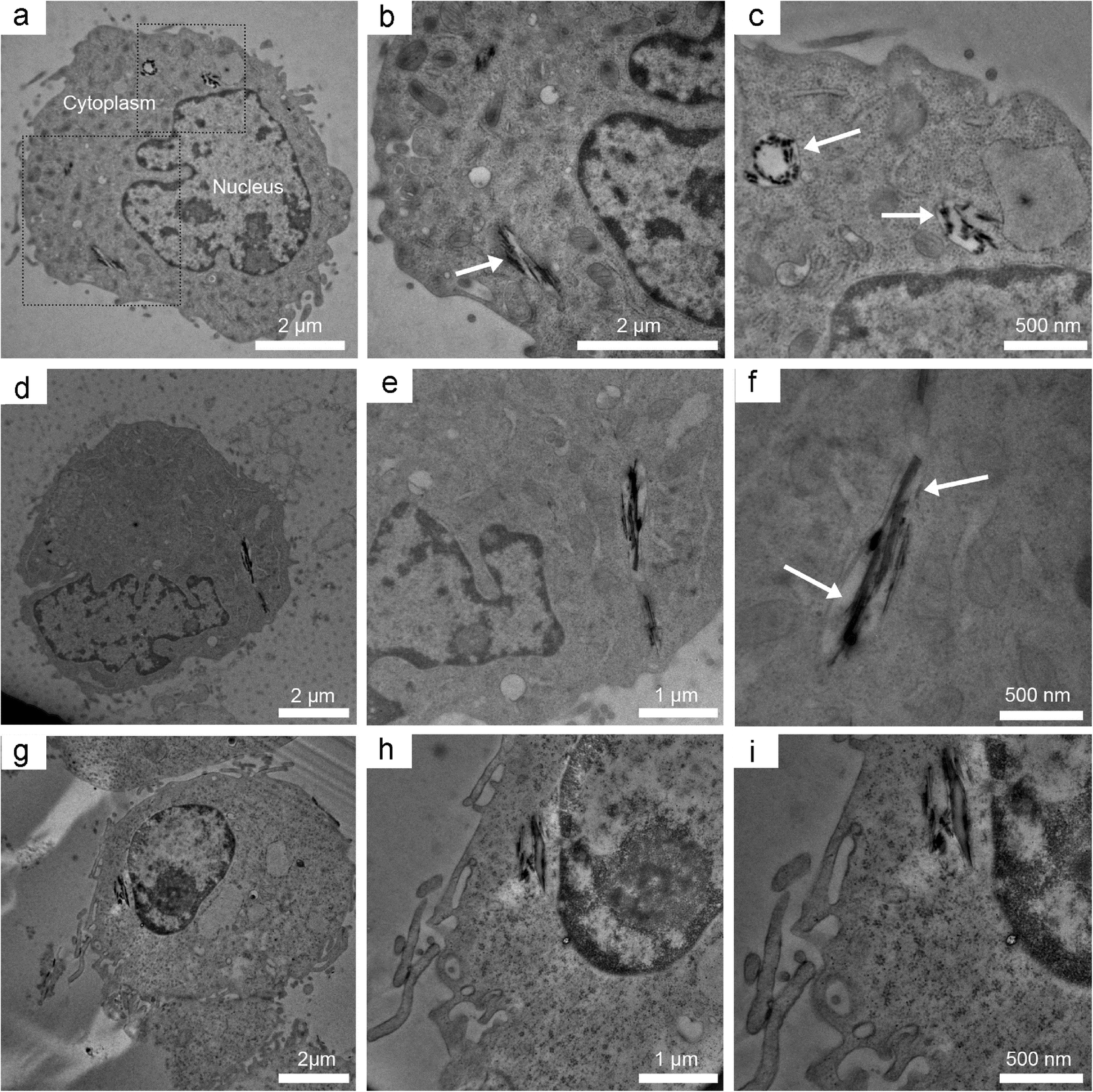
Sepiolite can be internalized spontaneously by eukaryotic cells, and endocytic and non-endocytic mechanisms were identified as the main internalization mechanisms (Castro-Smirnov Reference Castro-Smirnov2014). The spontaneous cellular internalization of sepiolite fibers was confirmed in V79 cells by laser confocal microscopy. Remarkably, time-lapse video-fluorescent microscopy revealed that mammalian cells were able to internalize sepiolite fibers spontaneously but were also able to eject them, and in this way, sepiolite nanofibers can be transported between adjacent cells. Observations by TEM revealed that the sepiolite fibers were surrounded by membranes or endosomes in the cytoplasm, suggesting an internalization mechanism through endocytosis (Fig. 8) (Castro-Smirnov Reference Castro-Smirnov2014).
TEM images of a set of three different V79 cells incubated with sepiolite nanofibers (a, d, and g) with progressive enlargement (×2) of internal sepiolite fibers in the respective images to the right (10 ng/μL of sepiolite in 5 × 10⁶ cells). The arrows indicate sepiolite nanofibers embedded in membranes (Castro-Smirnov Reference Castro-Smirnov2014). Images a, b, and c reproduced, with permission, from Castro-Smirnov et al. (Reference Castro-Smirnov, Ayache, Bertrand, Dardillac, Le Cam, Piétrement, Aranda, Ruiz-Hitzky and Lopez2017a)
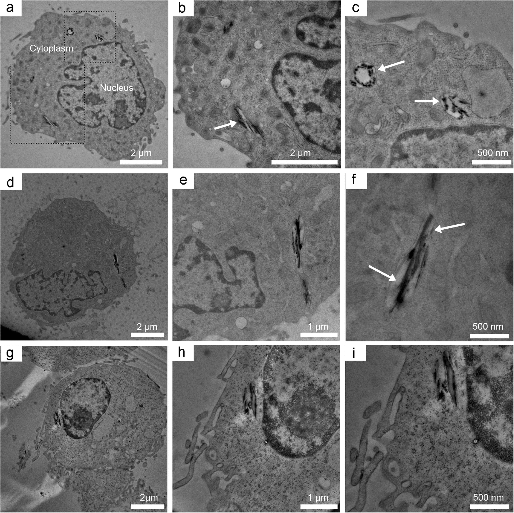
Specific endocytosis and macropinocytosis structures were observed at the membrane/sepiolite junction. Interestingly, some sepiolite fibers were not surrounded by endosomal membranes, suggesting an alternative pathway for endocytosis in the internalization of sepiolite (direct cytoplasmic membrane insertion). The involvement of macropinocytosis and clathrin-mediated endocytosis following incubation with sepiolite in the presence of endocytosis inhibitors were determined quantitatively using fluorescence-activated cell sorting (FACS) analysis. While chloroquine reduced by only 20% the sepiolite cellular internalization, amiloride inhibited it by 50%, showing that one of the main mechanisms of cellular internalization of sepiolite is macropinocitosis, in agreement with previous observations using TEM (Castro-Smirnov et al. Reference Castro-Smirnov, Ayache, Bertrand, Dardillac, Le Cam, Piétrement, Aranda, Ruiz-Hitzky and Lopez2017a).
In the aforementioned works, a detailed characterization of the (Sep/DNA) biohybrid materials was performed using diverse techniques and methodologies, proving that the DNA is adsorbed reversibly and efficiently on sepiolite nanofibers, previously disentangled and individualized as much as possible, through the silanol groups regularly located at the external surface of sepiolite by interactions that imply electrostatic attraction, hydrogen bonding, cation bridges, and van der Waals forces (Castro-Smirnov et al. Reference Castro-Smirnov, Piétrement, Aranda, Bertrand, Ayache, Le Cam, Ruiz-Hitzky and Lopez2016).
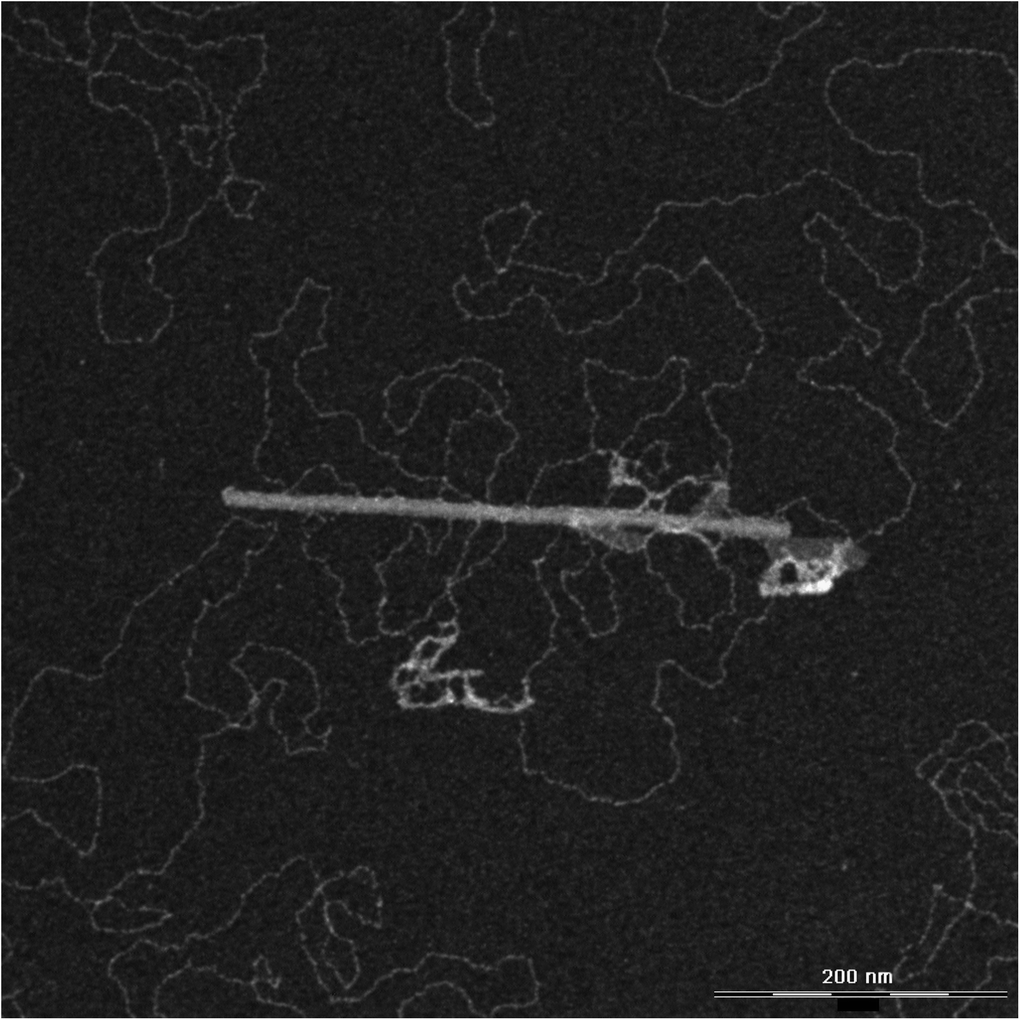
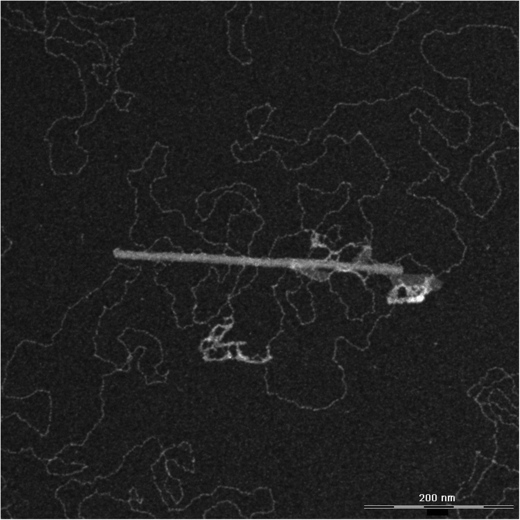
DNA could actually be adsorbed onto sepiolite spontaneously and polyvalent cations such as Mg2+, Ca2+, spermidine, and spermine stimulate strongly the DNA adsorption in a way that is correlated directly with the valence of the cations. Up to ~300 μg of DNA could be adsorbed per mg of sepiolite in the presence of the spermine tetravalent cation. Fourier-transform infrared spectroscopy (FTIR) confirmed that the interaction of DNA with sepiolite occurred through the external silanol groups of sepiolite (Castro-Smirnov et al. Reference Castro-Smirnov, Piétrement, Aranda, Bertrand, Ayache, Le Cam, Ruiz-Hitzky and Lopez2016). Moreover, analyses using TEM and atomic force microscopy (AFM) showed that sepiolite fibers assembled DNA biomolecules on their outer surface (Fig. 9).
TEM image of the sepiolite/DNA biohybrid, showing a sepiolite nanofiber with a plasmid DNA molecule adsorbed to its surface (image from O. Piétrement and F.A. Castro-Smirnov)
In addition, the reversibility of the DNA adsorption process onto sepiolite was confirmed, showing the possibility of recovering DNA which had been adsorbed previously on the fibrous clay. For this, the DNA-sepiolite biohybrid was re-suspended in the well known, standard, metal-ion chelating agent, ethylenediaminetetraacetic acid (EDTA). The displacement and sequestration of polycations favoring DNA binding to sepiolite allowed the recovery of DNA previously adsorbed on the clay surface through cation bridges. The quality of the DNA plasmids recovered by EDTA re-suspension was estimated by electrophoresis: the distribution of various DNA isoforms (supercoiled, open circle, linear) was unchanged during the sepiolite adsorption-desorption processes (Castro-Smirnov et al. Reference Castro-Smirnov, Piétrement, Aranda, Bertrand, Ayache, Le Cam, Ruiz-Hitzky and Lopez2016). This demonstrated that interaction with sepiolite does not affect the biological quality of DNA and even more, that the method of incubation of DNA with sepiolite nanoparticles and a chelating agent can be established as a new methodology for the extraction and purification of DNA, much cheaper than some available commercial systems (Castro-Smirnov Reference Castro-Smirnov2014).
Sepiolite is able to transfer small interfering RNA (FTIC labeled siRNA) efficiently into human cancer cells (A673 sarcoma cells) (Castro-Smirnov et al. Reference Castro-Smirnov, Piétrement, Aranda, Bertrand, Ayache, Le Cam, Ruiz-Hitzky and Lopez2016), with potential applications in gene silencing. The ability of the Sep/DNA biohybrid to transfer DNA into the nucleus of mammalian cells was evaluated by preparing the Sep/DNA using a specific DNA (the pCMV plasmid), which harbors a gene conferring resistance to G418 antibiotic cells. The efficiency of DNA transfer was then measured in cells through selection of G418-resistant cell colonies after the exposure of cells to Sep/DNA. Numerous resistant colonies were observed in V79 hamster cancer cells and U2OS human osteosarcoma cells after 10 days of incubation with the G418 antibiotic, demonstrating the ability of sepiolite to transfer an exogenous DNA into mammalian cells (Castro-Smirnov et al. Reference Castro-Smirnov, Piétrement, Aranda, Bertrand, Ayache, Le Cam, Ruiz-Hitzky and Lopez2016).
Another strategy has since been shown to increase the efficiency of sepiolite-mediated DNA transfection. This strategy consisted of disaggregating the sepiolite fibers more efficiently by sonication of the sepiolite suspension (sonicated sepiolite samples: SSEP), and the prior incubation of the cells with endocytosis inhibitors (chloroquine or amiloride) to favor the endosomal escape of nanofibers (Castro-Smirnov et al. Reference Castro-Smirnov, Rodriguez-Hoyos, Guzmán-Martínez, Lopez, Piétrement, Ayache, le Cam, Bertrand, Aranda and Ruiz-Hitzky2017b). Surprisingly, sonicated sepiolite complexes increased transfection efficiency by two orders of magnitude when applied to human cells in culture. In human cells and using biohybrids initially prepared using SSEP, chloroquine favors endosomal escape more effectively than the inhibition of the internalization of the bionanohybrid, thus increasing the transfection efficiency to 900 resistant colonies per microgram of DNA (Castro-Smirnov et al. Reference Castro-Smirnov, Rodriguez-Hoyos, Guzmán-Martínez, Lopez, Piétrement, Ayache, le Cam, Bertrand, Aranda and Ruiz-Hitzky2017b). Furthermore, a transfection efficiency similar to and only slightly less than that obtained using conventional transfection methods such as Jet PEI was achieved by combining the two protocols, chloroquine pretreatment plus SSEP/DNA (Castro-Smirnov et al. Reference Castro-Smirnov, Ayache, Bertrand, Dardillac, Le Cam, Piétrement, Aranda, Ruiz-Hitzky and Lopez2017a). This strategy is further complemented by the advantage sepiolite has in being cheaper than existing transfection agents and being non-toxic to cells.
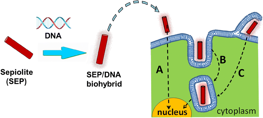
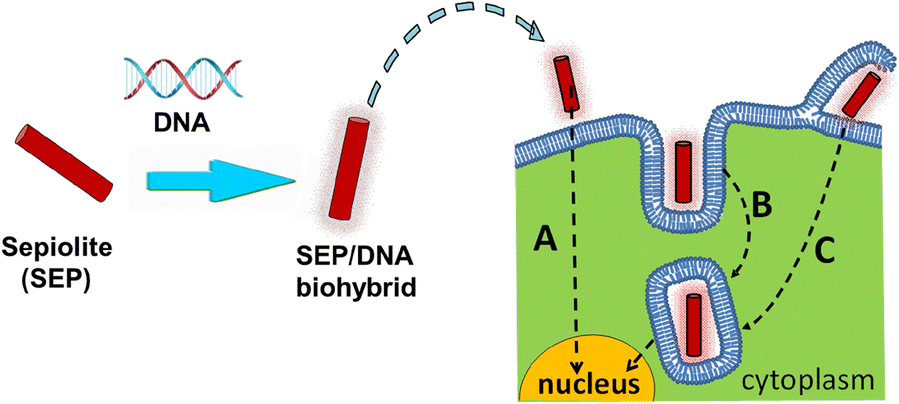
Overall, the abovementioned results indicate that sepiolite nanofibers can be internalized spontaneously and efficiently by mammalian cells through several processes, but mainly by clathrin-mediated endocytosis and macropinocytosis into cells, favoring the delivery of bound molecules such as DNA (Castro-Smirnov et al. Reference Castro-Smirnov, Ayache, Bertrand, Dardillac, Le Cam, Piétrement, Aranda, Ruiz-Hitzky and Lopez2017a). These processes are summarized in Fig. 10 which indicates some proposed pathways for internalization of the sepiolite/DNA biohybrid into the cell cytoplasm. After spontaneous release, the nucleic acid can finally be imported into the nucleus leading to its stable integration into the cell genome.
Schematic representation of sepiolite-mediated DNA transfection in mammalian cells. First, the sepiolite/DNA (SEP/DNA) biohybrid is prepared by adsorption of DNA on the sepiolite nanofibers; next, the Sep/DNA biohybrid is added to the cell culture media in which mammalian cells are incubated. SEP/DNA can be internalized spontaneously into cells either by direct insertion (a), or via clathrin-mediated endocytosis (b), or via macropinocytosis (c) (from (Castro-Smirnov et al. Reference Castro-Smirnov, Ayache, Bertrand, Dardillac, Le Cam, Piétrement, Aranda, Ruiz-Hitzky and Lopez2017a))
These results provide evidence, therefore, that biohybrids based on sepiolite nanofibers constitute an efficient transfection system for the vectorization (i.e. attachment to vectors) of different biological molecules in mammalian cells and particularly humans, with the aim of repairing or silencing a faulty gene or of incorporating a new genetic function, with potential applications in nanomedicine and nanobiotechnology (Castro-Smirnov et al. Reference Castro-Smirnov, Rodriguez-Hoyos, Guzmán-Martínez, Lopez, Piétrement, Ayache, le Cam, Bertrand, Aranda and Ruiz-Hitzky2017a).
Conclusions
The use of clays in the development of biohybrid materials for biomedical and pharmaceutical applications requires particular attention to the key role of the interfaces involved. In certain cases, direct assembly of the bioactive species with the clay can be achieved, as proved with the example of DNA vectorization using sepiolite fibrous clay reported above. In other cases, however, a prior modification of the clay surface by treatment with biopolymers or amphiphilic molecules, such as phosphatidycholine, is required to obtain biointerfaces on which further assembly of bioactive species was possible. A clear example that shows the importance of these biointerfaces is that which deals with the development of vaccines as the surroundings in which the virus is immobilized assure the maintenance of its bioactivity as immunogenic material, in this case also providing thermal and structural protection. Another relevant feature of biointerfaces is shown with the various examples described here, dealing with the development of new formulations for controlled drug delivery. Though many pharmaceutical drugs can be immobilized on clay minerals and LDH, their controlled release from the substrate can only be achieved and modulated after association with other compounds, such as biopolymers, which in certain cases also affords convenient processing and/or functionalities for action at specific sites and/or conditions.
Future activity in the clay-biohybrid materials area for use in integrative medicine will not only focus on the expected applications as traditional DDS of novel drugs for targeted therapies but also for molecular diagnostic tests with the aim of early detection and treatment. In fact, diverse analytical devices have already been developed, based on this type of biohybrid material, which show sensing ability introduced by both components, i.e. the clay substrate and the materials of biological origin. Thus, both rapid detection and diagnosis by in situ analysis of blood and other physiological fluids, biopsy tumors, infections of organs and tissues, etc. can be expected. Note that in a few decades, the genome will be sequenced at very low cost and so cancer, Alzheimer’s, and many other diseases will be largely preventable, which makes essential the development of new detection systems, perhaps some of them based on multifunctional biohybrids. This last type of material could be also of interest, as examples of it are very adaptable to pharmaceutical 3D printing for upcoming tailored treatments.
All of these approaches will potentially be addressed mostly to diseases of increasing incidence, e.g. cancers and manageable chronic diseases, involving organ (heart, lung, kidney, etc.) failures, brain and spinal cord injuries, obesity, ophtalmological and Parkinson’s, among others diseases, and, finally, contributing to a significant improvement in human health where clay-based biohybrid materials could make a valuable contribution.
ACKNOWLEDGMENTS
The authors acknowledge the financial support from MINECO, Spain (MAT2015-71117-R), and the EU COST Action Program (MP1202). B. Wicklein acknowledges MINECO, Spain, for the Juan de la Cierva grant (IJCI-2015-23886). The authors thank Dr. O. Piétrement and Dr. B.S. Lopez for fruitful discussions about sepiolite-DNA gene-transfer results.