INTRODUCTION
Babesia are tick-transmitted protozoa that comprise some of the most ubiquitous and widespread parasites of erythrocytes in humans and a wide range of wild and economically valuable domestic animals such as cattle and horses (Kuttler, 1988; Kjemtrup and Conrad, 2000; Homer et al. 2000; Wei et al. 2001). These single-celled organisms invade red blood cells and cause fatigue, aches, fever, chills, sweating, dark urine, enlarged spleen and anaemia. Infections can range from no serious symptoms to a fatal disease. The life-cycle of Babesia includes asexual and sexual stages in the vertebrate and tick host, respectively. Ticks transfer Babesia sporozoites during feeding and the sporozoites invade the host red blood cells. When Babesia gametocytes are ingested by a suitable tick host they develop into gametes, fuse to form a motile zygote that penetrates the peritrophic matrix (PM), enters the cells of the intestinal epithelium and divides into kinetes. The kinetes break free, enter the body cavity (haemocoel) by crossing the midgut and migrate to the salivary glands. Sporozoites invade the salivary glands to be injected into a vertebrate host when the tick ingests an infected bloodmeal (Kuttler, 1988). For transmission to occur, therefore, the Babesia parasite must complete an elaborate developmental programme in the hostile tick environment. However, the precise mechanisms by which ticks limit parasite development still remain to be elucidated.
In mosquito responses to midgut invasion by malarial parasites, microvillar proteins, peritrophic matrix and mosquito digestive enzymes appear important as barriers to parasite development (Shen et al. 1999; Sinden and Billingsley, 2001; Abraham and Jacobs-Lorena, 2004). Soft ticks also have a number of midgut barriers such as a peritrophic membrane (Grandjean, 1983), gut lysozymes (Kopacek et al. 1999) and antimicrobial peptides (Nakajima et al. 2002a,b) that limit or prevent invasion of parasites in non-vector species. The non-vector tick Ornithodoros moubata is easier to maintain and handle in laboratory experiments than vector ticks like Haemaphysalis longicornis because they are larger in size and have a short blood-feeding period. Therefore, if the Babesia parasite can develop in O. moubata, it may be useful for elucidating the genetic pathways involved in vector competence and clarifying the importance of the midgut barrier for development and transmission of Babesia parasites in ticks.
MATERIALS AND METHODS
Parasites
The United States Department of Agriculture (USDA) strain of Babesia equi was grown in horse erythrocytes in vitro as described by Avarzed et al. (1997). A B. gibsoni strain isolated from a hunting dog in Hyougo Prefecture Japan, designated strain NRCPD (Fukumoto et al. 2001a) was used for experimental infection of beagles.
Dogs
One-year-old female beagle dogs were confirmed to be free of natural B. gibsoni infection by detection of specific antibody prior to use in the experiments. All animal experiments were conducted in accordance with Standards for the Care and Management of Experimental Animals promulgated by the National Research Center for Protozoan Diseases, Obihiro University of Agriculture and Veterinary Medicine, Japan.
Ticks
Soft ticks, Ornithodoros moubata (Acari: Argasidae), were obtained from the National Institute of Animal Health (Tsukuba, Japan) and have been maintained at the National Research Center for Protozoan Diseases, Obihiro University of Agriculture and Veterinary Medicine for several generations by feeding on rabbits (Oryctolagus cunniculus) and maintained at 27±1 °C, 50–60% RH and total darkness. Feeding and rearing of ticks were performed as described by Chinzei (1983).
Artificial feeding
An artificial feeding study was used to introduce B. equi into the tick digestive tract. Adult female O. moubata were fed by an artificial membrane system using Parafilm M (Pechiney plastic packaging Menasha, WI 54952) similar to the Baudruche membrane described by Waladde et al. (1991) and Matsuo et al. (2004). The artificial meal consisted of B. equi-infected red blood cells with 12% parasitaemia suspended in the same volume of PBS.
Tick injection
Ticks were infected under the last coxa with 15 μl of B. equi- or B. gibsoni-infected RBC suspended in PBS with a 27-gauge intravenous needle.
Reverse transcriptase-polymerase chain reaction analysis (RT-PCR) and PCR
Total RNA was extracted with solution D, prepared by adding 2-mercaptoethanol to a stock guanidinium thiocyanate solution (Chomczynski and Sacchi, 1987) from injected and artificially fed ticks. Then 1 μg of total RNA from each sample was applied to a One Step RNA PCR kit (TAKARA, Otsu, Japan) according to the conditions recommended by the manufacturer with the following primers. Primers designed from 18S ribosomal RNA of B. equi were rE1F 5′-GTTTATTTGATGTTTGTTT-3′ and rE1R 5′-CCAAGCGCAGTCAACGAAA-3′ and for O. moubata ribosomal protein 18S were rOM 5′-GTTCCTTCC TTGATTGTCATGAG-3′ and rOM 5′-TCGGTTAGATGCACTGCTCGTCT-3′ as a loading control. Tick and blood samples collected from dogs were lysed in 0·1 M Tris-HCl (pH 8·0) containing 1% SDS, 0·1 M NaCl and 10 mM EDTA and treated with proteinase K (100 μg/ml) for 2 h at 55 °C. The DNA was extracted with phenol/chloroform and precipitated with ethanol, then dissolved in 25 μl of TE buffer and stored at 4 °C. PCR was performed with p18d3 and p18d4 oligonucleotide primers (5′-TCCGTTCCCACAA-CACCAGC-3′ and 5′-CGAATGAGGATGATG-AGGAGGA-3′) as targets for the 182 bp fragment of B. gibsoni P18 gene (Fukumoto et al. 2001b). DNA products were run on an agarose gel, stained with ethidium bromide and photographed.
Transmission electron microscopy (TEM)
The salivary glands and ovaries of ticks 7 days after injection with Babesia parasites were removed, fixed with cold 3% glutaraldehyde in sodium cacodylate buffer (pH 7·4) overnight at 4 °C, post-fixed with 1% OsO4 in the same buffer for 2 h at 4 °C after washing thoroughly with the same buffer, dehydrated in an ethanol series and embedded in quetol 651 resin (Nissin EM, Tokyo, Japan). Thin sections (approximately 80 nm thick) were cut on a Leica UCT ultramicrotome using a diamond knife and doubly stained with uranyl acetate and lead citrate before examination with a Hitachi H-7500 electron microscope.
Immuno-electron microscopy (IEM)
The ovaries were removed from ticks injected with infected RBC of B. equi and B. gibsoni, fixed in 4% paraformaldehyde with 0·1% glutaraldehyde in PBS overnight at 4 °C, washed thoroughly in PBS and embedded in 2% agarose. After dehydration with an ethanol series, the samples were embedded in LR Gold resin (Polysciences Inc., USA). Thin sections (about 80 nm thick) were cut on a Leica UCT ultramicrotome using a diamond knife and placed on nickel grids. Sections were exposed at room temperature (RT) for 30 min to 5% skim milk PBS as a blocking agent then incubated with anti-B. equi specific merozoite antigen 1 and EMA-1 (Kappmeyer et al. 1993) or EMA–2 (Knowles et al. 1997) antibodies overnight at 4 °C, and subsequently incubated with 10 nm gold-labelled goat anti-mouse IgG+IgM antibody (Amersham Biosciences, UK) at RT for 2 h. Normal mouse IgG was used instead of the primary antibodies as a negative control. Finally, these sections were counter-stained with uranyl acetate and lead citrate before examination with a H-7500 transmission electron microscope (HITACHI, Japan).
Tick transmission challenge on a dog
The ears of beagles infected with B. gibsoni by injection of infected blood were infested with 20 fourth instar nymphs (N4) and 20 adult O. moubata ticks. Ticks were recovered from the dogs 3 h after infestation when all ticks had dropped off. A thin blood smear from each dog was fixed with methanol for 2 min and stained with Giemsa solution for 30 min. The slides were then microscopically examined to calculate the peripheral parasitaemia.
Splenectomy of a dog
At day 20 after tick infestation the experimental dog was splenectomized under anaesthesia as described earlier by Fukumoto et al. (2000). All surgical procedures were conducted in accordance with the Guiding Principles for the Care and Use of Research Animals promulgated by Obihiro University of Agriculture and Veterinary Medicine.
ELISA
ELISA was carried out with B. gibsoni-specific antibodies according to the methods described previously (Fukumoto et al. 2004). Microplates (96 well) were coated with GST-Bg-TRAP (B. gibsoni-thrombospondin related adhesive protein) (Zhou et al. 2006) and GST (negative control) antigens at a concentration of 100 ng per well. The OD value of 0·1 was estimated as a cut-off value expressed as the reciprocal of the maximum dilution. The absorbance was shown in the distance of OD value of the antigen (GST-Bg-TRAP) well and the control antigen (GST) well.
RESULTS
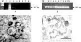
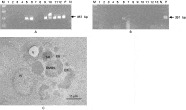
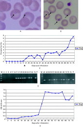
O. moubata ticks were fed blood infected with Babesia equi, which causes babesiosis in horses. The ticks ingested a bloodmeal, as evidenced by changes in tick size and form, but the parasites were quickly cleared as shown by the disappearance of B. equi specific ribosomal RNA (rRNA) from the ticks 5 days after feeding (Fig. 1A). The Babesia parasite rRNA was not detected in the babesia-fed ticks, suggesting that the parasites were passed in the feces or digested. The failure of Babesia parasites fed to O. moubata to establish an infection is likely a consequence of the parasite's inability to invade the gut cells or survive tick midgut-associated barriers such as the peritrophic membrane (Grandjean, 1983), and presence of lysozymes (Kopacek et al. 1999) and defensins (Nakajima et al. 2002a,b) in the midgut. Therefore, we hypothesized that if we could succeed in developing a way for the Babesia parasite to escape/avoid the midgut-associated immune barrier, a non-vector tick may become infected with Babesia. Moreover, it is very important to see whether O. moubata can transmit the Babesia parasite to a vertebrate host under such conditions. We conducted 2 experiments to test this hypothesis. First, B. equi parasite-infected horse blood from in vitro culture was prepared and injected directly into the haemocoel of adult female ticks to determine if the Babesia parasite can survive in the body of the non-vector tick. Next, infected ticks were allowed to feed on Babesia-free dogs to determine whether the parasite could be transmitted to the vertebrate host by the non-vector tick. In the first experiment B. equi-specific rRNA was detected up to 45 days after injection of the parasite (Fig. 1B). Very surprisingly, the rRNA was detectable even in eggs oviposited by injected ticks. These results suggest that the parasites can survive in the tick haemocoel and develop to be transmitted to the next generation through the egg stage. If the parasite can be transmitted transovarially the parasite oocysts must invade the tick ovary. This was confirmed by observing the Babesia parasites in the ovary by transmission electron microscopy (TEM). Numerous Babesia-like structures were ultrastructurally identified in the oocytes of ticks injected with Babesia-infected RBC (Fig. 1C). The surface and periphery of the oocyte cytoplasm of these structures reacted with both anti-EMA-1 and -2 antibodies confirming the presence of the Babesia parasites (Fig. 1D). The reactions against the anti-EMA-2 antibody were slightly stronger than against the anti-EMA-1 antibody. No Babesia-like structures were detected in the oocytes of normal ticks and no reaction against the antibodies was seen in normal ticks and the negative control. The second experiment was conducted with B. gibsoni-infected dog red blood cells to determine whether the O. moubata generated sporozoites were infectious to a vertebrate host. B. gibsoni-infected dog red blood cells were injected into the haemocoel of O. moubata and infection confirmed by PCR and TEM (Fig. 2A,B,C). No difference in the survival of O. moubata-injected ticks during babesial infection was observed. Then Babesia-free dogs were infested with 20 fourth instar nymphs (N4) and 20 adult females of O. moubata infected by inoculation with B. gibsoni-infected red blood cells. However, the B. gibsoni parasites were not detected by PCR until 2 weeks after infestation and the blood smears stained by Giemsa solution showed less than 1% parasitaemia until 3 weeks after infestation. However, after successful splenectomy of the dogs the parasitaemia increased gradually so that at 75 days the parasitaemia was as high as 8% (Fig. 3A,B,C). Moreover, the antibody production against B. gibsoni thrombospondin-related adhesive protein (Bg-TRAP) (Zhou et al. 2006) also gradually increased (Fig. 3E). These results indicate that the dogs became infected with B. gibsoni after infestation with B. gibsoni-infected O. moubata ticks. The dogs showed typical clinical symptoms for Babesia infection such as fever, progressive haemolytic anaemia and haemoglobinuria (data not shown).

Fig. 1. Detection of Babesia equi in Ornithodoros moubata ticks. (A) RT-PCR for detection of B. equi rRNA in O. moubata tick samples after artificial feeding with B. equi-infected red blood cells (8% parasitaemia). Lane M, Marker; Lane 1, Day 1; Lane 2, Day 3; Lane 3, Day 5; Lane 4, Day 9; Lanes 5–8, tick sample before feeding. (B) RT-PCR for detection of B. equi rRNA in O. moubata tick samples after injection of B. equi-infected red blood cells into haemocoel of ticks. Lane M, Marker; Lane 1, Day 1; Lane 2, Day 7; Lane 3, Day 15; Lane 4, Day 45; Lanes 5–9 are samples of eggs; Lane P, positive control (RNA from in vitro culture of B. equi); Lane N, negative control (tick injected with normal RBC). (C) Electron micrographs showing B. equi and (D) immuno-electron microscopy using anti-EMA-2 antibody against the parasites in the ovary of O. moubata 1 week after injection with infected RBC. DMBS, double membrane-bounded structure; ER, endoplasmic reticulum; N, nucleus; SB, spherical body.

Fig. 2. PCR for detection of Babesia gibsoni DNA in Ornithodoros moubata ticks. (A) Nymph and female samples. Lane M, Marker; Lanes 1–6, samples of N4 female 7 days after injection with B. gibsoni-infected dog blood. Lanes 1–4, negative; Lanes 5 and 6, positive. Lanes 7–12, samples of adult females 7 days after injection with B. gibsoni-infected dog blood. Lanes 7 and 8, negative; Lanes 9–12, positive. (B) Egg samples. Lane M, Marker; Lanes 1–13 are samples of eggs from females injected with B. gibsoni- infected dog blood. Lanes 6, 9 and 11, positive; Lane N, negative control – tick DNA injected with normal dog blood. Lane P, positive control – B. gibsoni DNA samples. (C) Electron micrographs showing B. gibsoni in the ovary of O. moubata 1 week after injection of infected RBC. DMBS, double membrane-bounded structure; ER, endoplasmic reticulum; N, nucleus; SB, spherical body.

Fig. 3. Transmission of Babesia gibsoni to dogs by Ornithodoros moubata ticks. (A and B) Light microscopy of Giemsa-stained thin blood smears. B. gibsoni infection in an experimental dog infested with B. gibsoni-infected O. moubata ticks. (C) Infectious course of B. gibsoni in an experimental dog infested with B. gibsoni-infected O. moubata ticks. [utrif ], Points to day of splenectomy. (D) Detection of B. gibsoni infection by PCR in an experimental dog. Lane numbers show days post-infestation with infected O. moubata ticks. (E) Detection of antibodies against the Bg-TRAP protein by ELISA in a dog experimentally infested with B. gibsoni-infected O. moubata ticks. Dotted line shows 0·1 cut-off value. The mean values from 3 individual wells are shown.
DISCUSSION
This is the first report to show that Babesia merozoites can develop in a non-vector tick and develop into sporozoites that are infective for vertebrate host animals. The results of this study indicate that the midgut may act as a major barrier for prevention of development of Babesia in non-vector ticks. Kopacek et al. (1999) have shown that O. moubata ticks have lysozymes in the gut that may function in the immune response. Nakajima et al. (2001, 2002a,b) have shown that defensins are secreted into the midgut lumen and have strong antimicrobial activity. These immune responses in the gut may play a major role in the prevention of Babesia invasion in the non-vector tick O. moubata. Defensins have also been isolated from various hard tick species (Johns et al. 2001; Fogaca et al. 2004; Lai et al. 2004). However, Sonenshine et al. (2005) showed that defensins are not secreted into the midgut of Dermacentor variabilis and concluded that antimicrobial activity in D. variabilis is primarily related to proteins derived from the host bloodmeal such as haemoglobin fragments (Fogaca et al. 1999; Nakajima et al. 2003, 2005). These studies indicate that there are important differences in the role of the midgut in the immune response of soft ticks and hard ticks. These differences may be important in determining the microorganisms that can be transmitted by certain tick species. This study indicates that the Babesia may be affected by these differences because if the parasite is injected into the haemocoel of the non-vector tick O. moubata, avoiding the midgut immune mechanisms, it can develop and be successfully transmitted to a vertebrate host. Further studies comparing midgut immune mechanisms in non-vector and vector ticks of Babesia as well as other infective microorganisms are needed.
Parasites that escape the midgut barriers then face two principal immune responses: the humoral factors present in the haemolymph that cause melanization and coagulation, and cellular responses of phagocytosis and encapsulation. As described for immunocytes of other arthropod species, most notably the insects (Gillespie et al. 1997), tick haemocytes participate in both cellular (Kryuchechnikov, 1991; Johns et al. 1998; Inoue et al. 2001; Ceraul et al. 2002) and humoral (Ceraul et al. 2003; Simser et al. 2004) immune responses in response to various types of challenges. In this study, the injected Babesia appeared to deal with the humoral factors present in the haemolymph such as phenoloxidases and plasma (Kadota et al. 2002) in O. moubata as with the host tick because Babesia was successfully transmitted to host dogs by O. moubata after injection. The cellular and humoral factors important in the immune responses of ticks are still poorly understood. Recently, very interesting molecules such as α–2-macroglobulin-like glycoprotein (TAM), have been reported from O. moubata haemolymph (Kopacek et al. 2000; Saravanan et al. 2003). Moreover, a significant up-regulation of TAM expression was observed in haemocytes, salivary glands and gut tissues of the ticks 24 h after a bloodmeal. The mechanisms controlling the transcription of the TAM gene are unclear but one activating factor may be an immune response to foreign particles (Saravanan et al. 2003). Research with O. moubata has numerous advantages available for investigation of not only Babesia-tick interactions but also tick vector immunology. By identifying factors and molecules that are essential to the survival of Babesia in the haemocoel of O. moubata, these experiments may contribute to the elucidation of factors in the host tick and the development of transmission-blocking vaccines against babesial infection. Further studies on the differences between Babesia transmission in vector and non-vector ticks may help to elucidate the mechanisms by which Babesia invades the tick and can be transmitted to vertebrate hosts. In addition, Babesia must overcome the immune responses of the host tick to be transmitted successfully. However, the understanding of tick innate immunity against pathogens especially the Babesia parasite is still in its infancy. These results indicate that O. moubata may provide a biological tool with numerous advantages available for investigation of not only Babesia-tick interactions but also tick vector immunology.
We thank Dr Fukumoto S. for kindly providing Babesia gibsoni parasite-infected blood and technical support. This study was supported by Grant-in-Aid for Scientific Research from the Japan Society for the Promotion of Sciences, by grants from the Bio-oriented Technology Research Advancement Institution (BRAIN) and by the 21st Center of Excellence Program of the Scientific Research from the Ministry of Education, Culture, Sports, Science, and Technology of Japan.