Implications
Dysregulation in the concentration of particular proteins has been used for the detection of pathological states; however, the obtained results could be of limited specificity for particular diseases. Glycoprotein macro and microheterogeneity determination are expected to improve disease specificity testing and could reflect disease-specific signatures even if individual protein concentrations remain similar. Presumed glycopattern alterations in pigs suffering from acute rectal prolapse have been observed in saliva, but not in serum in the present study. These results highlight the potential of saliva samples to provide additional information on disease condition and require intensive exploration in further studies.
Introduction
Glycosylation, mainly in the form of N- and O-linked glycans, is considered to be one of the most common forms of post-translational modification found on secreted and membrane proteins (Moremen et al., Reference Moremen, Tiemeyer and Nairn2012). As glycans seem to provide the first interface between the mammalian host and pathogens, glycosylation plays a crucial role in pathogenicity and invasion (Novotny et al., Reference Novotny, Alley and Mann2012). Changes in protein glycosylation have an important function also in the progression of human diseases (Fanayan et al., Reference Fanayan, Hincapie and Hancock2012; Almeida and Kolarich Reference Almeida and Kolarich2016; Loke et al., Reference Loke, Kolarich, Packer and Thaysen-Andersen2016). In fact, glycosylation patterns in chronic diseases can be highly aberrant as a consequence of changes in the expression or activity of glycosyltransferases or other factors affecting the glycan biosynthesis (Theodoratou et al., Reference Thaysen-Andersen, Thøgersen, Lademann, Offenberg, Giessing, Enghild, Nielsen, Brünner and Højrup2014). Glycoproteins represent an interesting subproteome that can be mined to discover putative robust, specific and selective diseases biomarkers (Ramachandran et al., Reference Ramachandran, Boontheung, Pang, Yan, Wong and Loo2008). Disease-specific glycoprofile changes of a specific protein or protein subset are considered to better reflect given physiologic condition compared with an overall dysregulation of a particular protein (Thaysen-Andersen et al., Reference Theodoratou, Campbell, Ventham, Kolarich, Pučić-Baković, Zoldoš, Fernandes, Pemberton, Rudan, Kennedy, Wuhrer, Nimmo, Annese, McGovern, Satsangi and Lauc2008). Although an up- or down-regulation in the level of specific markers could be observed in several pathological conditions, changes in the glycosylation patterns of these protein markers could prove disease specific, as reported for acute phase proteins (Kushner and Mackiewicz Reference Kushner and Mackiewicz1993; Almeida and Kolarich Reference Almeida and Kolarich2016). Thus, much effort has been put into extensive screening of glycoproteomic profiles of human biofluids such as serum, plasma, cerebrospinal fluid, urine and saliva (Ferreira et al., Reference Ferreira, Daniel-da-Silva, Alves, Duarte, Vieira, Santos, Vitorino and Amado2011).
Nowadays, saliva is gaining more interest as a source for clinical markers due to its potential to reflect systemic health conditions (Baldini et al., Reference Baldini, Giusti, Bazzichi, Lucacchini and Bombardieri2008). Numerous studies have sought to develop a complete catalogue of the proteins and peptides found in human saliva (Sondej et al., Reference Sondej, Denny, Xie, Ramachandran, Si, Takashima, Shi, Wong, Loo and Denny2009). Recent studies aim at a further comprehensive characterization of the post-translational modifications (Xu et al., Reference Xu, Bailey, Punyadeera and Schulz2014).
The specific enrichment and purification of glycoproteins/glycopeptides is essential before MS analysis as some glycoproteins are found in low abundance (Ongay et al., Reference Ongay, Boichenko, Govorukhina and Bischoff2012). Several methodologies have been used for the enrichment of glycoproteins such as lectin affinity purification, hydrazide-based chemical derivatization and hydrophilic affinity capture methods. A recent report has confirmed phenyl boronic acid (PBA)-based matrix as a valid non-destructive glycoprotein enrichment methodology for complex samples such as human whole saliva (Xu et al., Reference Xu, Bailey, Punyadeera and Schulz2014). However, no study about salivary glycoprotein profiles has been reported in any other species.
In this paper, PBA was used for salivary glycoprotein enrichment to obtain the overall salivary glycoprotein profile of healthy pigs for the first time. Moreover, the glycoprotein profiles obtained in healthy pigs and pigs with acute rectal prolapse were compared in order to identify possible disease-specific glycopatterns that could be related to the acute inflammatory condition. Proteins involved in those alterations were identified by MS analysis.
Material and methods
Animals and sample collection
A total of 10 pigs were included in the study. All pigs were males, conventional Duroc×(Landrace×Large White), which came from the same farm located in the Southeast of Spain. Five animals with rectal prolapse were selected. Five animals randomly selected from a pen in which animals had no clinical signs of disease at the time of sampling served as controls.
Whole saliva was collected at farm in the early morning by allowing the pigs to chew a sponge, clipped to a thin metal rod, for 1 to 2 min. Saliva was obtained by centrifugation of sponges inserted in specific tubes (Salivette tubes, Sarstedt, Nümbrecht, Germany) for 10 min at 3000×g. Blood was collected by veni-puncture of the jugular vein using vacutainer tubes (Becton Dickinson, New Jersey, USA) and was allowed to clot for 1 h at room temperature. To obtain serum, sample tubes were centrifuged for 15 min at 2000×g. Saliva and serum samples were stored at −80°C until analysis.
All procedures involving animals were approved by the Murcia University Ethics Committee and followed the recommendations of the European Convention for the Protection of Vertebrate Animals used for Experimental and Other Scientific Purposes (Council of Europe, ETS no. 123).
Quantification of total protein content and acute phase proteins
Total content of proteins of each saliva and serum sample was determined according to Bradford (Reference Bradford1976). Haptoglobin (Hp) was quantified by using a time-resolved immunofluorometric assay, which showed CV of 1.69% and 11.07% for intra and inter-assay precision, respectively (Gutiérrez et al., Reference Gutiérrez, Escribano, Fuentes and Cerón2009). C-reactive protein (CRP) quantifications were performed with a time-resolved immunofluorometric assay with CV for both intra and inter-assay imprecision below 10% (Gutiérrez et al., Reference Gutiérrez, Nöbauer, Soler, Razzazi-Fazeli, Gemeiner, Cerón and Miller2013a).
Glycoprotein enrichment
A glycoprotein enrichment kit, based on PBA matrix, was used (NuGelTM Glycoprotein enrichment PBA kit, Biotech Support Group, New Jersey, USA) according to the manufacturer’s instructions. In brief, 1 mg of serum or freeze-dried total saliva protein dissolved in 250 µl of binding buffer was added to 50 mg of PBA matrix, incubated for 10 min with continuous vortexing and centrifuged for 3 min at 12750 ×g. The unbound fraction was collected after centrifugation and stored at −20°C until analysis. Beads with glycoproteins were washed three times with 5 min incubation with 350 µl of wash buffer and a 2 min centrifugation at 12750 ×g. After a 10 min incubation with 300 µl of sorbitol elution buffer and a 3 min centrifugation at 12750 ×g, the glyco-enriched fraction was obtained. Two steps of dialysis against ammonium acetate buffer pH 6 were included within the enrichment procedure for saliva samples, using a mini dialysis kit 1 kDa (GE Healthcare Life Sciences, Munich, Germany). The first dialysis step was undertaken with saliva samples before the mixing of saliva and PBA matrix, and the second one was carried out on glyco-enriched fractions and unbound fractions. The glycoprotein enrichment procedure was performed in all saliva samples and in randomly selected serum samples from a healthy animal and a pig with rectal prolapse.
Two-dimensional gel electrophoresis and image processing
30 μg of freeze-dried whole saliva proteins or 15 μg of glyco-enriched or unbound fraction proteins were dissolved in rehydration butter and swollen into immobilized pH-gradient strips with 11 cm long non-linear gradients pH 3 to 11 (GE Healthcare Life Sciences, Munich, Germany). First-dimensional separation was carried out under reducing and denaturing conditions in a Multiphor II electrophoresis chamber (GE Healthcare Life Sciences, Munich, Germany). Isoelectrofocusing conditions included a multistep protocol in which voltage was increased in gradient mode with a maximum of 3500 V until 12 kVh at 20°C were reached. For the second dimension, strips were equilibrated for 10 min with 2% dithiothreitol (DTT) solution followed by 5 min with 2.5% of iodoacetamide solution, and SDS-PAGE was carried out at 15°C and 25 mA/gel for 4 to 5 h in homemade gradient 10 to 15% polyacrylamide gels of 140×140×1.5 mm. Gels were silverstained according to general protocols for obtaining overall patterns, whereas gels for MS analyses loaded with 30 µg total protein needed an MS compatible protocol (Miller et al., Reference Miller, Crawford and Gianazza2006).
Two-dimensional gel electrophoresis (2DE) gel images were digitalized using an ImageScanner II (GE Healthcare Life Sciences, Uppsala, Sweden) and evaluated by using specific software (Image Master 2D Platinum 7.0; GE Healthcare Life Sciences, Uppsala, Sweden). Analysis included spot detection, landmarking and spot matching of protein patterns of all gels in the set.
Sample preparation for MS identification
Spots of interest were excised manually from silverstained 2DE gels. After washing and destaining, spots were reduced with dithiothreitol and alkylated with iodoacetamide. In-gel digestion was performed with trypsin (Trypsin Gold, Mass Spectrometry Grade; Promega, Madison, WI, USA) with a final trypsin concentration of 20 ng/µl in 50 mM aqueous ammonium bicarbonate and 5 mM CaCl2. Digest proceeded for 8 h at 37°C (Shevchenko et al., Reference Shevchenko, Wilm, Vorm and Mann1996). Afterwards, peptides were extracted with three changes of 30 µl of 5% trifluoro acidic acid in 50% aqueous acetonitrile supported by ultrasonication for 10 min/change. Extracted peptides were dried down in a vacuum concentrator (Eppendorf, Hamburg, Germany). Dried peptides were concentrated and de-salted using Zip-Tips C18 (microbed) (Millipore, Billerica, MA, USA) according to the manufacturer’s instructions.
Tandem Matrix-assisted laser desorption/ionization Time-of-Flight (MALDI-TOF/TOF) MS and Database Search
De-salted peptides (0.5 µl) were spotted onto a disposable AnchorChip MALDI target plate pre-spotted with α-cyano-4-hydroxycinnamic acid (PAC target; Bruker Daltonics, Bremen, Germany). MALDI-TOF/TOF MS (Ultraflex II; Bruker Daltonics) was used for spectra acquisition in MS and MS/MS modes. Spectra processing and peak annotation were carried out using FlexAnalysis 3.0 and Biotools 3.2 (both Bruker Daltonics).
Processed spectra were searched via an in-house software server version 2.3 (Mascot, Matrix Science, Boston, MA, USA) and the software ProteinScape 2.1 (Bruker Daltonics) in the UniProt database (http://www.uniprot.org. Release 2014_07) using the following search parameters: global modification carbamidomethylation on cysteine; variable modifications oxidation on methionine; deamidation on asparagine and glutamine as well as formation of pyroglutamic acid; enzyme specificity trypsin; charge state z=1; MS tolerance 100 ppm; MS/MS tolerance 1 Da; one missed cleavage allowed; significance threshold P<0.05.
Western blot validation of proteins identified in uncommon positions in 2DE
Western blot analyses were performed both on one- and 2DE gels (protocols as reported above). A total of 15 µg of saliva, serum and glyco-enriched fractions from serum and saliva samples were applied for SDS-PAGE. 2DE was performed with saliva from a healthy animal and with serum and saliva from an animal suffering from rectal prolapse as reported above. After electrophoresis, proteins were transferred to polyvinylidene fluoride (PVDF) membranes at 25 V for 1 h by semi-dry blotting (Bio-Rad, Hercules, CA, USA) and tested using appropriate primary (Immunology Consultants Laboratory Inc., Newberg, OR, USA) and horseradish peroxidase (HRP)-conjugated secondary antibodies (Bio-Rad). The primary antibody used was an affinity purified rabbit polyclonal anti-Hp porcine immunoglobulin G (IgG) antibody that had been previously assessed for high reactivity with the corresponding porcine Hp (Gutiérrez et al., 2012). Detection of HRP activity was performed with an enhanced chemiluminescent substrate (Pierce™ ECL Western Blotting Substrate; Thermo Fisher Scientific, Rockford, IL, USA) and detected in a chemiluminescence imager (AI600; GE Healthcare Life Sciences, Uppsala, Sweden).
N-glycan analyses of gel-separated glycoproteins
Hp N-glycans were released and isolated from bands obtained after SDS-PAGE separation as described previously (Kolarich and Altmann, Reference Kolarich and Altmann2000). The released N-glycans were analyzed using a MALDI-TOF MS-based approach as described in detail elsewhere (Reiding et al., Reference Reiding, Blank, Kuijper, Deelder and Wuhrer2014).
Statistical analysis
Concentration differences of acute phase proteins (Hp and CRP) between healthy animals and pigs with rectal prolapse were statistically analyzed by using a non-parametric Mann Whitney test (GraphPad Prism 5; GraphPad software Inc., Suite, La Jolla, USA). The level of significance was set at P<0.05.
For the statistical analysis of any possible difference between classes (healthy pigs and pigs suffering from rectal prolapse) in the glyco-enriched fractions, the spot concentrations were calculated in ‘µg of spot/ml of saliva’ taking into account the total protein content of each saliva sample as reported before (Gutiérrez et al., 2013b). An unpaired t-test was used to evaluate spot concentration differences with the statistical software detailed above.
Results
Total salivary protein content and acute phase protein determinations
Mean total salivary protein content in healthy animals was 1.46 mg/ml (SD=0.23), and 2.39 mg/ml (SD=1.48) in pigs suffering from rectal prolapse, whereas levels of 68.44 mg/ml (SD=6.98) and 62.7 mg/ml (SD=10.90) were observed in serum samples from healthy pigs and pigs with rectal prolapse, respectively. No statistically significant differences in protein content were found between groups.
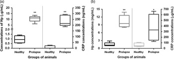
The levels of both acute phase proteins measured, Hp and CRP, were statistically significantly higher in the saliva of pigs suffering from rectal prolapse (11.21 μg/ml and 214.7 ng/ml for Hp and CRP, respectively) than in healthy animals (0.93 μg/ml and 0.50 ng/ml for Hp and CRP, respectively) (P=0.0079) (Figure 1a). The concentrations obtained in serum samples were also statistically significantly higher in pigs with rectal prolapse (9.50 mg/ml and 329.3 μg/ml for Hp and CRP, respectively) than in healthy pigs (1.25 mg/ml and 47.16 μg/ml for Hp and CRP, respectively), with levels of significance of P=0.0079 for Hp and 0.015 for CRP quantifications (Figure 1b).
Concentrations of haptoglobin (Hp) and C-reactive protein (CRP) in saliva samples (a; µg/ml or ng/ml) and serum samples (b; mg/ml or µg/ml) of healthy pigs and pigs suffering from rectal prolapse. The plot shows median (line within box), 25th and 75th percentiles (box), 5th and 95th percentiles (whiskers) and outliers (°). Asterisk represents the statistically significant differences from each group: *level of significance P<0.05; **level of significance P<0.01.

Glyco-enrichment results and MS identification
The percentage of protein recovered in the glyco-enriched fraction of saliva samples was similar in both groups of animals, yielding 8.35% (SD=1.74) in healthy pigs and 6.58% (SD=3.34) in animals suffering from rectal prolapse.
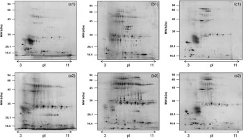
2DE was applied to follow the purification/enrichment process, as illustrated in Figure 2. Original patterns of a saliva sample from a healthy animal and a pig suffering from rectal prolapse before and after enrichment are displayed in Figure 2a. New spots were detected in both the enriched and the unbound fractions in comparison with the initial saliva sample as seen in Figure 2b and c.
Image of the (a) salivary protein patterns, (b) glyco-enriched fraction and (c) non-bound fraction after phenyl boronic acid enrichment protocol, obtained by two-dimensional gel electrophoresis, of samples from a healthy pig (upper images sub index 1) and a pig suffering from rectal prolapse (lower images sub index 2). MW=molecular weight.

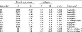
Statistical analysis revealed significant spot concentration differences (P<0.05) in a total of 34 matched spots when the glycoprotein enriched fractions from both groups of animals, healthy pigs and pigs suffering from rectal prolapse, were compared. From these, the 11 most intense spots were selected and analyzed by MS. Among them, two spots increased in healthy animals and nine were more abundant in pigs suffering from rectal prolapse (Table 1).
Detailed list of the spots differentially expressed in healthy animals or pigs suffering from rectal prolapse in glycoprotein enrichment fractions after two-dimensional gel electrophoresis (2DE) and MS identification

X=mean.
Statistical data obtained by using a 2DE software (ImageMasterTM 2D Platinum 7.0, GE Healthcare Life Sciences, Munich, Germany).
Mean: mean value (µg of protein in spot/ml of saliva).
List was sorted according to P-values.
1 Numbers correspond to spots reported in Figure 3.
2 Spot identification according to Table 2.
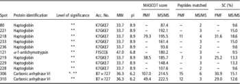
After MALDI-TOF/TOF MS analysis, all spots were identified with statistical significance in MS/MS mode with two to six peptides (Table 2). The identified proteins were Hp (eight spots), α-1-antichymotrypsin (one spot) and CA VI (two spots).
Protein identification of the spots shown in Figure 3 by MALDI-TOF/TOF MS

Acc. No.=accession number; SC=sequence coverage; PMF=identified by peptide mass fingerprint; MS/MS=identified by tandem mass spectrometry; pI, isoelectric points; MW=molecular weight.
*Statistically significant identification in PMF P<0.05; **Statistically significant identification by MS/MS P<0.05.
Hp was found exclusively in the glycoprotein profile of pigs suffering from rectal prolapse as several trains of spots with different molecular weights (MW) (~30 to 36 kDa) and alkaline isoelectric points (pI) (~8 and 9). These isoforms clearly separated into five horizontal series, exhibited different pI but same MW, and can also be regarded as three vertical lines of spots with similar pI but differing in MW (Figure 3). According to MS identifications all Hp spots correspond to the accession number K7GE7, which has a theoretical MW of 33.7 kDa and a pI of 8.9.
Detailed views of the spots differentially regulated from the glyco-enriched fraction patterns obtained by two-dimensional gel electrophoresis of samples from a healthy pig (a1) and a pig suffering from rectal prolapse (a2). Protein spots successfully identified by MALDI-TOF/TOF are marked with circles (numbering corresponds with data in Table 2). MW=molecular weight.

Spots identified as CA VI were the main spots of two adjacent horizontal spot trains with different pI (~6.7 to 7.3) and MW (~33 to 36 kDa) and were observed only in the glycoprotein profile of healthy animals. Results of MS showed spot identifications corresponding to the accesion number B7X727, which has a theoretical MW of 36.3 kDa and a pI of 6.2.
One spot identified as α-1-antichymotrypsin was detected at a pI of 8 and a MW of ~36 kDa in the glycoprotein profile of pigs suffering from rectal prolapse. The identification performed by MS analysis showed a correspondence to the accession number F1SCC6 with a theoretical MW of 47 kDa and a pI of 6.8.
Western blot validation of Hp spots in uncommon positions in 2DE
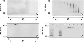
All newly detected Hp spots with alkaline pI (~8 and 9) in 2DE-stained immunopositive for porcine Hp according to 2DE Western blot analysis in saliva of a pig with acute rectal prolapse (Figure 4b). A similar experiment was negative with saliva samples from a healthy animal (Figure 4a), whereas a positive immunostaining was observed in serum from a pig with rectal prolapse (pI range 5 to 6; Figure 4c). Moreover, when the same analysis was performed in SDS-PAGE from non-treated serum and saliva samples, and also from glyco-enriched protein fractions of serum and saliva from both groups of animals, a positive signal could be seen as a band in the common position (38 kDa) in all serum samples, with higher intensity in samples from animals with rectal prolapse. In addition, only saliva and glyco-enriched protein fractions from saliva samples of animals with acute rectal prolapse showed immunoreactive bands with uncommon MW corresponding to those observed in the 2DE analysis (Figure 4d).
Detailed views of the area in which spots/bands positive for porcine haptoglobin (Hp) appear: two-dimensional gel electrophoresis Western blot analysis of saliva samples from a healthy animal (a) and of saliva (b) and serum (c) from a pig suffering from rectal prolapse; SDS-PAGE-Western blot analysis in saliva, serum and enriched fractions from both bodyfluids of a healthy pig and a pig with rectal prolapse (d). Lane 1: saliva from a pig with rectal prolapse, lane 2: saliva from a healthy pig, lanes 3 and 4 enriched fractions from the sample in lanes 1 and 2, respectively. Lane 5 serum sample from a pig with rectal prolapse, lane 6 serum from a healthy pig, lanes 7 and 8 enriched fractions from the serum sample in lanes 5 and 6, respectively. MW=molecular weight.

N-glycan analyses of gel-separated glycoproteins
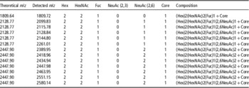
The glycosylation profiles of the Hp β-chain (obtained after SDS-PAGE separation) were determined from serum of a healthy animal and an animal with rectal prolapse and the N-glycan profiles analyzed by MALDI-TOF MS (Table 3). No significant qualitative or relative quantitative N-glycan pattern differences were found between both animal groups. A diantennary, doubly sialylated and core fucosylated N-glycan (Hex5HexNAc4FucNeuAc2) was the most abundant structure identified. Unfortunately, it was not possible to obtain sufficient, reliable N-glycan data for saliva Hp.
N-glycan compositions identified on serum haptoglobin (Hp) β-chain from samples of a healthy animal and an animal with rectal prolapse after an enzymatic deglycosylation of SDS-PAGE separated Hp

Hex=hexose; HexNAc=N-acetylhexosamine; Fuc=Fucose; NeuAc=N-acetyl neuraminic acid.
Thresholds for glycan list compilation included: score >2, fragmentation coverage >2% and intensity coverage >2%.
Discussion
Glycans can be present in various structural forms on the same protein, at the same glycosylation site, resulting in different glycoforms of the same molecule (Sondej et al., Reference Sondej, Denny, Xie, Ramachandran, Si, Takashima, Shi, Wong, Loo and Denny2009). Many diseases have been associated with glycan structure changes in humans including inflammatory diseases. However, to the authors’ knowledge, no related detailed studies have been reported until now in porcine medicine.
In the present study, the rectal prolapse model has been used as an acute inflammatory process to study possible overall salivary glycoprotein profile (obtained after boronic acid glycoprotein enrichment) modifications in pigs, which could be related to the inflammatory condition. The very large dynamic concentration range in which different glycoproteins occur in biological samples makes enrichment methodologies a good solution insofar as it does not compromise the reproducibility and quantification in comparative analyses (Novotny et al., Reference Novotny, Alley and Mann2012). We have selected the boronic acid enrichment as it seems to enrich a wide range of glycoproteins containing both N-linked and O-linked glycans with attractive characteristics such as simplicity of preparation and versatile surface modification (Wang et al., Reference Wang, Xia and Liu2013).
Acute phase protein values, Hp and CRP levels in serum and saliva samples, were used to corroborate the inflammatory state and the degree of inflammation. Both proteins appeared statistically significantly higher (P<0.05) in pigs with rectal prolapse in comparison with healthy pigs. The differences observed in CRP concentrations were larger in saliva than in serum samples, as reported before in animals suffering from porcine respiratory and reproductive syndrome outbreaks (Gutiérrez et al., Reference Gutiérrez, Escribano, Fuentes and Cerón2009).
Once the inflammatory model was evaluated, samples were subjected to an enrichment of glycoproteins protocol using PBA resin and the obtained enriched fractions were subjected to 2DE for salivary glycoprotein pattern comparison. Statistical analysis of spot data and MS identification showed three unique proteins that appeared dys-regulated in the salivary glycoprotein profile of healthy pigs v. pigs with rectal prolapse, namely α-1-antichymotrypsin, CA VI and Hp. From those proteins only Hp was further investigated as it appeared as multiple spots in an unusual position.
The complex pattern of Hp consisted in spots of high pI, around 8 to 9, and low MW, with a range of around 38 to 33 kDa, and was observed exclusively in the salivary glycoprotein profile of pigs with rectal prolapse. Previous studies performed in porcine saliva samples have detected Hp spots in positions similar to those reported in serum samples (Miller et al., Reference Miller, Wait, Sipos and Gemeiner2009; Marco-Ramell et al., Reference Marco-Ramell, Miller, Nöbauer, Möginger, Segalés, Razzazi-Fazeli, Kolarich and Bassols2014) with pI values of 5 to 5.6 and MW of 42 kDa, in healthy animals and in pigs with severe clinical signs of disease (Gutiérrez et al., 2013b). Two further analyses were carried out to get more detailed information about this uncommon Hp finding. First, Western blot analyses showed no altered Hp position in serum samples from healthy pigs and pigs with rectal prolapse and in saliva and glyco-enriched sample from healthy pigs. Nevertheless, the specific uncommon Hp pattern observed in 2DE in animals with rectal prolapse is already detectable in the untreated saliva samples, which was more evident in the glyco-enriched saliva sample. This could be considered as a clear sign that those spots are not created by sample pretreatment on the PBA matrix, but might be specific for the condition of the pigs instead.
The second more advanced approach was an N-glycan analysis of Hp from serum and saliva samples. Results confirm the serum Hp glycosylation pattern reported in previous porcine studies (Marco-Ramell et al., Reference Marco-Ramell, Miller, Nöbauer, Möginger, Segalés, Razzazi-Fazeli, Kolarich and Bassols2014), and the similarity of Hp β-chain glyco-modifications in healthy pigs and pigs with acute inflammation, as no inflammation-induced alterations in carbohydrate pattern were observed. However, this is the first time in which any possible Hp glyco-modifications are studied in porcine saliva and to date no conclusive results could be obtained, requiring further investigations to clarify the specific Hp glycopatterns associated to the inflammatory condition. The low amount of available saliva sample and the resulting low amounts of enriched Hp are the most reasonable explanation why the individual glyco-profiles could not be unambiguously determined from the boronic acid enriched saliva samples.
In humans, it has been largely reported that a shifting of Hp protein spots towards the cathode was caused by a reduction in terminal sialic acid residues, which may result in changes of the molecular conformation (Boonyapranai et al., Reference Boonyapranai, Tsai, Chen, Sriyam, Sinchaikul, Phutrakul and Chen2011). Moreover, post-translational modifications such as glycosylation and phosphorylation are known to influence the MW and pI of proteins (Ongay et al., Reference Ongay, Boichenko, Govorukhina and Bischoff2012). These modifications could be seen in 2DE analysis as a ‘train’ of spots of the same protein over a wide pH-range (Kolarich et al., Reference Kolarich, Weber, Turecek, Schwarz and Altmann2006). Therefore, we hypothesize that the uncommon Hp pattern could be caused by specific glyco-modifications as it has been reported in several inflammatory diseases in humans (Váradi et al., Reference Váradi, Mittermayr, Szekrényes, Kádas, Takacs, Kurucz and Guttman2013). This could include increased branching degree, abnormal fucosylation and Sialyl Lewis X levels.
The second protein observed as spot chains, with slight differences in MW and pI, was identified as porcine CA VI (as reported before in porcine saliva; Gutiérrez et al., 2013b). The electrophoretic heterogeneity could be related to post-translational modifications, such as glycosylation, or cleavage of saliva proteins (Song et al., Reference Song, Zhang, Liu, Ding, Wu, Gan and Yu2012). A decrease in CA VI below detection levels in diseased pigs could be the cause of the detection of CA VI only in healthy animals. This would be in line with reported decreased values of CA VI in saliva of diabetic patients (Border et al., Reference Border, Schwartz, Carlson, Dibble, Kohltfarber, Offenbacher, Buse and Bencharit2012), in pigs with marked clinical symptoms of disease (Gutiérrez et al., 2013b) and in urine of pigs with hyperplasic renal inflammation (Nishita et al., Reference Nishita, Yatsu, Watanabe, Ochiai, Ichihara, Orito and Arishima2014). However, another reason that should be further investigated could be an altered intracellular form of the protein similar to the one described in stressed mice (Sok et al., Reference Sok, Wang, Batchvarova, Kuroda, Harding and Ron1999).
An increase in one spot identified as α-1-antichymotrypsin was observed in the glycoprotein profile of pigs with rectal prolapse with an uncommon basic pI around 8 and MW of ~36 kDa. Previous studies classified porcine α-1-antichymotrypsin as a glycoprotein of 63 kDa composed of a single polypeptide chain (Stratil et al., Reference Stratil, Cízová-Schröffelová, Gábrisová, Pavlík, Coppieters, Peelman, Van de Weghe and Bouquet1995) and with an acidic pI in serum (Miller et al., Reference Miller, Wait, Sipos and Gemeiner2009) and in bronchoalveolar lavage fluid (BALF) of pigs (Kahlisch et al., Reference Kahlisch, Buettner, Naim and Gerlach2009) (~5.33 and 5.77, respectively). From the apparent MW of the spot observed in 2DE in the present study, we assume that we found a breakdown product of α-1-antichymotrypsin. Its presence in a basic pI may still be specific for the condition/disease and warrants further investigations.
In conclusion, our results demonstrate that saliva may be a sample type superior to serum when investigating protein profile alterations, particularly in combination with sample prefractionation as shown herein. Three glycoproteins, but predominantly Hp, were found consistently changed during an acute inflammatory condition such as rectal prolapse and further study should be undertaken to confirm the value of these findings and to clarify the underlying mechanisms.
Acknowledgements
Authors wish to express their gratitude to Dr Pablo Fuentes Pardo (Cefusa Corporation) for his involvement on animal sampling and Lennart Hoffman for technical assistance in Hp glycosylation analysis. D.K. acknowledges support by the European Union (Seventh Framework Programme ‘Glycoproteomics’ project, grant number PCIG09-GA-2011-293847) and the Max Planck Society.