Seckel syndrome is a very rare autosomal recessive disorder characterised by proportional short stature, low birth weight, dysmorphic facial appearance, and mental retardation. Due to its characteristic facial appearance, it is termed as “bird headed dwarfism”.
The syndrome was first described by Seckel in 1960.Reference Seckel1 Seckel syndrome has a genetic heterogeneity, to date, mutations in nine different genes have been reported.Reference Yigit, Brown and Kayserili2 Seckel syndrome-1 is caused by homozygous or compound heterozygous mutation in the ATR gene (601215) on chromosome 3q23. Other forms of Seckel syndrome include SCKL2 (606744), caused by mutation in the RBBP8 gene (604124) on chromosome 18q11; SCKL4 (613676), caused by mutation in the CENPJ gene (609279) on chromosome 13q12; SCKL5 (613823), caused by mutation in the CEP152 gene (613529) on chromosome 15q21; SCKL6 (614728), caused by mutation in the CEP63 gene (614724) on chromosome 3q22; SCKL7 (614851), caused by mutation in the NIN gene (608684) on chromosome 14q22; SCKL8 (615807), caused by mutation in the DNA2 gene (601810) on chromosome 10q21; SCKL9 (616777), caused by mutation in the TRAIP gene (605958) on chromosome 3p21; and SCKL10 (617253), caused by mutation in the NSMCE2 gene (617246) on chromosome 8q24.Reference Yigit, Brown and Kayserili2
Prominent facial features are microcephaly, large eyes, receding forehead, large and beak-like nose, and micrognathia. In addition to dysmorphic findings, cardiac, skeletal, haematologic, and endocrine system involvement has been seen.
We report two patients with Seckel syndrome with cardiac involvement which is very rare; one patient with dilated cardiomyopathy and the other with multiple ventricular septal defects. Dilated cardiomyopathy and isolated ventricular septal defect have not been previously reported in Seckel syndrome.
Case 1
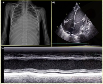
A 6-year-old boy was admitted to our department with symptoms of dyspnoea, altered mental status, and poor status. He had a history of cough and weakness for 3 days, exercise intolerance, poor appetite, and sweating for 6 months. He was previously diagnosed as Seckel syndrome with his phenotypic appearance by the Genetics Department. The patient was born from a 32-year-old, multigravida mother, with a birth weight of 1600 g. Second-degree consanguinity was reported between his parents. Physical examination showed that his general condition was poor. Body temperature: 38.2 °C, heart rate: 145 beats/min, respiratory rate: 50/min. He had tachypnoea and dyspnoea, intercostal and subcostal retractions. He had hyperdynamic precordium, tachycardia, gallop rhythm, low perfusion findings, weak pulses, and cold extremities. Liver was palpable 4 cm below the right subcostal margin. He had dysmorphic features; microcephaly (head circumference: 47.5 cm (<3 percentile), long face, high palate, and large eyes, and proportionate short stature (height: 103 cm (<3p) and body weight: 11 kg (<3p)). Laboratory tests showed elevated alanine aminotransferase (46.1 U/L), aspartate aminotransferase (87.8 U/L), lactate levels (3,6 mmol/L), international normalised ratio (1:8), procalcitonin (0.127 ng/ml), white blood count (13,680/µ'), N-terminal pro-B-type natriuretic peptide (4894 pg/m') with a normal level of troponin I. The possibility of underlying metabolic disease was ruled out by metabolic tests (urine and serum sample of organic acids, plasma ammonium level, acylcarnitine profile, plasma, and urine amino acids). Blood culture was negative, acute phase reactants were normal, viral serology serum samples and polymerase chain reaction were also negative. Chest X-ray showed cardiomegaly, pulmonary congestion, and bilateral pleural effusion (Fig 1a). Electrocardiography showed sinus tachycardia, left axis deviation, and left precordial ST depression. Echocardiography revealed global dilatation of both ventricles (Fig 1b), left ventricular end-diastolic dimension was 45 mm, (z score: +3.6) severe left ventricular dysfunction (ejection fraction was 22%) (Fig 1c) and also right ventricular dysfunction (tricuspid annular plane systolic excursion:12 mm), moderate mitral and tricuspid regurgitation, and dilated inferior caval vein and hepatic veins. Coronary computed tomographic angiography revealed normal coronary arteries. Patient was diagnosed with dilated cardiomyopathy and decompensated heart failure and hospitalised in ICU, Intravenous inotropes were started and mechanical ventilation was performed. Inotropes were tapered down by the 12th day and were transferred to paediatric ward on the 23rd day. He was discharged with oral furosemide, captopril, and carvedilol. About 6 months later, he was hospitalised with decompensated heart failure after an upper respiratory tract infection again in ICU, and unfortunately, the patient was lost despite all efforts.
a) Chest X ray of patient 1 in PA view reveals bilateral blunting of the costophrenic angle with cardiomegaly b) Apical four chamber view demonstrates marked dilation of all the chambers of the heart c) Reduced ejection fraction was measured with M-mode echocardiography.

Case 2
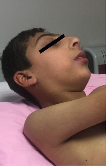
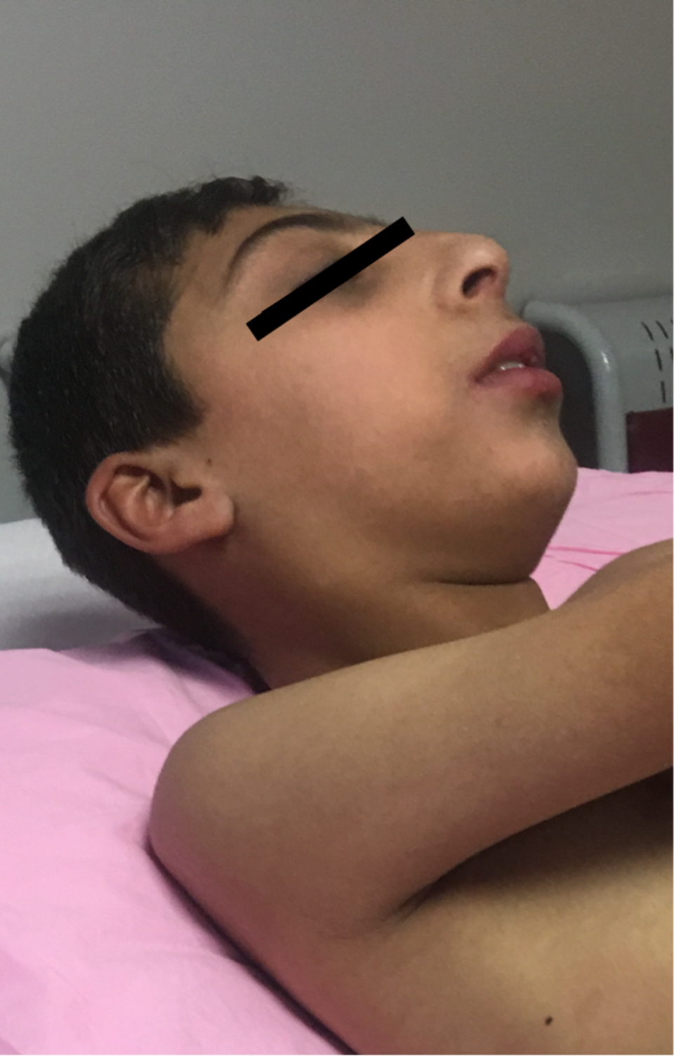
A 4-year-old boy was referred to our hospital for the evaluation of microcephaly, heart murmur, and exertional dyspnoea. At 37 weeks of gestation, he was born by caesarean section from a 26-year-old mother, with a birth weight of 2020 g. His parents were second-degree consanguineous. The patient had two brothers who died and had microcephaly, low stature, other dysmorphic findings, and mental retardation in family history. On physical examination, he had severe growth retardation, his body weight and height were below 3rd percentile (height: 93 cm, weight: 12.5 kg), had low-set ears, microcephaly (head circumference: 46.3 cm (<3 percentile)), prominent beak-like nose, large eyes, and severe mental retardation (Fig 2). Heart rate: 105 beats/minute, blood pressure: 90/60 mmHg, oxygen saturation by pulse oximetry: 94%. He had central cyanosis, clubbing, 2/6 pansystolic murmur on left sternal margin, loud second heart sound, and palpable liver 2 cm below the right subcostal margin. Electrocardiogram showed right axis deviation, right atrial dilatation, and right ventricular hypertrophy. Transthoracic echocardiography showed large apical multiple muscular ventricular septal defects, enlargement of the right chambers, right ventricular dysfunction, severe tricuspid regurgitation. Estimated systolic pulmonary artery pressure from tricuspid regurgitation was 80 mmHg. Subsequently, cardiac catheterisation was performed. Pulmonary artery pressures (systolic/diastolic/mean): 81/30/56 mmHg, aortic pressures (systolic/diastolic/mean): 85/49/63 mmHg, pulmonary vascular resistance was 7.5 Um2, systemic resistance: 9.6 Um2. Pulmonary vasoreactivity test with inhaled iloprost was negative. With the diagnosis of multiple large VSD, pulmonary hypertension, and Eisenmenger syndrome, he was inoperable and oral digoxin, captopril, furosemide, and bosentan (endothelin receptor antagonist for pulmonary hypertension) therapy were initiated. He was consulted by the Genetics Department. Peripheral chromosome analysis showed 46, XY, and he was diagnosed as Seckel syndrome by phenotypical findings. During the follow-up after 7 years, sildenafil and inhaled iloprost were added after the second heart catheterisation. Currently, the patient is 14 years old, in WHO FC III and on combination therapy with bosentan, sildenafil, and inhaled iloprost.
Facial features of patient 2 with a low-set ears, microcephaly and prominent beak-like nose.
Discussion
Seckel syndrome is a microcephalic primordial dwarfism, which is known to be inherited as an autosomal recessive.Reference Khetarpal, Das and Panigrahi3
Although its genetic role is not fully understood, it has been postulated that the genetic disorder is a result of the failure of cellular proliferation, differentiation, and apoptotic process in complex endocrine molecular mechanisms that regulate growth pathways.Reference Khetarpal, Das and Panigrahi3 Seckel syndrome is characterised by intrauterine growth retardation, microcephaly, micrognathia, mental retardation, and prominent beak-like nose.Reference Khetarpal, Das and Panigrahi3 Facial dysmorphy also includes large eyes, receding forehead, narrow face, high-arched palate, and low-set ear.Reference Faivre, Le Merrer and Lyonnet4
There is a clinical heterogeneity in Seckel syndrome. Diagnosis depends on clinical features; proportional dwarfism, microcephaly, facial features, and mental retardation.Reference Faivre, Le Merrer and Lyonnet4 Diagnosis of Seckel syndrome was established in our two patients by characteristic features like microcephaly, bird-headed proportional dwarfism, intrauterine onset growth retardation, and mental retardation.
In addition to the facial dysmorphic findings, cardiac, skeletal, dental, gastrointestinal, haematologic, and nervous system involvement have been reported. Cardiac involvement is very rare in Seckel syndrome and has been reported only in case reports (tricuspid atresia, tetralogy of Fallot, incomplete atrioventricular septal defect) in literature.Reference Arslan, Cimen and Guvenc5–Reference Ucar, Kilic and Dinleyici7 In two patients with Seckel syndrome, a permanent pacemaker was necessary due to AV block and severe sinus bradycardia with recurrent syncope.Reference Ramasamy, Satheesh and Selvaraj8,Reference Unal, Dogan and Ozkose9 Payne et al described a female patient with Seckel syndrome who suddenly died at 33 years of age presumably due to rupture of a previously documented abdominal aortic aneurysm.Reference Payne, Colnaghi and Rocha10 Silengo et al described two Italian sibs with a Seckel-like malformation syndrome characterised by intrauterine growth retardation, microcephaly, beaked nose, failure to thrive, and early death. One of the sibs died at 1 month with respiratory failure. Autopsy showed severe hypoplasia of the pulmonary artery and its branches, large atrial septal defect, and ventricular septal defect.Reference Silengo, Del Monaco and Linari11
In conclusion, we report two patients with Seckel syndrome with cardiac involvement which is extremely rare. Dilated cardiomyopathy and isolated ventricular septal defect have not been previously reported in Seckel syndrome. Cardiovascular valuation should be performed in all patients with Seckel syndrome. Early diagnosis of congenital and acquired heart diseases (e.g., arrhythmia) will reduce morbidity and mortality in these patients.
Acknowledgments
None.
Financial support
This research received no specific grant from any funding agency, commercial, or not-for-profit sectors.
Conflicts of interest
None.
Ethical standards
The authors assert that all procedures contributing to this work comply with the ethical standards of the relevant national guidelines on human medical regulations and with Helsinki Declaration of 1975, as revised in 2008. Written informed consent was obtained from all patient’s parents.