1. Scope of the review
Colicins (Cols) are highly specific protein antibiotics released by Escherichia coli that target and kill neighbouring E. coli during times of environmental stress as a means of reducing competition for resources. Normally plasmid-encoded, Cols are members of the bacteriocin family of antimicrobials, which include both peptide and protein antibiotics that are widespread among Gram-negative and Gram-positive bacteria. Long studied by microbial population ecologists in the context of their impact on the population dynamics of co-existing bacterial populations (Gardner et al. Reference Gardner, West and Buckling2004; Kirkup & Riley, Reference Kirkup and Riley2004; Riley, Reference Riley1998), the mode of action of Cols has proven fertile ground for bacteriologists since their discovery over 80 years ago. These studies have shown that while Cols can kill cells by a variety of mechanisms these generally fall into two cytotoxic classes; enzymatic Cols cleave either nucleic acid or peptidoglycan while pore-forming Cols depolarize the cytoplasmic membrane. Extensive biochemical, structural and biophysical work has been published for both classes. Given the number of studies on Cols, in comparison with bacteriocins that target other Gram-negative organisms such as Yersinia pestis or Pseudomonas aeruginosa, it is not surprising that they are among the best understood of the protein bacteriocins and not just from the perspective of their import across the cell envelope. Cols and their associated immunity (Im) proteins, which are the small antidote proteins that protect producing cells from the harmful cytotoxic effects of the Col, are often used as models for addressing fundamental questions in protein chemistry, particularly in the investigation of the protein-folding mechanism and in the dissection, design and directed evolution of protein–protein interaction specificity.
The present review focuses on recent discoveries on enzymatic Cols and their Im proteins and in particular nuclease Cols, which have been reviewed less in the literature than their pore-forming counterparts (Anderluh & Lakey, Reference Anderluh and Lakey2008; Cascales et al. Reference Cascales, Buchanan, Duche, Kleanthous, Lloubes, Postle, Riley, Slatin and Cavard2007; Iacovache et al. Reference Iacovache, Van Der Goot and Pernot2008; Ridley et al. Reference Ridley, Johnson and Lakey2010; Zakharov et al. Reference Zakharov, Kotova, Antonenko and Cramer2004b). As well as dealing with how these potent cytotoxins target and kill E. coli cells–the dogma in the field being that one molecule is sufficient to kill a cell–we also review some of the work in other areas of protein science where nuclease Cols and their Im proteins have proven tractable model systems. Although not dealing explicitly with pore-forming Cols, we make reference to work on these toxins where relevant to the wider question of how Cols translocate cytotoxic domains across the Gram-negative outer membrane (OM) as this is independent of cytotoxic activity. The reader is directed to the extensive review of Cascales et al. (Reference Cascales, Buchanan, Duche, Kleanthous, Lloubes, Postle, Riley, Slatin and Cavard2007) for topics not covered by the present article.
2. Structural biology of Cols and their Im proteins
Col production is generally induced via the SOS pathway in E. coli in response to stress. The toxin is then released to the extracellular environment through the action of a small bacteriocin-release protein although the mechanism by which this happens is poorly understood. The molecular weights of Cols vary substantially, ranging from 40 to 80 kDa, yet despite their very different structures all Cols are constructed from the same basic modular design: an N-terminal translocation (T) domain implicated in coordinating translocation across the OM, a central receptor-binding (R) domain, which localizes the toxin to the cell surface, and a C-terminal cytotoxic (C) domain, which is translocated across the OM and in the case of nuclease Cols also through the inner membrane (IM). Interestingly, the same three-domain arrangement is seen in proteins involved in the uptake of some filamentous bacteriophages into E. coli, to the extent that phage/Col chimaeras retain cytotoxic activity suggesting that there are likely to be parallels between the early stages of phage infection and Col translocation across the OM (Jakes et al. Reference Jakes, Davis and Zinder1988).
In order to avoid suicide all Col-producing E. coli produce an Im protein (~10 kDa). In the case of nuclease Cols, the immunity (Im) protein binds to the C-terminal cytotoxic domain and neutralizes its activity in the host (Kleanthous & Walker, Reference Kleanthous and Walker2001). Hence, these toxins are released to the environment as heterodimeric proteins necessitating removal of the Im protein during cell entry (Section 6). In the case of Cols that act from within the confines of the periplasm, and which are not active until they translocate into a cell, the Im protein is either localized to the IM through a lipoyl tether (peptidoglycan-cleaving Col) or is an integral IM protein (pore-forming Cols).
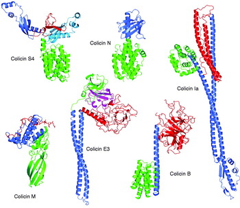
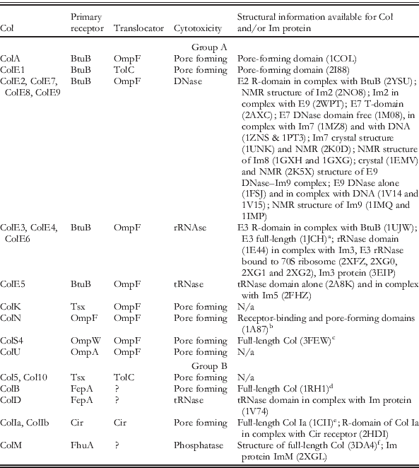
To-date, more than 20 Cols have been characterized (Table 1), with structural information for many deposited in the protein data bank (PDB). Full-length structures have been published for ColE3 (Soelaiman et al. Reference Soelaiman, Jakes, Wu, Li and Shoham2001), ColIa (Wiener et al. Reference Wiener, Freymann, Ghosh and Stroud1997), ColB (Hilsenbeck et al. Reference Hilsenbeck, Park, Chen, Youn, Postle and Kang2004), ColM (Zeth et al. Reference Zeth, Romer, Patzer and Braun2008) and ColS4 (Arnold et al. Reference Arnold, Zeth and Linke2009) (Fig. 1). In many instances, however, these structures lack electron density for part or all of the N-terminus of the toxin (e.g. the structure of ColN; Vetter et al. Reference Vetter, Parker, Tucker, Lakey, Pattus and Tsernoglou1998), with biophysical measurements (primarily nuclear magnetic resonance (NMR), circular dichroism (CD) and fluorescence spectroscopy) on several Cols (ColE9, ColN and ColA) consistent with these regions being intrinsically unstructured in solution (Collins et al. Reference Collins, Whittaker, Tozawa, Macdonald, Boetzel, Penfold, Reilly, Clayden, Osborne, Hemmings, Kleanthous, James and Moore2002; Deprez et al. Reference Deprez, Blanchard, Guerlesquin, Gavioli, Simorre, Lazdunski, Marion and Lloubes2002; Hecht et al. Reference Hecht, Ridley, Lakey and Moore2009b; Raggett et al. Reference Raggett, Bainbridge, Evans, Cooper and Lakey1998). The unstructured nature of Col N-termini is important for translocation into E. coli because these regions invariably contain one or more linear peptide signalling epitopes, which are destined to contact trans–periplasmic systems linked to the proton-motive force (PMF) across the IM (Sections 4 and 5). In addition to full-length structures, there are also structures available for isolated Col domains, most often the C-terminal cytotoxic pore-forming domain (e.g. ColA) or nucleases in complex with Im proteins (e.g. ColD DNase bound to ImD).
Structures of Cols S4 (PDB ID: 3FEW), N (PDB ID: 1A87), Ia (PDB ID: 1CII), M (PDB ID: 3DA4), E3 in complex with Im3 (PDB ID: 1JCH) and B (PDB ID: 1RH1) are shown in ribbon representation. Translocation (T-) domains are shown in red, receptor-binding (R-) domains in light and dark blue and cytotoxic (C-) domains in green, with the Im protein Im3 shown in magenta. The loosely structured residues of the TonB box (residues 2–8) are shown for Col M.

Table shows some of the major Cols that have been identified, their receptors and translocators, where known and, references to structural information for the Col and/or its Im protein

a ColE3 structure lacks the N-terminal 83 residues which are disordered (Soelaiman et al. Reference Soelaiman, Jakes, Wu, Li and Shoham2001).
b N-terminal 90 residues missing in the structure (Vetter et al. Reference Vetter, Parker, Tucker, Lakey, Pattus and Tsernoglou1998). T-domain (residues 17–66 ) unstructured, shown by CD, fluorescence and NMR (Hecht et al. Reference Hecht, Ridley, Lakey and Moore2009a; Raggett et al. Reference Raggett, Bainbridge, Evans, Cooper and Lakey1998).
c Residues 1–67 disordered in the structure (Arnold et al. Reference Arnold, Zeth and Linke2009).
d Residues 1–10 and 29–43 disordered but TonB box (11–28 ) visible (Hilsenbeck et al. Reference Hilsenbeck, Park, Chen, Youn, Postle and Kang2004).
e ColIa structure missing residues 1–22 but TonB box (23–27 ) visible (Wiener et al. Reference Wiener, Freymann, Ghosh and Stroud1997).
f Residues 2–35 (including Ton B box 2–8 ) present but lacking defined structured (Zeth et al. Reference Zeth, Romer, Patzer and Braun2008).
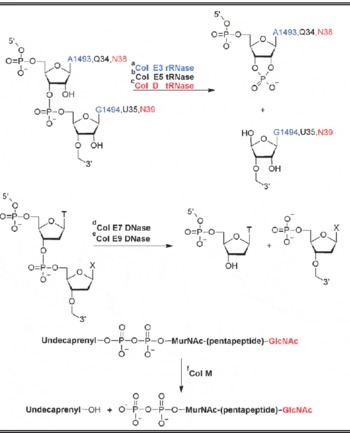
Although the nomenclature of Cols is somewhat ad hoc some systematic naming is to be found among this diverse group of proteins, most often reflecting the commonality of entry routes into cells and/or cytotoxic activities. For example, Cols E2–E9, which all utilize the BtuB receptor (Section 4), share a high degree of sequence identity in their Col translation domain (T-domain) and Col receptor-binding domain (R-domain) (70–90%), and hence have the same mode of entry into bacteria, but share no sequence similarity in their cytotoxic domains unless members of the same nuclease family. Interestingly, these Cols encompass the three different nuclease activities so far reported for Cols; deoxyribonucleases (DNases; ColE2, ColE7, ColE8 and ColE9), ribosomal-specific ribonucleases (rRNases; ColE3, ColE4 and ColE6), and a tRNA-specific ribonuclease (tRNase; ColE5). The structures of the different nuclease classes are quite distinct from each other implying that the entry routes of these Cols are insensitive to the toxic payload being carried. Consequently, the conserved T- and R-domains of Cols E2–E9 can be thought of as ‘targeting domains’ ensuring delivery of a variety of nucleases to the E. coli cytoplasm.
While all Cols share a characteristic three-domain organization their structures can differ significantly (Fig. 1). Some Cols adopt compact structures, such as Cols N, M and S4, while others are extended, such as Cols B, E3 and Ia. The C-terminal cytotoxic domains are generally small, globular domains, as in the bundle of α-helices in pore-forming Cols (built from two central hydrophobic helices surrounded by eight amphipatic helices) or the mixed α/β structures of nuclease Cols. The exception is Col M the cytotoxic domain of which accounts for more than half of molecule and is structurally similar to the open β-barrel of the Haemophilus influenzae Hir autoreceptor (Meng et al. Reference Meng, Surana, St Geme and Waksman2006). The structure of the central R-domain of Cols is particularly diverse in both size and shape, reflecting very different binding specificities for primary receptors. Cols B, E3 and Ia have extended α-helical R-domains. The ColB R-domain consists of single 74 Å long α-helix, while the ColE3 R-domain is a coiled-coil of two α-helices. ColIa has two long parallel α-helices separated by an additional small domain, formed by a two-stranded antiparallel β-sheet and three short α-helices. In the case of ColN, the R-domain consists of a twisted, seven-stranded β-sheet wrapped around an α-helix that extends into the pore-forming domain. The R-domain of ColM on the other hand contains six α-helices five of which surround a long central helix, α3 (Zeth et al. Reference Zeth, Romer, Patzer and Braun2008). The N-terminal T-domains of Cols are also structurally diverse. They can be small and disordered, as in the case of ColM (35 residues) and ColN (90 residues) or rather large with some unstructured regions, as in the case of Cols E3, B and Ia. The ColIa T-domain consists of two ~80 Å long antiparallel α-helices, that of ColE3 forms a jellyroll structure composed of three β-sheets flanked by two helical stretches while the ColB T-domain is composed of 17 β-strands and two α-helices that is topologically similar to ColE3. The structures for group A Cols ColE3 and ColN lack the first ~70–80 residues, consistent with these regions being disordered (Section 4). In other Cols, the N-terminus either lacks regular secondary structure (ColB and ColIa) or is largely unstructured (ColM) (Section 4).
The pore-forming Col S4 contains an unusual duplication of its R-domain (Pilsl et al. Reference Pilsl, Smajs and Braun1999). ColS4 has a compact structure with four distinct domains: an N-terminal T-domain, two central R-domains and a C-terminal pore-forming domain (Fig. 1). The first 67 amino acids of the T-domain are unstructured, the remainder consisting of a three-stranded β-sheet and short α-helix. The structures and sequences of the two R-domains are very similar (root mean square deviation (RMSD) <1 Å), composed of a three-stranded β-sheet followed by three α-helices. Mutational analysis has suggested that only one of the R-domains is necessary for cell-killing (Arnold et al. Reference Arnold, Zeth and Linke2009).
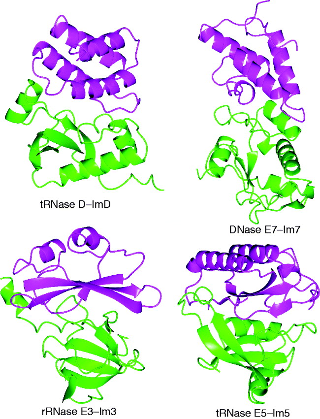
With the exception of ColM, enzymatic Cols form high-affinity complexes with their cognate Im proteins immediately after synthesis in the producing cell. Structures for a number of Im proteins, both in their unbound forms (Im2, Im3, Im7, Im8, Im9 and ImM) and in complex with Col catalytic domains (E7 DNase–Im7, E9 DNase–Im9, E3 rRNase–Im3, E5 tRNase–Im5 and D tRNase–ImD) have been reported (Fig. 2). The structures provide detailed insight into the mechanism of enzymatic Col inhibition by Im proteins (Section 9).
Structures for Col cytotoxic nuclease domains complexed with their cognate Im proteins. ColD tRNase–ImD (PDB ID: 1V74), ColE7 DNase–Im7 (PDB ID: 1MZ8), ColE3 rRNase–Im3 (PDB ID: 1E44) and ColE5 tRNase–Im5 (PDB ID: 2FHZ) are shown in ribbon representation, with the nuclease in green and Im protein in magenta.
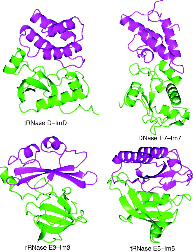
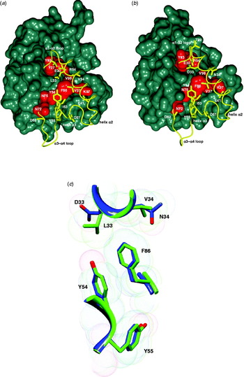
The first structural information on Im protein–Col complexes was reported for the DNase Cols E7 and E9 (Kleanthous et al. Reference Kleanthous, Kuhlmann, Pommer, Ferguson, Radford, Moore, James and Hemmings1999; Ko et al. Reference Ko, Liao, Ku, Chak and Yuan1999). DNase Im proteins share ~50% sequence identity and adopt an antiparallel four-helix bundle fold, with a characteristic short third helix. The ribosome-inactivating ColE3 Im protein Im3 adopts a four-stranded antiparallel β-sheet fold with two α-helices packed against one side of the sheet. The exposed face of the Im3 β-sheet interacts with an N-terminal α-helix and the first two β-sheets of the rRNase (Carr et al. Reference Carr, Walker, James, Kleanthous and Hemmings2000). Although the structure of the E3 rRNase–Im3 complex is significantly different from DNase–Im complexes, the mode of inhibition is the same; the inhibitor docks at an exosite adjacent to the active site that blocks substrate binding. The structure of the ColD Im protein ImD consists of four tightly packed α-helices. In contrast to other nuclease–Im protein complexes ImD inhibits nuclease activity by binding directly to the active site of ColD (Graille et al. Reference Graille, Mora, Buckingham, Van Tilbeurgh and De Zamaroczy2004). A positively charged groove on the surface of ColD that encompasses the active site interacts with a negatively charged patch on ImD, which mimics the backbone of the tRNA substrate. The tRNase Col ColE5 and its Im protein, Im5, form a compact complex with a large interaction area, characterized by a high degree of shape- and charge-complementarity (Luna-Chavez et al. Reference Luna-Chavez, Lin and Huang2006). Im5 adopts a mixed α-/β-fold comprising a four-stranded β-sheet and three α-helices. ColE5 and ColD have similar folds, but their RNA-binding sites as well as the location of their active sites are different. Even though the mechanism of inhibition of tRNase Cols by their Im proteins is similar the structures of Im5 and ImD differ significantly. Finally, the structure of the Im protein for ColM (Cmi or ImM) is composed of four α-helices and a four-stranded antiparallel β-sheet (Gerard et al. Reference Gerard, Brooks, Barreteau, Touze, Graille, Bouhss, Blanot, Van Tilbeurgh and Mengin-Lecreulx2010). ImM resembles a cystatin-fold and contains a disulfide bond between Cys31 and Cys107. Despite available structural information it remains unclear as to how ImM inhibits the activity of ColM.
3. Overview of Col import
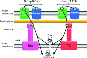
Cols translocate into E. coli by parasitizing a variety of membrane and periplasmic proteins in the cell envelope all of which serve important functions in the bacterium. Initial localization at the cell surface typically occurs through binding of the R-domain to an OM nutrient or vitamin receptor although porins can also function as Col receptors (Table 1; Section 4). Receptor-bound Cols then recruit an additional OM protein as a ‘translocator’ that the toxin uses to pass specific peptide epitopes to the periplasm, which are located within the disordered or unstructured regions of its T-domain and may even, it has been argued, be used to translocate the entire cytotoxic domain to the periplasm (Fig. 3). Porins such as OmpF and OmpC are most often used as translocator proteins. Other known translocators include TolC, which forms the exit tunnel for a multidrug efflux pump, and in at least one case it has been shown that a nutrient receptor can also serve as a translocator (Section 4).
Cols can be grouped according to which import route they take into an E. coli cell. Group A Cols contact the Tol system while group B Cols contact the Ton system. Both Col groups can deliver a cytotoxic pore-forming or nuclease to the cell. Each requires initial binding to a high-affinity receptor followed by recruitment of a translocator protein. The translocator is utilized to deposit one or more signalling epitopes to the periplasm that subvert the normal functions of the Tol or Ton systems, both of which are coupled to the PMF across the IM. Figure adapted from (Kleanthous, Reference Kleanthous2010) with permission from the Nature Publishing Group.

Col translocation across the E. coli OM requires contact between specific peptide-signalling epitopes within the T-domain of the receptor-bound Col and one of two periplasmic systems of the bacterium (Section 5). Group A Cols (such as ColE1–ColE9, ColA and ColN) contact Tol, while group B Cols (such as ColIa, ColB, ColD and ColM) contact Ton (Davies & Reeves, Reference Davies and Reeves1975a, Reference Davies and Reevesb). Both systems, which are evolutionarily related to the stator proteins MotA and MotB of the bacterial flagellar motor, are coupled to the PMF across the IM, acting as energy transducers for OM stability (Tol) and active transport of iron siderophores and vitamin B12 across the OM (Ton). While there is some involvement of PMF in Col toxicity it remains unclear as to how much of the translocation process is PMF dependent (Section 6).
The Ton system consists of three IM proteins TonB, ExbB and ExbD (Postle & Kadner, Reference Postle and Kadner2003). The central components of the Tol system are three IM proteins, TolA, TolQ and TolR, and two periplasmic proteins, TolB, a soluble protein, and Pal, which is anchored to the OM (Webster, Reference Webster1991). TolA and TonB are each thought to span the periplasm and, through associations with their IM partners, transduce the PMF into conformational transitions although what form these transitions take and how they ‘energize’ events at the OM are yet to be elucidated. Group A Col translocation generally requires an intact TolQRA complex with some Cols also requiring TolB. Pal is the only component of the Tol system not required by any known Col (Lazzaroni et al. Reference Lazzaroni, Dubuisson and Vianney2002), confirmed by a recent genome-wide survey that identified many previously unknown genes involved in Col uptake but not Pal (Sharma et al. Reference Sharma, Datsenko, Ess, Zhalnina, Wanner and Cramer2009). Although Pal is a central component of the Tol apparatus its lack of involvement in Col translocation likely reflects its function as an OFF-switch for Tol signalling (Bonsor et al. Reference Bonsor, Hecht, Vankemmelbeke, Sharma, Krachler, Housden, Lilly, James, Moore and Kleanthous2009b) (Section 5). Gene deletions of additional cytoplasmic (YbgC) and periplasmic (YbgF) proteins encoded within the tol operon show no discernible tol phenotype of OM instability (usually assayed by lack of bacterial growth in the presence of SDS) and are also not required for Col toxicity (Krachler et al. Reference Krachler, Sharma, Cauldwell, Papadakos and Kleanthous2010).
An intriguing feature of Col import, triggered by contact with Tol or Ton proteins, is that Cols can be readily engineered to translocate by the opposing pathway; for example, it has been shown that a group A Col can be converted into a group B Col merely by engineering the appropriate recognition epitopes (Pilsl & Braun, Reference Pilsl and Braun1998). This argues that import across the OM is likely to be by the same process for both group A and group B Cols involving contact with a system close to the underside of the OM that also traverses the periplasm and is linked to the PMF across the IM.
For pore-forming and peptidoglycan-cleaving Cols, the journey into the cell ends in the periplasm where they exert their cytotoxic activities. Nuclease Cols, however, continue on through the IM in order to reach their cytoplasmic nucleic acid targets. While this aspect of import has not been investigated extensively, studies suggest it may involve proteolytic processing and direct interaction between the nuclease domain and the cytoplasmic membrane followed by retro-translocation and refolding in the cytoplasm (Section 7).
4. Receptor binding and recruitment of OM translocator proteins
4.1 Receptor binding
The first step in the import of Cols into E. coli is binding to an OM receptor. Cols have evolved to parasitize a wide variety of OM proteins, including the vitamin B12 receptor BtuB used by E Cols ColE1–ColE9 and ColA, ferrichrome-iron receptor FhuA used by ColM, ferrienterobactin receptor FepA used by ColB and ColD, iron transport receptor Cir used by ColIa and ColIb, the nucleoside-specific channel-forming protein Tsx used by ColK, Col5 and Col10, and porins OmpF, OmpA and OmpW used by Cols N, S4 and U, respectively (Table 1). Structural and biophysical data are available for a number of these Col-receptor complexes. In the case of ColN (a group A Col), biochemical and electron microscopy (EM) studies indicate that the Col binds with a K d~1 μM (Bourdineaud et al. Reference Bourdineaud, Fierobe, Lazdunski and Pages1990b) to the outer surface of OmpF (Baboolal et al. Reference Baboolal, Conroy, Gill, Ridley, Visudtiphole, Bullough and Lakey2008). Two-dimensional (2D) crystals of OmpF obtained in the presence and absence of a fragment of ColN containing the receptor-binding and pore-forming domains (at 25 Å resolution) revealed electron density at the interface between two monomers of the OmpF trimer. Density for lipopolysaccharide (LPS), which is normally bound to OmpF, was absent in this co-crystal suggesting that Col-binding displaces LPS, a hypothesis confirmed by biochemical data (Baboolal et al. Reference Baboolal, Conroy, Gill, Ridley, Visudtiphole, Bullough and Lakey2008).
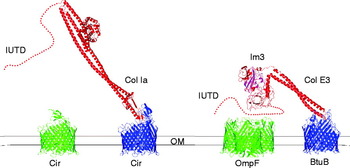
High-resolution crystal structures have been published for the R-domains of three Cols bound to their primary receptors; group A Cols ColE2 (PDB ID, 2YSU) (Sharma et al. Reference Sharma, Yamashita, Zhalnina, Zakharov, Datsenko, Wanner and Cramer2007) and ColE3 (PDB ID, 1UJW) (Kurisu et al. Reference Kurisu, Zakharov, Zhalnina, Bano, Eroukova, Rokitskaya, Antonenko, Wiener and Cramer2003) bound to BtuB, and ColIa, a group B Col, bound to Cir (PDB ID, 2HDI) (Buchanan et al. Reference Buchanan, Lukacik, Grizot, Ghirlando, Ali, Barnard, Jakes, Kienker and Esser2007) (Fig. 4). In both sets of complexes the Col-binding site overlaps that of the natural ligand. The buried surface areas of Cols E2 and E3 bound to BtuB are 1287 and 1533 Å2, respectively. The ColIa–Cir complex has a relatively small buried surface area of 990 Å2. Although the complexes of ColIa and ColE3 (and ColE2) are unrelated to each other their respective OM receptors show two striking similarities. First, both Cols bind at an ~45° angle relative to the membrane, which positions the T-domain and Col cytotoxic domain (C-domain) well away from the receptor and, second, Col-binding does not displace the central plug of the receptor. Transporters such as BtuB and Cir are 22-strand β-barrels with a large central pore that is sealed by a soluble plug domain. The plug has to be displaced in order to allow entry of the cognate ligand of the receptor in a PMF-dependent process involving contact between the TonB box of the receptor and periplasmic TonB. The TonB box is an N-terminal, periplasmically located peptide sequence which signals to TonB when the ligand is bound to the receptor. A further striking feature of Col binding to these receptors is that there is little or no change to their TonB boxes. Binding of Cols E2 and E3 to BtuB causes ordering of the N-terminal Ser4–Pro5 residues of the TonB box of the receptor but no changes were evident in the ColIa–Cir complex structure (Buchanan et al. Reference Buchanan, Lukacik, Grizot, Ghirlando, Ali, Barnard, Jakes, Kienker and Esser2007; Kurisu et al. Reference Kurisu, Zakharov, Zhalnina, Bano, Eroukova, Rokitskaya, Antonenko, Wiener and Cramer2003; Sharma et al. Reference Sharma, Yamashita, Zhalnina, Zakharov, Datsenko, Wanner and Cramer2007). Conformational changes do occur in the receptors, however, but are localized to the extracellular regions. In the complexes of Cols E2 and E3 there are alterations in the conformations of surface loops while in the complex of ColIa–Cir a lid domain in the receptor opens in order for the Col to bind.
Structures of Cols Ia and E3–Im3 bound to their primary receptors, Cir and BtuB, respectively, depicting the recruitment of translocator proteins through their intrinsically unstructured translocation domains (IUTDs). Following binding to its primary receptor (PDB ID: 2HDI and 1CII), ColIa binds a second copy of the Cir protein (PDB ID: 2HDF) although the mechanism involved is unknown. After binding to BtuB (PDB ID: 1UJW and 1JCH), the ColE3–Im3 complex recruits OmpF (PDB ID: 3O0E) through specific OmpF-binding sites (OBSs). See Figs 5–7 for details.

It has been suggested that interactions between the R-domain of enzymatic Cols and BtuB±OmpF lead to unfolding of the Col although this has proven controversial. The suggestion is based on (i) some disordering of the ends of the coiled-coil R-domain in the ColE3-bound structure (Kurisu et al. Reference Kurisu, Zakharov, Zhalnina, Bano, Eroukova, Rokitskaya, Antonenko, Wiener and Cramer2003); (ii) changes in the CD spectrum for the complex relative to the unbound proteins (Kurisu et al. Reference Kurisu, Zakharov, Zhalnina, Bano, Eroukova, Rokitskaya, Antonenko, Wiener and Cramer2003); (iii) changes in measured distances between the cytotoxic and T-domain of the Col (Fig. 1) as measured by fluorescence resonance energy transfer (FRET), these effects dependent on the presence of the translocator OmpF in the OM (Zakharov et al. Reference Zakharov, Sharma, Zhalnina and Cramer2008). The suggestion that the R-domain becomes disordered in its distal ends on binding BtuB contrasts the conclusions of Housden et al., who found that the thermodynamics of binding between ColE9 and BtuB (K d~2 nM), as measured by isothermal titration calorimetry (ITC), were unchanged in a disulfide-locked mutant of the Col (Housden et al. Reference Housden, Loftus, Moore, James and Kleanthous2005); detailed discussion on the thermodynamics of Col complexes have been reviewed recently (Housden & Kleanthous, Reference Housden and Kleanthous2011). The disulfide, positioned within the coiled-coil and distal to the receptor, was designed to block opening of the coiled-coil following receptor binding and does indeed abolish Col toxicity unless reduced (Penfold et al. Reference Penfold, Healy, Housden, Boetzel, Vankemmelbeke, Moore, Kleanthous and James2004a). These observations are consistent with the coiled-coil of the R-domain undergoing significant structural changes but that these likely occur post receptor binding, for example, during translocation across the OM (Section 5).
Since the plug domains of BtuB and Cir bound to Col R-domains show little or no change in conformation, this tends to rule out a translocation path through the primary receptor. A similar conclusion was reached for ColB binding its receptor FepA using fluorescein maleimide labelling of unique cysteine residues (Smallwood et al. Reference Smallwood, Marco, Xiao, Trinh, Newton and Klebba2009). As the central channels of Col receptors do not appear to be the routes of translocation to the periplasm this explains why additional OM proteins have to be recruited. This assertion is consistent with the way in which the T- and C-domains of enzymatic E Cols and ColIa are presented well away from their primary receptors, in a bid to recruit a translocator (Fig. 4). There has been much recent progress in our understanding of how such translocators are recruited and indeed how they are then used to deliver essential signalling epitopes to the periplasm.
4.2 Translocator recruitment and the mechanism of T-domain translocation to the periplasm
Col recruitment of a translocator protein in the E. coli OM is through interactions with regions of the Col that comprise the intrinsically unstructured translocation domain (IUTD), which has been shown for ColE3 and ColE9 and more recently ColIa (Housden et al. Reference Housden, Loftus, Moore, James and Kleanthous2005; Jakes & Finkelstein, Reference Jakes and Finkelstein2010; Kurisu et al. Reference Kurisu, Zakharov, Zhalnina, Bano, Eroukova, Rokitskaya, Antonenko, Wiener and Cramer2003) (Fig. 4). An exception to this rule is ColN, one of the smallest Cols known, which is thought to use the same OmpF to which it is bound as the translocation route for its IUTD (El Kouhen et al. Reference El Kouhen, Fierobe, Scianimanico, Steiert, Pattus and Pages1993). In most instances, however, it is likely that an additional OM protein needs to be recruited as the translocator. In the case of ColIa, an additional copy of the Cir protein is recruited (Jakes & Finkelstein, Reference Jakes and Finkelstein2010), whereas enzymatic Cols such as ColE3 and ColE9 recruit OmpF or OmpC to the receptor-bound Col (Housden et al. Reference Housden, Loftus, Moore, James and Kleanthous2005; Kurisu et al. Reference Kurisu, Zakharov, Zhalnina, Bano, Eroukova, Rokitskaya, Antonenko, Wiener and Cramer2003; Zakharov & Cramer, Reference Zakharov and Cramer2004). Two translocation routes of Col IUTDs to the periplasm via porins have been postulated; translocation through the porin lumen or via the lipid–protein interface (Baboolal et al. Reference Baboolal, Conroy, Gill, Ridley, Visudtiphole, Bullough and Lakey2008; Housden et al. Reference Housden, Wojdyla, Korczynska, Grishkovskaya, Kirkpatrick, Brzozowski and Kleanthous2010; Yamashita et al. Reference Yamashita, Zhalnina, Zakharov, Sharma and Cramer2008). The following sections detail work on Cols ColE3 and ColE9, which explain how porin translocators are recruited and that, at least for these Cols, the lumen of the porin is clearly the route by which a specific signalling epitope within the IUTD is delivered to the periplasm.
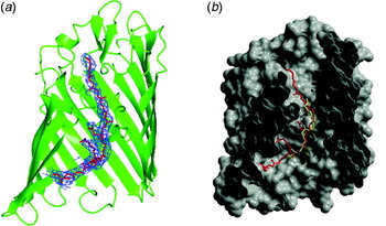
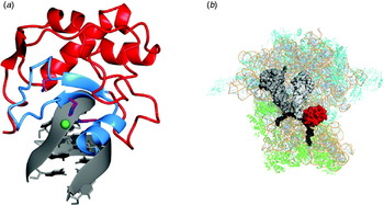
Housden et al. first showed that Col IUTDs are used to recruit a translocator following binding of the Col to its receptor in the E. coli OM (Housden et al. Reference Housden, Loftus, Moore, James and Kleanthous2005). Using the ColE9–Im9 complex in which the Im protein was His-tagged (and where the Col was disulfide-locked to prevent cell-killing), they demonstrated that the entire ColE9–Im9–BtuB–OmpF complex could be affinity-purified from OM extracts but that recruitment of OmpF was lost when the 83-residue IUTD was deleted. Consistent with this region being sequestered post receptor-binding, Zhang et al. reported that unique enterokinase cleavage sites engineered in the first 61 residues of the IUTD became inaccessible to enzymatic cleavage after binding to BtuB in the OM (Zhang et al. Reference Zhang, Vankemmelbeke, Holland, Walker, James and Penfold2008). Housden et al. went on to delineate the regions of the ColE9 IUTD involved in binding OmpF using a combination of deletion analysis and ITC (Housden et al. Reference Housden, Wojdyla, Korczynska, Grishkovskaya, Kirkpatrick, Brzozowski and Kleanthous2010). They showed that the primary OmpF binding site (OBS1), K d~2 μM, comprises residues 2–18 of the Col (the N-terminal methionine is often missing in ColE9). They also identified a second, weaker, OmpF binding site (OBS2) further along the IUTD between residues 54 and 63, which binds OmpF with a K d~25 μM. Importantly, binding of OBS2 is blocked by the presence of OBS1 suggesting that they share the same binding site. Moreover, the two OBSs flank the 16-residue TolB-binding epitope (TBE; comprising residues 32–47) in the centre of the IUTD, previously identified by Loftus et al. (Reference Loftus, Walker, Mate, Bonsor, James, Moore and Kleanthous2006) (Fig. 5; Section 5). Mutational analysis revealed that many ColE9 OBS1 side chains are involved in stabilizing the complex with OmpF although none are essential for binding or toxicity. In addition, these studies suggested that conformational flexibility of the epitope (which is rich in glycine residues) and a free N-terminus are major factors in binding to the porin. Cell-killing assays demonstrated that at least one OBS is required for antibiotic activity. Housden et al. also showed that the relative importance of the OBS sites is different in cells expressing either OmpF or OmpC, emphasizing that the two porins do not behave identically in terms of Col translocation. A luminal translocation route to the periplasm was revealed by the 3·1 Å crystal structure of the OmpF–OBS1 complex in which the peptide was bound in an extended, crescent-shaped conformation (Fig. 6a). OBS1 spans the membrane bilayer in its complex with OmpF, traversing the central constriction point of the porin channel that generally restricts molecules of >600 Da passing across the OM (Fig. 6b). Moreover, the orientation of OBS1 is with its N-terminus facing the periplasm and C-terminus exposed to the extracellular surface where the rest of the receptor-bound Col would be located.
Sequence alignment of N-terminal IUTD sequences of ColA, ColE9 and ColN showing the presence of the various OmpF, TolB (TBE) and TolA (TAB) binding epitopes.

Structure of the OmpF-ColE9 OBS1 (PDB ID: 3O0E) and OmpF·ColE3 IUTD (PDB ID: 2ZLD) complexes. (a) OmpF monomer shown in green ribbon (OmpF is trimeric in its native state) with ColE9 OBS1 peptide (chain L, residues 2–16) modelled into the electron density map (2F o–F c, shown at 1σ sigma cut-off) within the lumen. Note how the Col traverses the constriction point at the centre of the porin. (b) Superposition of the Col peptide structures from the complex of the ColE3 IUTD, where ~7 residues of unassigned sequence could be seen, and the structure of ColE9 OBS1 bound to OmpF, shown as a molecular surface cut-away to reveal the lumen of the porin. The OBS1 peptide is shown in red and the fragment from the ColE3 IUTD in yellow. Figure adapted from Housden et al. (Reference Housden, Wojdyla, Korczynska, Grishkovskaya, Kirkpatrick, Brzozowski and Kleanthous2010) with permission from the National Academy of Sciences (USA).

A translocation route for the ColE9 IUTD to the periplasm through the lumen of the porin is also supported by a separate series of studies on the ColE3 IUTD from the Cramer laboratory; the two enzymatic Cols share ~90% sequence identity in their 83-residue IUTD. Focusing on the voltage-gated channels displayed by OmpF in planar-lipid bilayer experiments, Zhaharov et al. demonstrated that the IUTD of ColE3 blocks OmpF channel activity (Zakharov et al. Reference Zakharov, Eroukova, Rokitskaya, Zhalnina, Sharma, Loll, Zgurskaya, Antonenko and Cramer2004a), suggesting that the polypeptide blocks the porin lumen. They subsequently obtained a 3·0 Å resolution structure of OmpF crystallized in the presence of the ColE3 IUTD which showed the presence of density corresponding to ~7 amino acids although the identity of these residues, which did not pass the constriction point in OmpF, could not be ascertained (Yamashita et al. Reference Yamashita, Zhalnina, Zakharov, Sharma and Cramer2008) (Fig. 6b). The subsequent structure from Housden et al. confirmed these residues as likely corresponding to the OBS1 sequence of the IUTD (Housden et al. Reference Housden, Wojdyla, Korczynska, Grishkovskaya, Kirkpatrick, Brzozowski and Kleanthous2010).
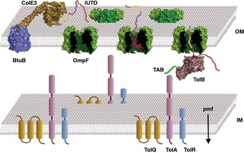
The investigations of the ColE3 and ColE9 IUTDs are consistent with the disordered domain recruiting OmpF through a sequential binding mechanism in which OBS1 first docks into the porin lumen followed by diffusion of the polypeptide toward the periplasm (possibly guided by the overhanging receptor-bound Col), subsequent docking of OBS2 in the lumen and presentation of the Col's TBE for binding TolB (Fig. 7). The initial driving force for binding of the OBSs to the OmpF lumen, which is cation selective, is likely to be electrostatic since both have overall positive charge. The intervening sequence between the two OBSs does not bind OmpF – the TBE is negatively charged and does not bind OmpF in ITC experiments, plus this region contains two proline residues and prolines were shown to abolish peptide binding to the OmpF lumen (Housden et al. Reference Housden, Wojdyla, Korczynska, Grishkovskaya, Kirkpatrick, Brzozowski and Kleanthous2010).
Overview of early OM events during cell entry of Group A nuclease Cols. Following binding to BtuB (vitamin B12 transporter, blue) through its R-domain (ColE3–Im3 bound to BtuB is shown), the Col–Im protein complex (gold and yellow, respectively) recruits OmpF (green) through its IUTD (shown as spheres), first docking the OBS1 epitope (red spheres) into the lumen of the porin followed by OBS2 (yellow spheres). The Col's TBE (magenta spheres) then binds TolB (pink) in the periplasm. Col mimics interactions of the lipoprotein Pal (not shown), but unlike Pal enhances the recruitment of TolA (magenta cylinders) through the disordered N-terminal TAB of TolB (green line). TolA is coupled to the PMF through its associations with TolR (cyan cylinders) and TolQ (orange cylinders) in the IM. Contact between TolB and TolA in the periplasm triggers release of the Im protein to the external medium in a PMF-dependent step that initiates Col translocation into the cell.

An important new development for probing Col import is the use of single molecule methods. Spector et al. have shown using Total Internal Reflection Fluorescence (TIRF) microscopy that the lateral diffusion of the ColE3-bound BtuB complex is relatively unhindered in the E. coli OM but that of OmpF is restricted, implying collision between receptor and translocator must occur to enable Col translocation (Spector et al. Reference Spector, Zakharov, Lill, Sharma, Cramer and Ritchie2010). It is likely that future single molecule methods such as TIRF will feature prominently in the elucidation of Col import into bacterial cells.
5. Recruitment of periplasmic proteins to form a translocon complex at the OM
The term ‘translocon complex’ has been coined to describe the self-assembled supramolecular assembly formed between the Col and its receptor, translocator and periplasmic binding partners (Kurisu et al. Reference Kurisu, Zakharov, Zhalnina, Bano, Eroukova, Rokitskaya, Antonenko, Wiener and Cramer2003), by analogy with the translocons of secretory systems (Johnson & van Waes, Reference Johnson and Van Waes1999; Keenan et al. Reference Keenan, Freymann, Stroud and Walter2001; Pohlschroder et al. Reference Pohlschroder, Prinz, Hartmann and Beckwith1997). Formation of the translocon is thought to trigger Col translocation across the OM. Translocon assembly requires the presentation of specific signalling epitopes located within the IUTD in the periplasm via translocator proteins, as described in Section 4. For the group B Col ColIa, which uses the Cir protein as receptor and translocator, a TonB box is delivered that is presumed to mimic the interactions of the TonB box of nutrient receptors with TonB. How the IUTD of ColIa delivers its TonB box through the translocator Cir protein remains to be established. Much more is known about the interactions of IUTD signalling epitopes of group A Cols with their periplasmic binding partners where molecular mimicry has been shown to play an important role in the establishment of the translocon complex. The following sections summarize this research, which includes work on the IUTDs of both pore-forming (ColA and ColN) and enzymatic (ColE9) Cols.
5.1 ColE9
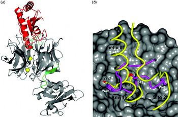
Enzymatic Cols E2–E9 contain a 16-residue signalling epitope within their IUTDs called the TBE that binds TolB with low μM affinity. Col TBE residues are essential for antibiotic activity; mutations abolish Col toxicity in vivo and TolB binding in vitro (Bouveret et al. Reference Bouveret, Rigal, Lazdunski and Benedetti1998; Hands et al. Reference Hands, Holland, Vankemmelbeke, Fraser, Macdonald, Moore, James and Penfold2005). TolB is a 44-kDa protein that has an N-terminal α-/β-domain and C-terminal, six-bladed β-propeller domain that binds the OM lipoprotein Pal with sub-μM affinity as part of the Tol system in the bacterial periplasm. Crystal structures have revealed that both the ColE9 TBE and Pal bind to the same canyon binding site on the β-propeller domain, distal to the N-terminal domain (Bonsor et al. Reference Bonsor, Grishkovskaya, Dodson and Kleanthous2007; Loftus et al. Reference Loftus, Walker, Mate, Bonsor, James, Moore and Kleanthous2006) suggesting that the Col ‘competitively recruits’ TolB from its complex with Pal (Loftus et al. Reference Loftus, Walker, Mate, Bonsor, James, Moore and Kleanthous2006). The soluble domain of Pal, comprised a four-stranded β-sheet and four α-helices, contacts the canyon in two sites (Fig. 8a) (Bonsor et al. Reference Bonsor, Grishkovskaya, Dodson and Kleanthous2007). Helix III of Pal interacts with TolB through residues 110–130 (Site 1) while a neighbouring loop accompanied by a β-strand interacts with TolB through residues 144–163 (Site 2) (Fig. 8b). The intrinsically unstructured ColE9 TBE does not adopt regular secondary structure on binding TolB, but rather an unusual conformation resembling a distorted paperclip (Fig. 8). Importantly, Glu42 and Trp39 of the ColE9 TBE mimic interactions made by Glu116 and Tyr117, respectively, of Pal with TolB at Site 1 while Trp46 of the ColE9 TBE is buried within a Site 2 pocket on TolB that has no equivalent in the Pal complex due to accompanying conformational changes (Fig. 8b). This latter contact has important implications for TolB signalling to TolA, which is discussed below. In contrast, no conformational changes occur in TolB as a result of ColE9 TBE binding, which is also relevant to the mechanism of Col translocation.
(a) Superposition of the complexes of E. coli TolB (grey) with the ColE9 TBE (cyan, PDB ID 1IVZ) and Pal (red, PDB ID 2HQS). Two Ca2+ ions (yellow) are present in the ColE9 TBE structure while the TolB TAB (green), 22Glu-Ser33, is only visible in the Pal-bound structure. (b) Comparison of the TolB-binding site canyon (grey) where both the ColE9-TBE (magenta) and Pal (yellow) dock. Only helix III (Site 1, residues 110–130) of Pal and its neighbouring loop and β-strand (Site 2, residues 144–163) are shown for clarity. ColE9 TBE residues Glu42 and Trp39 mimic hydrogen bond interactions made by Pal residues Glu116 and Tyr117, respectively. ColE9 Trp46, on the other hand, binds in a deep pocket in Site 2 on the TolB surface for which there is no equivalent in the Pal complex due to conformational changes that occlude the pocket. Binding of ColE9 Trp46 into Site 2 shifts the conformational equilibrium of TolB so that the TolB TAB undergoes an order–disorder transition and so becomes available for binding TolA (See Fig. 7).

The buried solvent-accessible surface area in the TolB–Pal complex is significantly greater than the surface area buried in the ColE9–TolB complex (2600 versus 1600 Å2), although the latter shows greater complementarity (Loftus et al. Reference Loftus, Walker, Mate, Bonsor, James, Moore and Kleanthous2006). TolB undergoes significant conformational changes in both its domains on binding Pal, while Pal undergoes only limited change in conformation; helix III of Pal unwinds by 1·9 Å due to steric and electrostatic clashes with TolB residues. Both the Pal and ColE9 TBE complexes with TolB are stabilized by a significant number of hydrogen bonds, which are mostly intermolecular in the Pal complex but intramolecular in the ColE9 TBE complex. The outcome of these structural differences between the Pal and ColE9 TBE complexes with TolB is a 20-fold difference in their affinities, but these affinities are tuned by the presence of divalent metal ions such as Ca2+ that bind within the tunnel running through the β-propeller domain of TolB (Fig. 8a). The affinity of Pal for TolB in the absence of metal ions is 47 nM, as measured by ITC (Loftus et al. Reference Loftus, Walker, Mate, Bonsor, James, Moore and Kleanthous2006), with binding involving a single protonation event (Bonsor et al. Reference Bonsor, Grishkovskaya, Dodson and Kleanthous2007). Moreover, binding is enthalpically and entropically favoured, consistent with the significant hydrophobic contact and extensive hydrogen bonding between the two proteins. Under the same metal-free conditions, the ColE9–TBE binds TolB with an affinity of 1 μM where the association is enthalpically favourable but entropically unfavourable in accordance with the significant loss of conformational entropy upon binding as the intrinsically disordered ColE9 TBE folds. In the presence of 1 mM Ca2+ ions, the affinity for Pal is reduced 2-fold (~100 nM), becoming equivalent to the affinity of ColE9 under the same conditions (Loftus et al. Reference Loftus, Walker, Mate, Bonsor, James, Moore and Kleanthous2006). This 10-fold increase in the affinity of ColE9–TBE for TolB is largely explained by electrostratic interactions. In the presence of Ca2+ ions, the lower region of the TolB canyon has an overall positive charge, which favours association of the negatively charged ColE9–TBE. It is reasonable to conclude that divalent metal ions, which are abundant in the E. coli cell envelope, would have to be bound in order for the ColE9–TBE to competitively recruit TolB from its complex with Pal. Binding of TolB by the Col would also be favoured by virtue of Pal's other binding interaction in the periplasm, which is to peptidoglycan (Parsons et al. Reference Parsons, Lin and Orban2006). TolB and peptidoglycan binding are mutually exclusive of each other (Bouveret et al. Reference Bouveret, Benedetti, Rigal, Loret and Lazdunski1999) and so free TolB would invariably be available for Col binding.
The conformational changes within the C-terminal β-propeller of TolB that result from Pal binding are transmitted to the N-terminal α-/β-domain of TolB through motions of loops and β-strands of the C-terminus. These changes are relayed to the N-terminal domain of TolB through the hinge connecting the two domains and specifically between a β-strand of the β-propeller domain and residues Glu22–Ser33, the N-terminal most residues of TolB (Bonsor et al. Reference Bonsor, Grishkovskaya, Dodson and Kleanthous2007). Residues Glu22–Ser33 are disordered in crystal structures of TolB as well as in the ColE9 TBE–TolB complex, but become ordered in the Pal complex, binding in a cleft between the N- and C-terminal domains of TolB that becomes exposed when Pal is bound (Fig. 8a) (Bonsor et al. Reference Bonsor, Hecht, Vankemmelbeke, Sharma, Krachler, Housden, Lilly, James, Moore and Kleanthous2009b). Deletion of TolB resides Glu22–Ser33 results in a tol OM instability phenotype characteristic of a tolB deletion, showing that this region is indispensable for in vivo functionality of the Tol system, and renders E. coli cells resistant to Cols, indicating their importance to OM translocation. Taken together, these studies reveal that TolB is an allosteric protein whereby Pal and the ColE9–TBE are opposing allosteric effectors modulating the accessibility of the Glu22–Ser33 sequence at its N-terminus.
The role of the TolB N-terminal residues Glu22–Ser33 was revealed when it was shown using cross-linking and ITC by Bonsor et al. that this epitope is the binding site for the C-terminal domain of TolA, which we refer to as the TolA box (TAB) (Fig. 7). TolA has three domains, an N-terminal transmembrane helix (TMH; domain I), a central domain postulated to form a coiled-coil that spans the periplasm (domain II) and a small globular C-terminal domain (domain III or TolAIII). Binding between TolAIII and TolB through the TAB is weak (K d~43 μM) and endothermic in ITC experiments indicating an entropically driven interaction (Bonsor et al. Reference Bonsor, Hecht, Vankemmelbeke, Sharma, Krachler, Housden, Lilly, James, Moore and Kleanthous2009b). Pal abolishes binding between TolB and TolAIII in ITC experiments while the ColE9 TBE enhances binding 4-fold and switches the thermodynamics of binding from endothermic to exothermic. These effects can be explained by the structural and biophysical properties of the two complexes. Pal binding to TolB sequesters the TAB to the surface of TolB while ColE9 TBE binding renders the TAB disordered and available for TolAIII binding. Hence, even though the ColE9–TBE mimics interactions made by Pal, the effect of its binding to TolB has a different conformational outcome. The key difference between the complexes is the hydrophobic contact made by ColE9–TBE Trp46 in Site 2 on the surface of TolB. The deep pocket into which Trp46 inserts is only accessible in TolB in the absence of conformational changes, which unlike Pal the Col does not induce. Moreover, these data suggest that TolB is in conformational equilibrium between these two structural states, with Pal and the ColE9–TBE stabilizing opposing states (Bonsor et al. Reference Bonsor, Hecht, Vankemmelbeke, Sharma, Krachler, Housden, Lilly, James, Moore and Kleanthous2009b). Not only does this interpretation correspond to the changes observed in binding thermodynamics between TolB and its protein ligands but is also supported by solution NMR experiments; the TolB TAB sequence is only discernible in 1H-15N-HSQC (heteronuclear single-quantum coherence) spectra as an intrinsically disordered polypeptide when TolB is bound to the ColE9–TBE (Bonsor et al. Reference Bonsor, Hecht, Vankemmelbeke, Sharma, Krachler, Housden, Lilly, James, Moore and Kleanthous2009b).
Based on these observations a model for the hijacking and activation of the Tol system by ColE9 has been proposed in which the Col manipulates the allosteric transition of TolB. In the Pal-bound state, the TolB TAB is folded against the surface of the protein disfavouring binding to TolA, signalling the OFF-state for the system (Fig. 8a). In the ColE9–TBE-bound state, the TolB TAB is disordered and available to bind TolA, signalling an ON-state for the Tol system (Fig. 7). Hence, the recruitment of TolB to the ColE9 translocon results in this surface bound toxin being able to make a connection to the PMF through protein–protein interactions that promote contact between TolB in the periplasm and TolA in the IM (Bonsor et al. Reference Bonsor, Hecht, Vankemmelbeke, Sharma, Krachler, Housden, Lilly, James, Moore and Kleanthous2009b). The potential outcome of this contact is explored further in Section 6.
Studies of Col parasitization of the Tol system have highlighted two important functional characteristics of this trans-periplasmic complex. First, the order–disorder transition of the TolB TAB that lies at the heart of the interaction between TolB and TolA is reminiscent of the order–disorder transitions of OM receptor TonB boxes and TonB (Cadieux et al. Reference Cadieux, Phan, Cafiso and Kadner2003; Wiener, Reference Wiener2005), suggesting that the same basic signalling mechanism underpins the Ton and Tol systems of Gram-negative bacteria (Kleanthous, Reference Kleanthous2010). Second, these studies cast doubt on the generally accepted view of Tol function, which is that of a PMF-driven machine ensuring appropriate juxtaposition of the inner and OMs. Components of the Tol system are known to be recruited to septation sites in E. coli (Gerding et al. Reference Gerding, Ogata, Pecora, Niki and De Boer2007), consistent with Tol function being predominantly expressed during cell division. A role as a transmembrane tether necessitates interaction between Pal in the OM and TolA in the IM, originally suggested by in vivo cross-linking experiments (Cascales et al. Reference Cascales, Gavioli, Sturgis and Lloubes2000, Reference Cascales, Lloubes and Sturgis2001). Moreover, it has been argued that this periplasmic interaction is regulated by cellular PMF (Cascales et al. Reference Cascales, Gavioli, Sturgis and Lloubes2000, Reference Cascales, Lloubes and Sturgis2001). However, extensive biochemical and biophysical analysis using purified Pal and TolA failed to demonstrate such an interaction in vitro (Bonsor et al. Reference Bonsor, Hecht, Vankemmelbeke, Sharma, Krachler, Housden, Lilly, James, Moore and Kleanthous2009a). An indirect interaction between Pal and TolAIII via TolB is also unlikely given that Pal diminishes/abolishes the interaction between TolA and TolB (see above). It remains to be elucidated how and for what physiological purpose Tol signalling is deployed during cell division.
5.2 ColN
The N-terminal 90 residues of the pore-forming Col ColN comprise its IUTD (Raggett et al. Reference Raggett, Bainbridge, Evans, Cooper and Lakey1998; Vetter et al. Reference Vetter, Parker, Tucker, Lakey, Pattus and Tsernoglou1998). Unlike ColE9, ColN does not require TolB for Col antibiotic activity (and does not bind TolB in vitro), but rather contacts TolA directly through its own TAB sequence, residues Asn42–Gly68 (Gokce et al. Reference Gokce, Raggett, Hong, Virden, Cooper and Lakey2000). There is no sequence similarity between the TABs of TolB and ColN (Fig. 5). The structure for the TolAIII–ColN TAB complex is not yet known, but FRET and NMR experiments indicate that the TAB epitope undergoes a disorder–order transition and that its two tryptophan residues (Trp44 and Trp46) are buried on binding TolAIII (Anderluh et al. Reference Anderluh, Hong, Boetzel, Macdonald, Moore, Virden and Lakey2003; Raggett et al. Reference Raggett, Bainbridge, Evans, Cooper and Lakey1998), reminiscent of ColE9–TBE binding TolB. The ColN–IUTD interaction with TolAIII has been studied extensively by the Lakey and Moore laboratories by mutagenesis, surface plasmon resonance (SPR), ITC and NMR. Intact ColN binds TolAIII with a K d of 18 μM, with this affinity increasing 20-fold when the T-domain in isolation is used (Raggett et al. Reference Raggett, Bainbridge, Evans, Cooper and Lakey1998). These differences in binding suggest that the TAB sequence may be sequestered in intact ColN, possibly through interactions with other parts of the Col. This interpretation has been borne out by heteronuclear NMR experiments which show the IUTD contacts the R-domain of the Col through what has been coined ‘self-recognition’ (Hecht et al. Reference Hecht, Ridley, Boetzel, Lewin, Cull, Chalton, Lakey and Moore2008). NMR data also indicate that parts of the IUTD may in fact be partially structured (Hecht et al. Reference Hecht, Ridley, Boetzel, Lewin, Cull, Chalton, Lakey and Moore2008), with binding to TolAIII requiring the disassembling of this structure prior to binding (Hecht et al. Reference Hecht, Ridley, Lakey and Moore2009b).
A critical part of the TAB (61SYHITFH67) is thought to adopt a β-conformation upon complexation with TolAIII (Hecht et al. Reference Hecht, Ridley, Lakey and Moore2009b). The involvement of a tyrosine residue (Tyr62) within the TAB suggests the binding interaction may be analogous to the binding motifs of other TolAIII binding proteins such as the N1 domain of g3p, which contacts TolAIII as part of the infection process of bacteriophage fd. Indeed, comparison of the crystal structure of TolAIII–N1 g3p (Lubkowski et al. Reference Lubkowski, Hennecke, Pluckthun and Wlodawer1999) with chemical shift mapping data, using a 13C/15N-labelled TolAII–III binding unlabelled ColN T-domain, have suggested that not only are the binding sites of g3p and ColN overlapping on TolAIII but that similar conformational changes may be induced in the Tol protein (Hecht et al. Reference Hecht, Ridley, Lakey and Moore2009b).
5.3 ColA
The IUTD of ColA requires both TolB and TolA for cytotoxic activity (Benedetti et al. Reference Benedetti, Frenette, Baty, Knibiehler, Pattus and Lazdunski1991), with biophysical experiments indicating that both are bound by distinct and recognizable Tol binding sites; an N-terminal TBE (residues Lys9–Glu18) (Zhang et al. Reference Zhang, Vankemmelbeke, Baardelang, Paoli, Penfold and James2009b) and a C-terminal TAB (residues 58–90 (Cascales et al. Reference Cascales, Buchanan, Duche, Kleanthous, Lloubes, Postle, Riley, Slatin and Cavard2007; Deprez et al. Reference Deprez, Blanchard, Guerlesquin, Gavioli, Simorre, Lazdunski, Marion and Lloubes2002; Hecht et al. Reference Hecht, Zhang, Li, Penfold, James and Moore2010; Journet et al. Reference Journet, Bouveret, Rigal, Lloubes, Lazdunski and Benedetti2001) (Fig. 5). Interestingly, the ColA–TBE, which bears strong sequence similarity to the ColE9–TBE is close to the N-terminus of the Col, similar to the position of the ColE9 OBS1. This raises the interesting possibility that the 20 first residues of the ColA N-terminus may serve a dual role, both recruiting OmpF and contacting TolB during Col translocation.
The ColA–TBE encompasses the N-terminal half of the 16-residue ColE9–TBE. With fewer amino acids involved in binding it is not surprising that the affinity of the ColA–TBE–TolB complex is significantly weaker than its ColE9 counterpart; K d 1·6 μM, in the presence of Ca2+ ions, compared to 90 nM for ColE9 (Loftus et al. Reference Loftus, Walker, Mate, Bonsor, James, Moore and Kleanthous2006; Zhang et al. Reference Zhang, Vankemmelbeke, Baardelang, Paoli, Penfold and James2009b). The crystal structure of the ColA–TBE bound to TolB shows a similar mode of binding to the TolB β-propeller as ColE9 (Zhang et al. Reference Zhang, Vankemmelbeke, Baardelang, Paoli, Penfold and James2009b). The RMSD for the core elements of the ColA and ColE9 TBEs bound to TolB is 0·39 Å, with a greater number of hydrogen bonds and hydrophobic interactions observed in the larger ColE9–TBE complex, consistent with its higher affinity. In particular, the key hydrogen bond interactions of Glu42 and Trp39 of the ColE9–TBE, which mimic the interactions of Pal with TolB (see above), are emulated in the ColA–TBE–TolB complex by Glu18 and Trp15 of ColA, respectively.
The ColA TAB binds TolAIII in SPR experiments with a K d~0·5 μM (Derouiche et al. Reference Derouiche, Zeder-Lutz, Benedetti, Gavioli, Rigal, Lazdunskil and Lloubes1997). However, the structural delineation of the complex has proven elusive because of the unusual characteristics of the complex in solution. Using NMR, Deprez et al. reported that binding of ColA to TolAIII caused the domain to unfold, confirmed by subsequent NMR studies (Hecht et al. Reference Hecht, Ridley, Lakey and Moore2009b). The physiological relevance of this effect has been questioned by Hecht et al. who found that no unfolding was observed if ColA T-domain constructs were used that only contained the binding site for TolAIII (constructs by Deprez et al. contained binding sites for both TolA and TolB) or if TolB was added to the ColA T-domain–TolAIII complex (Hecht et al. Reference Hecht, Zhang, Li, Penfold, James and Moore2010). These latter experiments also confirmed that a ternary complex of ColA T-domain–TolB–TolA could be formed in vitro (Hecht et al. Reference Hecht, Zhang, Li, Penfold, James and Moore2010), first shown by antibody overlay experiments (Bouveret et al. Reference Bouveret, Rigal, Lazdunski and Benedetti1998). Hecht et al. have also shown through NMR chemical shift mapping that the binding sites for all TolAIII protein partners thus far characterized (N1 domain of g3p, ColN TAB and ColA TAB) are distinct but overlap each other. It remains to be seen if this binding region coincides with that of the cognate binding partner for TolAIII in the bacterial periplasm, the TAB of TolB.
The question arises as to why ColA requires contact with two Tol proteins when contact with TolA or TolB is sufficient to catalyse translocation for other Cols such as ColN and ColE9? This requirement likely reflects the twin problems of (i) weaker TolB binding than that required to competitively recruit TolB from its higher-affinity complex with Pal (which is supported by analytical gel filtration experiments (Zhang et al. Reference Zhang, Vankemmelbeke, Baardelang, Paoli, Penfold and James2009b); and (ii) the inability of ColA to allosterically promote contact between TolB and TolA. Although this has not been formally addressed in biophysical experiments, the structure of the ColA–TBE–TolB complex shows that there is no interaction equivalent to that of Trp46 of the ColE9–TBE (Figs 5 and 8), which inserts into a deep pocket in Site 2 on the TolB surface (Section 5). Occupation of this site by the tryptophan is important for manipulation of the conformational equilibrium in TolB and hence for enhancing binding between TolB and TolAIII (Bonsor et al. Reference Bonsor, Hecht, Vankemmelbeke, Sharma, Krachler, Housden, Lilly, James, Moore and Kleanthous2009b). Without such an interaction it is unlikely the ColA T-domain could promote the interaction between TolA and TolB.
Some studies have suggested that translocating Cols can contact other Tol proteins in addition to TolB and TolA. For example, pull-down and immunoprecipitation assays have suggested that the group A Col ColK simultaneously contacts TolB, TolA, TolQ and TolR (Barneoud-Arnoulet et al. Reference Barneoud-Arnoulet, Gavioli, Lloubes and Cascales2010b), although the specific binding epitopes on the Col for the IM proteins TolQ and TolR have not been elucidated. It is also unclear as to how ColK contacts TolB since it does not contain a recognizable TBE sequence.
6. Col translocation across the OM and the role of the PMF
How Cols translocate cytotoxic domains across the OM following assembly of their translocons is not understood. Some of the contributing processes to this event have emerged, however, from work on both pore-forming and nuclease Cols. Significant conformational changes to the Col are thought to be associated with OM translocation, which is unsurprising given that these are folded proteins bound to the external surface of the cell. Urea denaturation experiments and disulfide bond engineering have both suggested that ColA undergoes large-scale structural changes during OM translocation (Benedetti et al. Reference Benedetti, Lloubes, Lazdunski and Letellier1992; Duche et al. Reference Duche, Baty, Chartier and Letellier1994). The use of an internal disulfide bond, which locks the coiled-coil R-domain of ColE9 closed and so blocks Col entry into cells and cytotoxicity (Section 4), is also consistent with the Col experiencing significant changes in structure in order for translocation to occur (Penfold et al. Reference Penfold, Healy, Housden, Boetzel, Vankemmelbeke, Moore, Kleanthous and James2004b). Moreover, cell-killing and protease protection studies on Col-treated cells have suggested that translocated Cols not only span the cell envelope but that part of the toxin remains exposed to the external surface during cell-killing (Benedetti et al. Reference Benedetti, Lloubes, Lazdunski and Letellier1992; Beppu et al. Reference Beppu, Kawabata and Arima1972; Cramer et al. Reference Cramer, Zhang, Schendel, Merrill, Song, Stauffacher and Cohen1992; Nose & Mizuno, Reference Nose and Mizuno1968; Zhang & Cramer, Reference Zhang and Cramer1992). Notwithstanding these important pieces of information, there is currently very little structural understanding of what a translocated Col looks like or indeed how much of the Col moves across the membrane in order to deliver the cytotoxic domain to the periplasm/cytoplasm. It is not inconceivable that answers to these questions will emerge in the future since isolation of part of the translocon for ColE9, containing the Col, BtuB and OmpF, has already been described (Housden et al. Reference Housden, Loftus, Moore, James and Kleanthous2005).
An area of controversy in the Col field concerns the energy dependence of the OM translocation step. Since the OM is not an energized system, cellular processes requiring the input of energy, such as the active transport of iron through siderophore receptors, rely on connection to PMF-driven machines, the Ton system in the case of nutrient uptake. The direct relevance of cellular PMF to Col uptake through Ton or Tol has proven difficult to ascertain. Indeed, early studies (Bourdineaud et al. Reference Bourdineaud, Boulanger, Lazdunski and Letellier1990a) suggested that in the case of ColA the PMF was not involved in translocation. Recently, however, Im protein-bound nuclease Cols have provided direct evidence for the involvement of the PMF in the OM translocation step. Im proteins bind Col nuclease domains with femtomolar binding affinities (Section 9), which raises the key question of how a complex that has a dissociation half-life of the order of several days at 37°C is dissociated at the cell surface on a timescale commensurate with cell-killing (τ <15 min). Studies have now shown that not only is the Im protein released at the cell surface but this is a PMF-dependent process.
Using an SOS–lux reporter system, James and co-workers demonstrated that translocation of the ColE9 DNase to the cytoplasm (detected by DNA damage-induced luminescence) takes the same amount of time whether the Im protein is bound or not (Vankemmelbeke et al. Reference Vankemmelbeke, Healy, Moore, Kleanthous, Penfold and James2005), suggesting Im protein dissociation is not rate-limiting for cell-killing. Using an EGFP–Im2 fusion as well as Western blotting, the Lloubes lab showed that the Im protein from the ColE2–Im2 complex dissociates on the same timescale as the kinetics of cell-killing and that the full translocon machinery is needed for the separation of Col from Im protein (Duche et al. Reference Duche, Frenkian, Prima and Lloubes2006). Using a similar approach but with Alexa594-labelled Im9 in complex with disulfide-locked ColE9, James and co-workers have shown that Im dissociation at the cell surface requires reduction of the disulfide implying translocon assembly is not sufficient for Im release but that Col unfolding is a pre-requisite for complex dissociation (Zhang et al. Reference Zhang, Vankemmelbeke, Holland, Walker, James and Penfold2008). Critically, they demonstrated that ionophores which destroy the cellular PMF reduce Im release from the Col complex at the cell surface and that a mutation within the proton-conducting path of the TolQRA complex, which disrupts Tol function in vivo (Goemaere et al. Reference Goemaere, Cascales and Lloubes2007), has a similar effect (Vankemmelbeke et al. Reference Vankemmelbeke, Zhang, Moore, Kleanthous, Penfold and James2009). Finally, Bonsor et al. have shown that it is the contact between TolAIII and the TAB sequence of TolB within the periplasm that is the necessary step for enzymatic Col Im release (Bonsor et al. Reference Bonsor, Hecht, Vankemmelbeke, Sharma, Krachler, Housden, Lilly, James, Moore and Kleanthous2009b).
Current models suggest that the PMF across the cytoplasmic membrane is transduced into conformational transitions within TolA through the TolQRA complex. Group A nuclease Cols parasitize this system by promoting contact between TolA and TolB thereby promoting Im protein dissociation at the cell surface. Several studies have begun to investigate conformational changes within the IM TolQRA complex (which has an estimated stoichiometry of 4–6:2:1; Cascales et al. Reference Cascales, Lloubes and Sturgis2001) and in particular the role of PMF in transmembrane interactions. The Lloubès lab have deployed disulfide cross-linking to demonstrate that dimerization occurs between TolR molecules through their TMHs (Zhang et al. Reference Zhang, Goemaere, Thome, Gavioli, Cascales and Lloubes2009a) and periplasmic C-terminal α-helices (Goemaere et al. Reference Goemaere, Cascales and Lloubes2007). Cellular PMF regulates the rotation of the TMH pairs (Zhang et al. Reference Zhang, Goemaere, Thome, Gavioli, Cascales and Lloubes2009a) and conformational changes experienced by each C-terminal helix (Goemaere et al. Reference Goemaere, Cascales and Lloubes2007). The latter is also regulated by specific residues in the TolQR ion-conducting pathway, comprised of one TolR and two TolQ TMHs. Self-association of TolQ has also been demonstrated by cysteine scanning mutagenesis (Zhang et al. Reference Zhang, Goemaere, Seddiki, Celia, Gavioli, Cascales and Lloubes2011). Presumably, it is the rotation of Tol complex TMHs transduced into a conformational change in TolA that affects its interaction with the TolB/nuclease Col IUTD complex at the OM and provides the signal for Im protein dissociation at the external surface of the cell, although how this occurs is not known.
The Cramer lab has reported that release of the Im protein Im3 from its complex with ColE3 leads to significant destabilization/unfolding of the nuclease and that the rRNase domain occludes voltage-gated OmpF channels in planar lipid bilayer experiments. The two observations have been combined to suggest the destabilized rRNase is translocated through the OmpF pore in a similar fashion to the IUTD of the Col (Zakharov et al. Reference Zakharov, Zhalnina, Sharma and Cramer2006) (Section 4). While attractive as a mechanism for translocating the nuclease to the periplasm this model remains problematic for several reasons. First, in other work Walker et al. found that the ColE3 rRNase domain free of Im3 is rather a stable protein (T m~48°C), which has a single co-operative unfolding transition and near UV-CD and tryptophan emission fluorescence spectra all indicative of a folded domain (Walker et al. Reference Walker, Lancaster, James and Kleanthous2004a). Second, if this were the translocation route of the nuclease it would be expected to be used by other nuclease Cols similarly destabilized by removal of their Im protein. Col DNases, however, are even more thermodynamically stable than the E3 rRNase domain (in their metal bound states, van den Bremer et al. Reference Van Den Bremer, Keeble, Jiskoot, Spelbrink, Maier, Van Hoek, Visser, James, Moore, Kleanthous and Heck2004) and there is currently no evidence that DNases block OmpF pores. Third, while OmpF occlusion can be clearly linked to translocation of the ColE3 (and ColE9) IUTD (Section 4) it remains to be shown if this also reflects translocation of the entire cytotoxic domain to the periplasm.
7. Col nuclease domain translocation across the IM and the role of proteolytic processing
While a number of studies have suggested proteolytic cleavage of Cols occurs, the linkage of such processing to the import mechanism itself is often less clear-cut. Cleavage by bacterial proteases has been shown to take place at the cell surface and in the periplasm. For example, the OM protease OmpT was reported to cleave group A Cols A, E1, E2 and E3 (Cavard & Lazdunski, Reference Cavard and Lazdunski1990). Recently, ColE2 cleavage by OmpT was studied in more detail (Duche et al. Reference Duche, Issouf and Lloubes2009). In vivo assays established that a region of the ColE2–Im2 complex is proteolytically processed at the cell surface by OmpT leading to the release of the DNase domain. Moreover, BtuB was found to be necessary for the cleavage to take place, and experiments with cells overproducing OmpT were more sensitive to ColE2–Im2 treatment than Col alone. The results also suggested that Im2 protects ColE2 from proteolytic cleavage at the cell surface. It has been demonstrated recently that while the ColD and ColE3 RNases undergo OmpT proteolytic cleavage at the cell surface, ΔompT cells retain their sensitivity to both Cols, which discounts the involvement of OmpT in the import and cytotoxicity of group A or B nuclease Cols (Chauleau et al. Reference Chauleau, Mora, Serba and De Zamaroczy2011). Cleavage in the periplasm has been shown for a number of Cols. The group A Col ColE7 is cleaved at the junction between the R- and DNase domains (residue 447), and this appears to be required for import to the cytoplasm and for cytotoxicity (Shi et al. Reference Shi, Chak and Yuan2005). In other studies, the IM leader protease LepB cleaves the group B Col ColD in a step that is also reported to be essential for cytotoxicity (de Zamaroczy & Buckingham, Reference De Zamaroczy and Buckingham2002; de Zamaroczy et al. Reference De Zamaroczy, Mora, Lecuyer, Geli and Buckingham2001). However, a lepB-resistant mutant remains sensitive to other Cols (B, E1, E2 and E3) implying that this is a very specific effect (see below).
While the mechanism by which the nuclease domain of group A and group B nuclease Cols translocate across the IM is yet to be resolved, evidence is emerging which suggests a direct interaction of the nuclease with the membrane may play a role. The nuclease domains of both ColE3 (rRNase) and ColE9 (DNase), which are highly basic proteins, are significantly destabilized by electrostatically driven interactions with anionic phospholipids, causing them to become molten-globule-like, with this effect inhibited by Im protein binding (Mosbahi et al. Reference Mosbahi, Walker, Lea, Moore, James and Kleanthous2004, Reference Mosbahi, Walker, James, Moore and Kleanthous2006). The physiological relevance of these interactions to the IM translocation step is inferred from two observations. The first relates to one of the consequences of such associations for Col DNases, which form random, non-voltage-gated channels in planar lipid bilayer experiments. DNase channel activity is abolished by an internal disulfide bond within the nuclease domain that also abolishes Col toxicity (Mosbahi et al. Reference Mosbahi, Lemaitre, Keeble, Mobasheri, Morel, James, Moore, Lea and Kleanthous2002). The second observation comes from studies investigating the relationship between Col nuclease electrostatics and cell-killing behaviour of an E. coli strain where the negative charge of the cytoplasmic membrane could be regulated. Walker et al. demonstrated that a direct correlation exists between the net positive charge on the DNase and the level of cell-killing in such a strain, with more positively charged DNases being more effective antibiotics (Walker et al. Reference Walker, Mosbahi, Vankemmelbeke, James and Kleanthous2007).
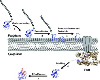
The potential for Col nucleases to form complexes with anionic phospholipid membranes such as the E. coli IM raises the critical question of how the Col might subsequently translocate to the cytoplasm? One possibility that has been considered is some form of retro-translocation through the Sec or Tat secretion pathways. However, mutations in these pathways fail to stop Col activity suggesting that they are not the route to the cytoplasm (Walker et al. Reference Walker, Mosbahi, Vankemmelbeke, James and Kleanthous2007). FtsH has been identified as a potential candidate for the retro-translocation step (Fig. 9). FtsH is an essential AAA+ ATPase/protease in the E. coli IM that degrades misfolded membrane proteins after first dislocating the protein to the cytoplasm in an ATP-dependent process (Ito & Akiyama, Reference Ito and Akiyama2005). Testing an array of different Cols with various cytotoxic activities and different import routes, in combination with FtsH deletion and point mutants, Walker et al. showed that FtsH is required for the cytotoxicity of both group A and group B nuclease Cols (rRNase, tRNase and DNase) (Walker et al. Reference Walker, Mosbahi, Vankemmelbeke, James and Kleanthous2007). Hence, the requirement for FtsH is independent of the import route across the OM. FtsH was, however, not required for pore-forming and peptidoglycan-cleaving Cols, which function within the periplasm. Moreover, both protease and ATPase activities of FtsH are required for nuclease Col toxicity.
Hypothetical model depicting retro-translocation of a Col nuclease domain across the E. coli cytoplasmic membrane. Although the model incorporates observations on several Cols, as described in the text, the figure highlights work on group A nucleases such as ColE3, ColE7 and ColE9. Step 1, electrostatically driven interaction between the Col nuclease domain and the anionic phospholipid bilayer of the cytoplasmic membrane; Step 2, destabilization of the nuclease domain (the DNase of ColE9 is shown) to form a molten-globule; Step 3, retro-translocation of the destabilized nuclease domain via the cytoplasmic AAA+ ATPase domain of FtsH followed by proteolytic processing. FtsH normally degrades proteins down to peptides, the mechanism by which Col nucleases avoid complete degradation remains unknown. Step 4, refolding of a single Col nuclease in the cytoplasm; Step 5, folded and functional Col nuclease attacks its cellular nucleic acid target, in this instance the E. coli chromosome.

The essential role of FtsH in the processing and translocation of ColD and ColE3 RNases has recently been confirmed by the de Zamaroczy lab who showed that FtsH is required for the proteolytic processing of these nuclease domain as they enter the cytoplasm (Chauleau et al. Reference Chauleau, Mora, Serba and De Zamaroczy2011). In the case of ColD, FtsH processing is mediated by LepB although this appears not to be a catalytic requirement but a structural one. However, this is not the case for ColE3 the processing of which by FtsH is LepB independent. These distinct requirements for LepB may be a result of the nuclease domain of ColE3 being more positively charged, and hence better able to insert in phospholipid bilayers, than ColD (Chauleau et al. Reference Chauleau, Mora, Serba and De Zamaroczy2011).
Taken together, these experiments suggest FtsH plays an important role in the IM retro-translocation of membrane-associated Col nuclease domains, mistaking the Col as a misfolded membrane protein (Fig. 9). The FtsH-mediated mechanism is similar to the retro-translocation of bacterial toxins such as the cholera toxin across the ER membrane through the ER-associated protein degradation pathway. If this is the route across the IM for the nuclease domain it would only require one nuclease molecule to avoid being degraded by the protease and its subsequent refolding in the cytoplasm in order for cell-killing to proceed. There is currently, however, no evidence that Col nucleases can avoid being degraded by FtsH, although a number of studies have shown that they are readily refolded from their denatured states (Ogawa et al. Reference Ogawa, Tomita, Ueda, Watanabe, Uozumi and Masaki1999; Walker et al. Reference Walker, Moore, James and Kleanthous2003; Wallis et al. Reference Wallis, Reilly, Barnes, Abell, Campbell, Moore, James and Kleanthous1994).
8. Mechanism of action of enzymatic cytotoxic domain
Four enzymatic activities have been identified in Cols; DNases that target cytoplasmic DNA (ColE2 and ColE7–ColE9), rRNases that target 16S ribosomal RNA (ColE3, E4 and E6), tRNases that target specific tRNAs (ColE5 and D) and a phosphatase that targets peptidoglycan precursors in the periplasm (ColM) (Table 2). The following sections summarize the structural biochemistry for each enzymatic class and how these toxins are thought to elicit cell death.
The catalytic activities of enzymatic Cols.

b Ogawa et al. (Reference Ogawa, Inoue, Yajima, Hidaka and Masaki2006).
c Tomita et al. (Reference Tomita, Ogawa, Uozumi, Watanabe and Masaki2000).
d Doudeva et al. (Reference Doudeva, Huang, Hsia, Shi, Li, Shen, Cheng and Yuan2006).
e Pommer et al. (Reference Pommer, Cal, Keeble, Walker, Evans, Kuhlmann, Cooper, Connolly, Hemmings, Moore, James and Kleanthous2001).
f El Ghachi et al. (Reference El Ghachi, Bouhss, Barreteau, Touze, Auger, Blanot and Mengin-Lecreulx2006).
8.1 DNase Cols
Col DNases are divalent metal ion-dependent enzymes that randomly degrade DNA (Table 2). Sequence similarity in the C-terminal 134 amino acid domains of the four E Col DNases that have been identified to date (E2 and E7–E9) ranges from 70 to 80%. The ColE7 and E9 DNase domains in complex with DNA and metal ions have been reported which provide a structural foundation for understanding the mechanism of dsDNA hydrolysis (Doudeva et al. Reference Doudeva, Huang, Hsia, Shi, Li, Shen, Cheng and Yuan2006; Hsia et al. Reference Hsia, Chak, Liang, Cheng, Ku and Yuan2004; Mate & Kleanthous, Reference Mate and Kleanthous2004) (Fig. 10a). The core of the active site of Col DNases is the H-N-H motif located at the C-terminus of the protein sequence. This ~34-amino-acid-long motif contains four conserved histidine residues some of which chelate a single metal ion to catalyse DNA hydrolysis. The H-N-H motif is widespread in metal-finger nucleases across all kingdoms of life (Gorbalenya, Reference Gorbalenya1994; Keeble et al. Reference Keeble, Maté, Kleanthous, Belfort, Derbyshire, Stoddard and Wood2005; Shub et al. Reference Shub, Goodrich-Blair and Eddy1994). They are divided into four sub-groups: bacterial toxins, homing endonucleases, restriction/repair enzymes and helicases/reverse transcriptases, with homing enzymes being the largest subgroup (Galburt & Stoddard, Reference Galburt and Stoddard2002). The H-N-H motif is also structurally related to the ‘ββα-Me’ motif (Kuhlmann et al. Reference Kuhlmann, Moore, James, Kleanthous and Hemmings1999) found in such nucleases as I-PpoI from Physarum polycephalum, Serratia nuclease from Serratia marcescens, Vvn from Vibrio vulnificus and the Holliday junction-resolving enzyme T4 endo VII.
(a) ColE9 DNase domain bound to the minor groove of dsDNA (PDB ID: 1V14) highlighting the active site H-N-H motif (cyan), along with two of the motif's characteristic histidine residues, and its bound metal ion. (b) ColE3 rRNase domain (red) bound to the A-site of the E. coli 70S ribosome in which is also bound mRNA (black) and P- (light grey) and E-site (dark grey) tRNAs (PDB ID: 2XFZ and 2XG0).

The ColE9 DNase has been shown to preferentially cleave dsDNA after thymine, but with little cleavage specificity exhibited for ssDNA and ssRNA (Pommer et al. Reference Pommer, Cal, Keeble, Walker, Evans, Kuhlmann, Cooper, Connolly, Hemmings, Moore, James and Kleanthous2001). Optimal activity of E9 DNase against dsDNA has been reported in the presence of millimolar concentrations of Mg2+ or Ca2+ while transition metals such as Ni2+ and Co2+ are preferred for ssDNA hydrolysis. Cleavage of ssRNA appears not to be metal dependent. Somewhat different results have been obtained for the closely related ColE7 DNase that is reported to cleave dsDNA in the presence of Zn2+ (which by contrast is inhibitory for the E9 DNase) and ssDNA in the presence of Zn2+ and Ni+ (Doudeva et al. Reference Doudeva, Huang, Hsia, Shi, Li, Shen, Cheng and Yuan2006).
The structure of the E9 DNase bound to dsDNA in the presence of Zn2+ or Mg2+ ions (Fig. 10a) shows that the architecture of the E9 DNase active site does not change upon binding to the minor groove of substrate DNA (Mate & Kleanthous, Reference Mate and Kleanthous2004). However, the DNA becomes distorted, which positions a single scissile phosphodiester bond close to the divalent cation in the active site. Widening and bending of the dsDNA groove has also been observed in other structures of similar enzymes containing the V-shaped active site characteristic of the H-N-H motif, such as I-PpoI and Vvn, as well as in the E7 DNase (Hsia et al. Reference Hsia, Chak, Liang, Cheng, Ku and Yuan2004). Interestingly, the same nucleic acid interactions are made regardless of which metal ion is bound. The Mg2+ ion is coordinated by two histidine residues within the H-N-H motif (His102 and His127 in E9 DNase, numbering of isolated domain), while Zn2+ binding also recruits a third histidine, His131, which is thought to explain the lack of enzymatic activity of the E9 DNase in the presence of zinc. It has been proposed that His103 serves as a general base, with the role of the Mg2+ ion being to polarize the scissile phosphate, stabilize the transition state (TS) and activate the hydrolytic water molecule (Pommer et al. Reference Pommer, Cal, Keeble, Walker, Evans, Kuhlmann, Cooper, Connolly, Hemmings, Moore, James and Kleanthous2001).
It is yet to be established how Col DNases result in cell death since the single-strand nicks to dsDNA caused by the enzyme could be repaired by the DNA repair machinery of the cell. Indeed, DNase Cols are known to induce the SOS response in E. coli that repairs DNA damage (Walker et al. Reference Walker, Rolfe, Thompson, Moore, James, Hinton and Kleanthous2004b). Cell death likely results either because the nicking activity of the Col DNase overwhelms the repair machinery of the cell or random nicking of the genome occasionally produces lethal double-strand breaks.
8.2 rRNase Cols
Col E3, the first of the rRNase Cols to be isolated (Herschman & Helinski, Reference Herschman and Helinski1967), cleaves a single phosphodiester bond between A1493 and G1494 in helix 44 of 16S rRNA at the decoding (A-site) in the 30S subunit (Table 2) (Senior & Holland, Reference Senior and Holland1971). The exact mechanism by which this cleavage inhibits translation has until recently proven enigmatic. Detailed investigations from the Rodnina lab (Lancaster et al. Reference Lancaster, Savelsbergh, Kleanthous, Wintermeyer and Rodnina2008), using a variety of steady-state and pre-steady approaches, showed that cleavage affects predominantly the elongation step of translation, while the rate of initiation slows down and becomes less efficient. ColE3 cleavage inhibits peptide bond formation and translocation but its main perturbation is on the decoding step of elongation, consistent with its cleavage of the phosphodiester bond adjacent to the critical decoding nucleotides A1492 and A1493. In addition, ColE3-mediated cleavage reduces the stability of the codon–recognition complex and the rate of accommodation of amino acyl-tRNA in the A-site decreases ~200-fold. Accumulation of those two effects results in production of short peptides by ColE3-cleaved ribosomes that ultimately leads to cell death.
The 3·2 Å resolution structure of the E3 rRNase bound to Thermus thermophilus 70S ribosome in complex with mRNA and tRNAs (Fig. 10b) sheds light on the high specificity and mechanism of RNA cleavage (Ng et al. Reference Ng, Lang, Meenan, Sharma, Kelley, Kleanthous and Ramakrishnan2010a). The ColE3 rRNase binds to the decoding A-site of the 30S subunit with a topology similar to the anticodon stem-loop of tRNA, contacting one 30S subunit protein, S12, via its N-terminus. When bound to its Im protein, the N-terminal residues of the E3 rRNase form an α-helix, while in the ribosome they form a pseudo-β-sheet that makes extensive hydrogen bonding with S12 residues. No major global changes occur in the 70S ribosome as a result of E3 rRNase binding and cleavage. However, the decoding site adopts an open conformation characteristic of an empty 30S subunit in which nucleotide G530 is in a syn conformation and helix 69 of the 23S rRNA undergoes a minor change in conformation. The crystal structure of the E3 rRNase bound to the 70S ribosome represents the post-cleavage state since the bond between A1493 (3′-terminal 2′,3′-cyclic phosphate) and G1494 (free 5′ hydroxyl group) was hydrolysed even though mutants inactive in cell-killing assays were used in the crystallization experiments. While the 3′-16S rRNA product fragment does not change conformation, A1492 and A1493 in the 5′ end get pushed towards the E3 rRNase with which they form a number of interactions. Based on the structure and previously published data, a cleavage mechanism was proposed in which His58 and Glu62 form a catalytic general acid–base pair, while Asp55 and Glu60 provide additional stabilization of the TS (Ng et al. Reference Ng, Lang, Meenan, Sharma, Kelley, Kleanthous and Ramakrishnan2010a). The structure of the E3 rRNase–70S product complex is consistent with the biochemical data on the effects of 16S rRNA cleavage; slowing down/halting protein synthesis is due to increased flexibility in the decoding centre with consequent defects in the acceptance of amino acyl-tRNAs.
8.3 tRNase Cols
Cols E5 and D cleave single phosphodiester bonds in the anticodon loops of specific tRNAs which ultimately arrests protein synthesis (Table 2). ColE5 cleaves the Q(G)34pU35p dinucleotide at the anticodon triplet of tRNATyr, tRNAHis, tRNAAsn and tRNAAsp (Ogawa et al. Reference Ogawa, Inoue, Yajima, Hidaka and Masaki2006), while ColD cleaves between nucleotides 38 and 39 in at least 3 of the 4 tRNAArg (ICG, U*CU,CCU) (Tomita et al. Reference Tomita, Ogawa, Uozumi, Watanabe and Masaki2000). Cols E5 and D have similar folds; however, their substrate recognition mechanism and active sites are different. They contain mainly β-sheets and are structurally similar to the toxin RelE and the ribonucleases 1. ColE5, which is unique among ribonuclease in not having any histidine residues, cleaves the phosphodiester bond between Q34 and U35 on the 3′ side of the wobble position (Ogawa et al. Reference Ogawa, Tomita, Ueda, Watanabe, Uozumi and Masaki1999). Based on the crystal structure, mutational analysis and docking experiments, tRNA cleavage has been proposed in which Asp46 acts as a general base, activating the 2′-OH group at the position 34, while Arg48 stabilizes the TS (Lin et al. Reference Lin, Elias and Huang2005). ColD has one histidine residue (His611) the mutation of which abolishes ColD cytotoxic activity (Tomita et al. Reference Tomita, Ogawa, Uozumi, Watanabe and Masaki2000). Based on the structure of ColD in complex with ImD, two other residues, Lys608 and Lys610, were proposed to be involved in the cleavage reaction (Graille et al. Reference Graille, Mora, Buckingham, Van Tilbeurgh and De Zamaroczy2004). Both ColD and ColE5 give rise to the same reaction products, which are 5′-hydroxyl and 2′,3′-cyclic phosphate (Ogawa et al. Reference Ogawa, Tomita, Ueda, Watanabe, Uozumi and Masaki1999; Tomita et al. Reference Tomita, Ogawa, Uozumi, Watanabe and Masaki2000).
8.4 Peptidoglycan hydrolase Col
ColM affects peptidoglycan biogenesis by hydrolysing lipid intermediates I and II between C55 and pyrophosphoryl groups thereby inhibiting peptidoglycan and O-antigen synthesis (Table 2). Products of the cleavage reaction are undecaprenol (C55-OH) and either 1-pyrophospho-MurNAc-pentapeptide or 1-pyrophospho-MurNAc(pentapeptide)-GlcNAc (El Ghachi et al. Reference El Ghachi, Bouhss, Barreteau, Touze, Auger, Blanot and Mengin-Lecreulx2006). Based on previously published inactivating point mutants, along with sequence alignment data of Col M homologues, Barreteau et al. identified 20 putative active side residues (Barreteau et al. Reference Barreteau, Bouhss, Gerard, Duche, Boussaid, Blanot, Lloubes, Mengin-Lecreulx and Touze2010). Enzymatic activity of single alanine mutants at these sites was examined using in vivo and in vitro assays. Five residues (Asp229, His235, Asp226, Tyr228 and Arg236), which are all essential for ColM activity, cluster at the surface of the Col and likely comprise the ligand-binding and catalytic sites.
It has been shown that the antibacterial activity of ColM depends on the periplasmic chaperone FkpA (Hullmann et al. Reference Hullmann, Patzer, Romer, Hantke and Braun2008). Periplasmic chaperones aid in folding of OM proteins and prevent aggregation of misfolded periplasmic proteins. ColM is inactive against fkpA E. coli cells, as well as four fpkA mutants. Denatured ColM regains its cytotoxic activity when incubated with purified FpkA, suggesting FkpA helps in ColM refold in the periplasm following translocation through OM (Hullmann et al. Reference Hullmann, Patzer, Romer, Hantke and Braun2008). Conversely, other groups have shown that the isolated C-terminal enzymatic domain does not require FkpA for activity in vivo (Barneoud-Arnoulet et al. Reference Barneoud-Arnoulet, Barreteau, Touze, Mengin-Lecreulx, Lloubes and Duche2010a; Barreteau et al. Reference Barreteau, Bouhss, Gerard, Duche, Boussaid, Blanot, Lloubes, Mengin-Lecreulx and Touze2010).
9. Protein–protein interaction studies of the Col endonuclease–Im protein complex
Im proteins inhibit the nuclease domains of Cols through two distinct modes of action; direct occlusion of the enzyme active site (tRNase-specific Im proteins Im5 and ImD) or indirect occlusion of nucleic acid binding through association with an Im protein exosite (IPE) adjacent to the active site (DNase-specific Im9 and rRNase-specific Im3, for example). Below we review the extensive biophysical, structural and computational analyses of Col DNase–Im protein complexes, which have proven to be a powerful model system for investigating specificity in protein–protein interactions. We also review less extensive work on the ColE3 rRNase–Im3 complex.
9.1 Kinetic and thermodynamic analyses of Col DNase–Im protein association
The complexes between Col DNases and their cognate Im proteins are ultra-high affinity and so the exploration of the corresponding binding constants has been performed predominantly through kinetic studies employing stopped-flow tryptophan fluorescence. Both ColE2 DNase–Im2 (Li et al. Reference Li, Keeble, Giffard, James, Moore and Kleanthous2004) and ColE9 DNase–Im9 (Wallis et al. Reference Wallis, Moore, James and Kleanthous1995) complexes associate by a diffusion-limited, bimolecular collision (of the order of 108–109 M−1 s−1 at pH 7, 25°C and in 200 mM salt) resulting in the formation of an encounter complex NI*, where N is the nuclease and I the inhibitor (Eq. 1):
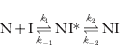
Electrostatics play a significant role in the formation of NI* as demonstrated by the strong dependence of the association rates (k 1) on the ionic strength. The encounter complex subsequently collapses to the stable complex NI (Eq. 1) in a second slower phase which, it has been argued, represents rigid body rotation of the Im protein on the DNase surface (Keeble & Kleanthous, Reference Keeble and Kleanthous2005). There are no gross changes in conformation in either protein on forming the complex. In the case of the ColE3 rRNase–Im3 complex (Walker et al. Reference Walker, Moore, James and Kleanthous2003), a single phase is observed in stopped-flow tryptophan fluorescence experiments, attributed to the rapid bimolecular association between the two proteins which is not accompanied by any conformational changes. The resulting kinetically derived equilibrium dissociation constants for nuclease–Im protein complexes are all in the femtomolar region (0·07−20×10−15 M), depending on the ionic strength (Li et al. Reference Li, Keeble, Giffard, James, Moore and Kleanthous2004; Walker et al. Reference Walker, Moore, James and Kleanthous2003; Wallis et al. Reference Wallis, Moore, James and Kleanthous1995).
Arrhenius analysis of the observed rate constants for the ColE9 DNase–Im9 complex revealed that low activation barriers for the bimolecular association and the subsequent conformational rearrangement are favoured both by low enthalpy and positive entropy values while the dissociation of the encounter complex is slow mainly due to large negative activation entropy (Wallis et al. Reference Wallis, Moore, James and Kleanthous1995). A similar trend is seen in the case of the ColE3 rRNase–Im3 complex for the bimolecular association and dissociation steps (Walker et al. Reference Walker, Moore, James and Kleanthous2003). A large positive entropy change favours the observed femtomolar binding constant as revealed by Van't Hoff analysis of the kinetically determined K d values in the case of the ColE9–Im9 complex. This entropy change must be associated with changes in the solvation entropy of the two proteins upon association (Wallis et al. Reference Wallis, Moore, James and Kleanthous1995). On the other hand, a large negative enthalpy change favours the formation of the ColE3 rRNase–Im3 complex, which is associated with a significant number of intermolecular hydrogen bonds formed upon association of the two proteins (Carr et al. Reference Carr, Walker, James, Kleanthous and Hemmings2000; Walker et al. Reference Walker, Moore, James and Kleanthous2003).
Kinetically determined enthalpy changes that occur upon formation of the ColE9 DNase–Im9 and ColE3 rRNase–Im3 complexes have been verified by ITC (Keeble et al. Reference Keeble, Kirkpatrick, Shimizu and Kleanthous2006; Walker et al. Reference Walker, Moore, James and Kleanthous2003). Calorimetric analysis of all four cognate DNase–Im complexes shows that binding is enthalpically driven (−33·1, −30·3, −24·2 and −10·5 kcal/mole, respectively). In the case of the ColE2, ColE7, ColE8 DNase complexes binding is entropically disfavoured (–13·5, −10·5 and −4·4 kcal/mol, respectively), while for the ColE9 DNase–Im9 complex binding is entropically favoured (+8·1 kcal/mole) (Keeble et al. Reference Keeble, Kirkpatrick, Shimizu and Kleanthous2006). These observations are consistent with the greater polarity of the Im protein complexes of ColE2, ColE7 and ColE8 DNases and in contrast to the ColE9 DNase–Im9 interface, which is more hydrophobic. In accordance with the observed large negative values (ranging between −0·45 and −0·82 kcal mol−1 K−1) for the association heat capacity, binding of the ColE2, ColE7 and ColE8 DNases with their cognate Im partners is accompanied by a strong rigidification of one or both counterparts while in the case of the ColE9 DNase–Im9 complex association is coupled to a significant expulsion of interfacial water (Keeble et al. Reference Keeble, Kirkpatrick, Shimizu and Kleanthous2006).
9.2 A dual-recognition mechanism underpins binding specificity in Col DNase–Im protein complexes
Exosite binding of Im proteins has driven an evolutionary arms race between Col-producing bacteria, allowing extensive sequence variation at the Col DNase–Im interface (Kleanthous & Walker, Reference Kleanthous and Walker2001). The manifestation of this diversification is the ten-orders-of-magnitude range of binding affinities that have been recorded among cognate and non-cognate Col DNase–Im protein complexes (Li et al. Reference Li, Keeble, Giffard, James, Moore and Kleanthous2004). This, in turn, has provided a powerful model system with which to explore the determinants of binding specificity more generically in protein–protein interactions.
The majority of studies aimed at investigating Col DNase–Im protein complexes have focused primarily on how the Im protein binds and recognizes the DNase specifically. These studies have proposed that a dual-recognition mechanism underpins binding specificity wherein the bulk of complex stabilization emanates from conserved hydrogen bonds and hydrophobic interactions between Col DNase IPE residues and a hotspot of conserved Im residues in helix α3 (shown in Fig. 11a for the ColE9 DNase–Im9 complex). These contacts are present in both cognate (ColE7 DNase–Im7 (Ko et al. Reference Ko, Liao, Ku, Chak and Yuan1999) and ColE9 DNase–Im9 (Kuhlmann et al. Reference Kuhlmann, Pommer, Moore, James and Kleanthous2000; Montalvao et al. Reference Montalvao, Cavalli, Salvatella, Blundell and Vendruscolo2008)) and non-cognate (ColE9 DNase–Im2) (Li et al. Reference Li, Hamill, Hemmings, Moore, James and Kleanthous1998, Reference Li, Keeble, Giffard, James, Moore and Kleanthous2004) complexes. The stability enhancement in the case of the cognate complexes stems from interactions between charged or hydrophobic residues of Im helix α2 (shown in Fig. 11a for the ColE9–Im9 complex) and residues of identical chemical nature in the DNase IPE (Kuhlmann et al. Reference Kuhlmann, Pommer, Moore, James and Kleanthous2000). In the case of the non-cognate complexes, the presence of residues of non-identical chemical nature between Im helix α2 and the DNase destabilizes the complex, which play a critical role in determining the 7–10-orders-of-magnitude difference in affinities between cognate and non-cognate complexes (Kuhlmann et al. Reference Kuhlmann, Pommer, Moore, James and Kleanthous2000; Wallis et al. Reference Wallis, Leung, Osborne, James, Moore and Kleanthous1998). In order to confer specificity to the DNase, only a small number of Im helix α2 residues need vary without concomitant alteration of helix α3 core interactions, which facilitates the diversification and evolution of Im specificity without compromising the ability of the Im protein to inactivate the toxin (Kuhlmann et al. Reference Kuhlmann, Pommer, Moore, James and Kleanthous2000; Meenan et al. Reference Meenan, Sharma, Fleishman, Macdonald, Morel, Boetzel, Moore, Baker and Kleanthous2010).
Dual-recognition underpins binding specificity in Col DNase–Im protein complexes. (a) Cognate complex of the ColE9 DNase–Im9 complex (K d~10−14 M). In ColE9 DNase (green), the IPE residues PDB ID 1EMV (Asn75, Phe86, Lys97, Val98; isolated domain numbering) (red) form a central belt at the binding interface where mutation to alanine has the largest impact on the binding free energy (ΔΔG). At the centre of this belt Phe86 stacks with the conserved Im9 helix α3 (purple) residue Tyr54 and is surrounded by the variable Im9 helix α2 hydrophobic residues (purple) Leu33, Val34 and Val37 and Ile53. Additional stabilizing interactions are made by Im9 helix α3 residues Asp51 and Tyr55. Further specificity interactions made between E9 DNase Lys97 and Val98 with Im9 residues Glu41 and Val34, respectively. E9 DNase Arg54, at the periphery of the IPE, forms a salt bridge with Glu30 of Im9 helix α2. Asn75, the only completely conserved interfacial residue on the DNase IPE, participates in a water-mediated hydrogen bond to the Im9 backbone. Also shown are residues within two critical loops of Im9 (α1–α2, α3–α4), which are thought to contribute to binding specificity. (b) The non-cognate ColE9 DNase–Im2 complex (K d~10−7 M) (PDB ID 2WPT). The key interactions from Im2 helix α3 (Tyr54, Tyr55 and Asp51) that surround the E9 DNase IPE residue Phe86 are maintained, including the stacking interaction with Tyr54. The strength of Im protein helix α3 hotspot (which also involves several interfacial water molecules, not shown) results in unfavourable packing between Phe86 and Asp33 and Asn34 causing frustration at the interface. Also evident are the changes in loop conformation in Im2 relative to Im9 (α1–α2, α3–α4 loops) that affect the interactions of other E9 DNase IPE residues, particularly Tyr83 and Asn72. (c) Comparison of cognate and non-cognate packing interactions at the core of the ColE9 DNase–Im protein interface. The figure emphasizes the chemical frustration that results from the juxtaposition of charged and polar residues in Im2 (Asp33 and Asn34, respectively) with E9 DNase Phe86, which destabilize the stability of the complex.

9.3 The cognate ColE9 DNase–Im9 complex
An alanine scan of ColE9 DNase IPE interfacial residues, where the ΔΔG of Im9 binding between the ColE9 mutants and the wild-type (WT)-ColE9 was determined computationally and experimentally using stopped-flow fluorescence kinetics, revealed that the effect of the IPE residues on Im9 binding is not as extensive as that of the hotspot residues in Im9 (Keeble et al. Reference Keeble, Joachimiak, Mate, Meenan, Kirkpatrick, Baker and Kleanthous2008; Kuhlmann et al. Reference Kuhlmann, Pommer, Moore, James and Kleanthous2000). The scan showed four ColE9 DNase IPE residues (Asn75, Phe86, Lys97 and Val98; isolated domain numbering) create a specificity hotspot for the DNase which form a central belt at the interface of the ColE9–Im9 complex (Fig. 11a). Phe86 is coordinated by the conserved Tyr54 and Tyr55 of Im9 helix α3 and the specificity Im9 helix α2 residues Val34 and Val37 while Lys97 and Val98 interact with Glu41 and Val34 of Im9 helix α2, respectively (Kuhlmann et al. Reference Kuhlmann, Pommer, Moore, James and Kleanthous2000). The F86A ColE9 DNase mutant exhibited the greatest ΔΔG for Im9 binding of all four hotspot alanine variants. Structural and double-mutant cycle (DMC) analyses (experimental and computational) for the F86A mutant binding Im9 (V34A, V37A, Y54A and Y55A) and Im2 mutants (D33A, N34A, V37A, Y54A and Y55A) were performed. This confirmed the importance of Tyr54 and Tyr55 in the stability of cognate and non-cognate complexes alike but also showed minimal coupling between Phe86 and Im9 residues (with the specificity residues exhibiting greater interaction than Tyr54 and Tyr55). Negative DMC values were obtained for Im2 specificity residues relative to Phe86, the alanine substitutions of these Im2 residues relieving the instability conferred to the ColE9 DNase–Im2 complex by the WT residues (see below). Phe86 is conserved between the DNase of ColE2 and ColE9 (the corresponding specificity residue in ColE7 and ColE8 is a lysine) while Asn75 is conserved among all DNases. The rest of the hotspot IPE residues are variable. Accordingly, Col DNase–Im specificity is dictated by the conserved elements of the IPE and buffered by variable residues (Keeble et al. Reference Keeble, Joachimiak, Mate, Meenan, Kirkpatrick, Baker and Kleanthous2008).
The conformation and dynamics of the ColE9 DNase–Im9 cognate complex and the individual proteins have recently been studied through MD simulations (Baron et al. Reference Baron, Wong, De Oliveira and Mccammon2008). In this study, the conformational entropy of Im9 was demonstrated to be substantially decreased upon binding the Col, with the flexibility of the latter only increasing at the binding interface. In accordance with the crystal structure of the ColE9 DNase–Im9 complex, the MD simulations were consistent with conformational sampling of residues 54–64 in Im9 (involving the α2–α3 loop) and helix α3 along with the conserved hotspot residues Tyr54 and Ty55 at the binding interface.
9.4 The non-cognate ColE9 DNase–Im2 complex
Further insight into the dual-recognition mechanism underpinning Col DNase–Im protein complex specificity emerged from the recent crystal structure of the non-cognate ColE9 DNase–Im2 complex (Meenan et al. Reference Meenan, Sharma, Fleishman, Macdonald, Morel, Boetzel, Moore, Baker and Kleanthous2010) (Fig. 11b). Previous attempts at obtaining a structure for a non-cognate complex, which typically have K d's in the mM–nM range, had been hampered by the rotational dynamics which are thought to be intrinsic to non-cognate complexes. Meenan et al. employed NMR-based docking restraints as a guide to place cysteine residues across the protein–protein interface in order to trap and crystallize the E9 DNase–Im2 complex, which is seven-orders-of-magnitude weaker binding than the E9 DNase–Im9 complex and eight-orders-of-magnitude weaker than the E2 DNase–Im2 complex. Surprisingly, the resulting high-resolution structure showed an interface devoid of a covalent bond; the engineered disulfide merely stabilized the crystal lattice, with the non-cognate interface formed between neighbouring, covalently attached complexes.
The binding interface for the non-cognate E9 DNase–Im2 complex localizes to the ColE9 DNase IPE (Phe86), with Im2 anchored through the conserved helix α3 residues Tyr54, Tyr55 and Asp51, in good agreement with the structure of the cognate ColE9 DNase–Im9 complex. Tyr54 (Im2) and Phe86 (ColE9 DNase) are aromatically stacked and present helix α2 N-terminal residues of Im2 to the interface in a fashion similar to that seen in the cognate complex (Fig. 11b). Computational alanine scanning of non-cognate interfacial residues using the program Rosetta design confirmed the conserved hotspot residues Tyr54, Tyr55 and Phe86 are all highly stabilizing in the non-cognate complex (Meenan et al. Reference Meenan, Sharma, Fleishman, Macdonald, Morel, Boetzel, Moore, Baker and Kleanthous2010).
Despite these similarities the non-cognate complex is significantly less stable than the cognate due to a series of destabilizing contacts that dictate the binding specificity. A hydrophobic interaction in the cognate complex between E9 DNase Phe86 and the specificity helix α2 residues Leu33 and Val34 of Im9 is switched to a charged-hydrophobic interaction in the ColE9 DNase–Im2 complex (Fig. 11b). The resulting unfavourable packing between E9 DNase Phe86 with Asp33 and Asn34 of Im2 is highly destabilizing as evidenced by the enhancement in binding afforded by alanine mutations (Keeble et al. Reference Keeble, Joachimiak, Mate, Meenan, Kirkpatrick, Baker and Kleanthous2008) and the even greater enhancement (up to 10 000-fold) in binding when Asp33 and Asn34 are substituted for their Im9 counterparts, leucine and valine (Keeble et al. Reference Keeble, Joachimiak, Mate, Meenan, Kirkpatrick, Baker and Kleanthous2008; Meenan et al. Reference Meenan, Sharma, Fleishman, Macdonald, Morel, Boetzel, Moore, Baker and Kleanthous2010). The destabilizing interactions of Im2 Asp33 and Asn34 are maintained in the non-cognate complex at significant energetic cost through the highly stabilizing interactions surrounding ColE9 DNase Phe86 at the centre of the interface. These are largely through the conserved residues of helix α3 of Im2 and aided by many conserved hydrogen bonds mediated by interfacial water molecules. As a consequence, the non-cognate complex experiences ‘frustration’ through the juxtaposition of non-optimal contacts (Fig. 10c) (Meenan et al. Reference Meenan, Sharma, Fleishman, Macdonald, Morel, Boetzel, Moore, Baker and Kleanthous2010). It is the relief of this frustration that provides the large enhancements in binding that result from single mutations at the Col DNase–Im protein interface. The effect of such a single mutation in vivo can be appreciated by the data in Fig. 12, which show the relationship between binding affinity of ColE9 DNase–Im protein complexes and the degree of cell protection afforded by the same Im protein when expressed in E. coli (Li et al. Reference Li, Keeble, Giffard, James, Moore and Kleanthous2004). Not only do these data emphasize the wide range of binding affinities observed in Col DNase–Im protein complexes, but they also strikingly show how a single mutation within the specificity helix α2 (Asp33 to Leu33 in Im2) results in a sufficiently large improvement in binding free energy as to confer resistance towards the Col and hence survival of the bacterial culture.
Dual-recognition underpins evolution and diversity of Col DNase–Im protein complex protein–protein interaction specificity. Adapted from Li et al. (Reference Li, Keeble, Giffard, James, Moore and Kleanthous2004) with permission from Elsevier. The figure shows the relationship between in vivo survival of E. coli K-12 cells expressing imm9, non-cognate Im genes or im9 chimeras/mutant genes when challenged with ColE9 and the equilibrium K d for the same Im protein binding the ColE9 DNase domain in vitro. Col resistance was scored for each construct using an overlay plate assay in the presence (filled symbols) or absence (open symbols) of the inducer IPTG. Examples of the same construct ±IPTG are indicated with squares and triangles. Cells become completely resistant towards ColE9 even in the absence of inducer (due to leaky expression from the pTrc99a plasmid used in these experiments) when harbouring an Im protein with an affinity for the E9 DNase of greater than 10−10 M, but are eradicated when the affinity falls below 10−6 M. Hence, the evolution of novel ColE9 Im protein specificity in nature likely occurs within this affinity window (pink shading). The frustration of side-chain interactions at the heart of the dual-recognition mechanism primes the interface for high-affinity binding so that a single mutation (in this instance an Asp-to-Leu mutation at position 33 of Im2) yields an Im protein that completely protects the cell from the Col (see Fig. 11c) (Meenan et al. Reference Meenan, Sharma, Fleishman, Macdonald, Morel, Boetzel, Moore, Baker and Kleanthous2010). Further enhancements in binding come from additional residues in the Im protein specificity helix α2, contributions from loop regions and interfacial water molecules (see text for details).

The significance of the α1–α2 and α3–α4 loops in contributing to Im2 binding specificity was also highlighted by the ColE9 DNase–Im2 crystal structure. Thr27 in the Im9 α1–α2 loop and ColE9 DNase Tyr83 form hydrogen bonds but they are not aligned optimally in the non-cognate complex because of an intervening water molecule. The same occurs between Asp62 of the α3–α4 loop and ColE9 DNase Asn72. An important consequence of the non-optimal placement of the α1–α2 loop is that this affects the interactions with the E9 DNase hotspot residue Tyr83 (Fig. 11a, b) (Kuhlmann et al. Reference Kuhlmann, Pommer, Moore, James and Kleanthous2000; Meenan et al. Reference Meenan, Sharma, Fleishman, Macdonald, Morel, Boetzel, Moore, Baker and Kleanthous2010). Although not directly involved in binding the ColE9 DNase, residues within the α1–α2 loop of Im9 have been shown to be involved in defining Im9 specificity relative to Im7 (see below). The flexibility of the Im9 helix-spanning loops and the significance of their configuration to ColE9 DNase binding were also demonstrated in an MD simulation study of the ColE9 DNase–Im9 complex (Baron et al. Reference Baron, Wong, De Oliveira and Mccammon2008). Apart from the destabilizing interference in the Im2 loop configuration, water molecules help offset several unfavourable interactions at the interface, centred around variable specificity residues. For this reason, more water is found at the interface of the non-cognate complex. It has been suggested that the exclusion of this water from the cognate interface would lead to a higher affinity through the gained desolvation entropy, which is consistent with the available calorimetric data (Meenan et al. Reference Meenan, Sharma, Fleishman, Macdonald, Morel, Boetzel, Moore, Baker and Kleanthous2010). Essentially complementing the observations of the ColE9 DNase–Im9 cognate complex, the ColE9 DNase–Im2 non-cognate complex further illustrates that the dual recognition mechanism in this protein–protein interaction relies on highly stabilizing interactions emanating from the conserved elements of the Col DNase–Im interface that are modulated by the surrounding variable residues of Im helix α2. The structure further shows the importance of interhelical loop configurations and buried interfacial water molecules in encoding Im protein specificity (Meenan et al. Reference Meenan, Sharma, Fleishman, Macdonald, Morel, Boetzel, Moore, Baker and Kleanthous2010).
9.5 Evolution and design of Col DNase–Im protein complexes
Protein drugs are finding increasing use in medicine but their potential for off-target effects can have disastrous consequences (Hale, Reference Hale2006; Kiely & Johnson, Reference Kiely and Johnson2002; Oldham & Dillman, Reference Oldham and Dillman2008). Hence, a major objective in the study of protein–protein interactions is the ability to engineer the affinity and binding specificity of interacting pairs. In recent years, directed evolution and computational design have both had significant impact in this area (Khersonsky et al. Reference Khersonsky, Roodveldt and Tawfik2006; Pokala & Handel, Reference Pokala and Handel2001; Tokuriki & Tawfik, Reference Tokuriki and Tawfik2009). The rational design of novel specificities in protein complexes relies on the inspection of the protein structure and its in silico perturbation through the introduction of substitutions at specific locations which are then computationally screened for the change in complex stability (Kortemme et al. Reference Kortemme, Joachimiak, Bullock, Schuler, Stoddard and Baker2004). Directed evolution enables specificity alterations to emerge in in vitro evolution experiments that mimic natural evolution and is based on the use of an appropriate selection pressure (Roodveldt et al. Reference Roodveldt, Aharoni and Tawfik2005). Both approaches have been applied in the exploration of novel protein–protein interaction specificities in Col DNase–Im protein complexes, which are ideally suited for these applications for a number of reasons. First, the affinities of cognate complexes are high (femtomolar) while the level of discrimination between cognate and non-cognate pairs can approach ~10-orders of magnitude in binding affinity (Li et al. Reference Li, Keeble, Giffard, James, Moore and Kleanthous2004), essentially matching the spectrum of K d's observed in nature. Second, crystal structures of cognate (ColE9 DNase–Im9 and ColE7 DNase–Im7), and non-cognate (ColE9 DNase–Im2) provide a structural framework for understanding binding specificity (Ko et al. Reference Ko, Liao, Ku, Chak and Yuan1999; Kuhlmann et al. Reference Kuhlmann, Pommer, Moore, James and Kleanthous2000; Meenan et al. Reference Meenan, Sharma, Fleishman, Macdonald, Morel, Boetzel, Moore, Baker and Kleanthous2010). Third, the cytotoxicity of Cols provides an assay (both in vivo and in vitro) for screening, selecting and analysing viable alterations in specificity (Kortemme et al. Reference Kortemme, Joachimiak, Bullock, Schuler, Stoddard and Baker2004; Levin et al. Reference Levin, Dym, Albeck, Magdassi, Keeble, Kleanthous and Tawfik2009). Fourth, the metal dependence of Col DNase activity provides a route by which nuclease activity can be readily activated (Levin et al. Reference Levin, Dym, Albeck, Magdassi, Keeble, Kleanthous and Tawfik2009). Recent advances using both approaches are discussed.
In vitro compartmentalization (IVC) was used by the Tawfik lab to evolve Im9 towards Im of the ColE7 DNase to which Im9 binds ~6-orders of magnitude more weakly than to ColE9 (Bernath et al. Reference Bernath, Magdassi and Tawfik2005; Levin et al. Reference Levin, Dym, Albeck, Magdassi, Keeble, Kleanthous and Tawfik2009; Li et al. Reference Li, Keeble, Giffard, James, Moore and Kleanthous2004). In each round of selection ~1010 Im9 variants were generated (with a single gene/droplet of the emulsion) and compartmentalized with the ColE7 DNase, which was activated by the addition of Co2+ ions after the Im genes were transcribed and translated. Active inhibitors encoded by genes that survived the ColE7 nuclease activity were recovered and PCR amplified. Im9 variants exhibiting increased ColE7 inhibition relative to WT-Im9 were isolated after eight rounds of mutation and selection (Bernath et al. Reference Bernath, Magdassi and Tawfik2005; Levin et al. Reference Levin, Dym, Albeck, Magdassi, Keeble, Kleanthous and Tawfik2009). In order to increase the affinity of the evolving Im9 towards ColE7 while also reducing the cross-reactivity with ColE9 an inactive variant of the ColE9 DNase, bearing the H103A active site mutation (Section 8), was added for three additional rounds of selection (Levin et al. Reference Levin, Dym, Albeck, Magdassi, Keeble, Kleanthous and Tawfik2009). The inactive ColE9 DNase provided a negative selection for ColE9 DNase cross-binders thereby enhancing ColE7 DNase specificity. The authors of this study analysed emerging specificity variants and managed to solve structures for some of these to provide a molecular picture of the evolutionary process.
Of the selected ColE7 DNase binding ‘nevIm7’ variants only one mutation in the variable region of Im9 helix α2 (V34D) increased directly the affinity of Im9 towards ColE7 (Fig. 13a, b). Asp34 (the cognate residue in Im7) interacts with Lys528 and Lys537 in ColE7 switching a hydrophobic interaction in the ColE9 DNase–Im9 complex into an electrostatic one in the newly evolved ColE7 DNase–nevIm7 complex (Levin et al. Reference Levin, Dym, Albeck, Magdassi, Keeble, Kleanthous and Tawfik2009). The only other mutations that increase the affinity towards ColE7 were in residues of the α1–α2 loop (N24D, D26N, T27A and S28T) lying close to the variable region (Fig. 13a, b). These, however, were not previously seen to be involved in binding and the substituted amino acids were not identical to those in Im7. In the structures of Im9 and Im7 in complex with their cognate enzymes the Im proteins are rotated 19o around the Tyr54–Tyr55 (Im9) or Tyr55–Tyr56 (Im7) hotspot (Kuhlmann et al. Reference Kuhlmann, Pommer, Moore, James and Kleanthous2000). In the case of nevIm7, the emerging α1–α2 loop mutations (N24D, D26N, T27A and S28T) resulted in an intermediate configuration between these two rotameric forms (Fig. 13b). This intermediate configuration resulted in a 10–30-fold affinity increase of nevIm7 towards the ColE7 DNase relative to Im9 by reduction of the dissociation rate constant as observed by stopped-flow fluorescence kinetic measurements (Levin et al. Reference Levin, Dym, Albeck, Magdassi, Keeble, Kleanthous and Tawfik2009). Notably, even two of these α1–α2 loop mutations could confer the observed affinity increase although none of them is involved directly in binding. Asn24 has a critical role in this switch; when mutated to aspartic acid in nevIm7 it becomes part of the α1–α2 loop whereas in Im9 it forms the C-terminus of α1 helix (Fig. 13a, b). The altered configuration leads to latent interactions between pre-existing residues (salt bridges between Glu30 and Asp51 of Im9 with Arg520, Gln532, Thr531 of the ColE7 DNase) which are not possible for wild-type (WT) Im9 α1–α2 loop residues (Levin et al. Reference Levin, Dym, Albeck, Magdassi, Keeble, Kleanthous and Tawfik2009). These data complement the studies of Meenan et al. on the non-cognate ColE9 DNase–Im2 complex, described above, where again α1–α2 loop residues are implicated in contributing to binding specificity (Fig. 13b) (Meenan et al. Reference Meenan, Sharma, Fleishman, Macdonald, Morel, Boetzel, Moore, Baker and Kleanthous2010).
Directed evolution and design of Im protein specificity. (a) Secondary structure diagram (left) and the structure of Im9 (right, PDB ID 1EMV) showing positions of altered residues resulting from IVC experiments aimed at evolving the specificity of Im9 for the ColE7 DNase and as observed in the mutant nevIm7 (black circles, yellow balls and sticks). See text for details. (b) Superposition of the ColE7 DNase-bound structures of WT Im7 (grey, PDB ID 7CEI) and nevIm7 (purple, PDB ID 3gkl) highlighting the positions affecting Col binding that emerged from directed evolution experiments and the restructuring of the nevIm7 α1–α2 loop. When bound to their cognate DNases Im9 and Im7 are related by a 19o rigid body rotation, with the nevIm7 binding configuration intermediate between these two extremes. Specificity residues spanning the α1–α2 loop and helix α2 (green) and the hotspot residues (Tyr55 and Tyr56, cyan) in helix α3 of Im7 are highlighted. Alterations in residues that occur during the first five rounds of selection in the α1–α2 loop and helix α2 (Asp24, Ala27, Thr28 and Asp34) are shown in yellow. Ile37 (orange) and Gly41 (cyan) emerged after 8 and 12 rounds of selection, respectively. Also shown in the figure are the three positions, Thr51 (T51Q) and Ile68 (I68W) in helices α3 and α4 (magenta), respectively, and Asp35 (D35Y) in helix α2, where computational design of Im7 led to altered Col E7 DNase specificity.

The combination of V34D and the four α1–α2 loop mutations resulted in higher affinity for the nevIm7 towards the ColE7 DNase but also decreased the stability of the new Im proteins (determined by urea denaturation experiments and intrinsic tryptophan fluorescence). Extending the selection for ColE7 DNase by three more rounds resulted in the V37I mutation (Fig. 13b), which improved packing between helices α1 and α3 and recovered this lost stability. The nevIm7 variants isolated up to this point all exhibited equal affinity for both ColE7 and ColE9; in other words, they reduced ColE9 DNase and increased ColE7 DNase binding but with no increase in selectivity. Following the incorporation of the negative selection step the selectivity for ColE7 increased relative to ColE9 by 30–70-fold. Variants emerging from these rounds contained the E41G mutation (Fig. 13b) which when combined with V37I led to molecules of adequate stability that conferred Im to ColE7 in in vivo cell-killing assays (Levin et al. Reference Levin, Dym, Albeck, Magdassi, Keeble, Kleanthous and Tawfik2009).
The Baker lab have deployed computational redesign of the ColE7 DNase–Im7 interface as a test of how robust such computational methods can be in generating novel (and predictable) specificities at protein–protein interfaces. In a first approach, the binding interface was redesigned based on a computational second-site suppressor strategy (using the molecular modelling program ROSETTA), where rotamers of amino acid side chains were modelled on the fixed polypeptide taken from the crystal structure of the ColE7 DNase–Im7 complex such that the effect of a destabilizing mutation on one protein was compensated by the alteration of interacting residues on its counterpart (Kortemme et al. Reference Kortemme, Joachimiak, Bullock, Schuler, Stoddard and Baker2004). Redesign of the interface was accomplished through polarity and steric switches both in combination and in isolation. The DNase inhibition properties of the designed variants was estimated in vivo in E. coli cells and also through an in vitro endonuclease assay while the binding between them (cognate and non-cognate) was estimated via SPR and fluorescence measurements (Kortemme et al. Reference Kortemme, Joachimiak, Bullock, Schuler, Stoddard and Baker2004). The structural accuracy of the prediction method was validated subsequently by the crystal structure of the redesigned steric switch (RMSD 0·62 and 0·5 Å for the interface residues and the Cα backbone, respectively). In all cases, the specificity of the designed proteins towards their homologous designed counterpart (cognate binding) was increased relative to the WT or the other designed counterparts (non-cognate binding). However, the designed cognate complexes were significantly weaker than the WT ColE7 DNase–Im7 complex and the affinity differences between the designed cognate and non-cognate complexes are much lower than the corresponding differences between the WT complexes. These discrepancies were attributed to the role of interactions mediated by interfacial water molecules, which play an essential role in both stabilizing these complexes and moulding specificity and were not modelled by the applied energy function (Kortemme et al. Reference Kortemme, Joachimiak, Bullock, Schuler, Stoddard and Baker2004; Kuhlmann et al. Reference Kuhlmann, Pommer, Moore, James and Kleanthous2000; Meenan et al. Reference Meenan, Sharma, Fleishman, Macdonald, Morel, Boetzel, Moore, Baker and Kleanthous2010).
In a follow-up study by Joachimiak et al. binding energies were calculated for the cognate ColE7 DNase–Im7 complex taking account of all rigid-body conformations around the 19° pivot centring on Tyr54–Tyr55 (or Tyr55–Tyr56) in Im7 and Im9, respectively (Joachimiak et al. Reference Joachimiak, Kortemme, Stoddard and Baker2006). The design in this case involved the introduction of key mutations predicted to increase the binding affinity both with and without a second-site suppressor approach. After structural determination of the complexes of the corresponding ColE7 DNase and Im7 variants their binding was estimated via SPR and fluorescence measurements and optimized again for specificity (Joachimiak et al. Reference Joachimiak, Kortemme, Stoddard and Baker2006). A complex of the two proteins bearing seven mutations (K528Q, T539Q, N516T and K525R in ColE7 and D35Y, T51Q and I68W in Im7) was designed (Fig. 13b). The corresponding crystal structure revealed one site where a specifically designed hydrogen bond network (included in the applied energy function) coincided with the native hydrogen bond network between Gln528, Gln539 of the ColE7 DNase and Tyr35 and Gln51 of Im7. A second site comprised direct interactions between Thr516 and Gln517 of the ColE7 DNase with Trp68 of Im7 while Arg525 replaced the salt bridge made by the WT lysine. The designed protein pair resulted in a 300-fold higher binding selectivity for the cognate versus the non-cognate complexes and an affinity of 5·5×10−12 M, which is two orders of magnitude weaker than the binding affinities of cognate Col DNase–Im protein complexes (Li et al. Reference Li, Keeble, Giffard, James, Moore and Kleanthous2004). As in the case of the directed evolution experiments, the ColE7 DNase–Im7 computational redesign did not boost specificity dramatically over non-cognate complexes. However, the design demonstrated how interfacial residues and interface hydration leads to the modulation of specificity and affinity between the Col DNase IPE and Im proteins (Joachimiak et al. Reference Joachimiak, Kortemme, Stoddard and Baker2006).
10. Im protein folding mechanism and its link to function
The presumption that proteins attain their 3D-fold via a two-state process involving only the unfolded (U) and native (N) states is almost certainly an oversimplification even for small proteins (Jackson, Reference Jackson1998). A simple two-state folding process would require no transitional intermediates existing en route to the N state and that folding take place cooperatively with all the productive collisions leading to native interactions occurring simultaneously (Baldwin et al. Reference Baldwin, Frieden and Rose2010). Regarding the U state as a large ensemble of featureless interchangeable conformers with small energy barriers between them (Tanford, Reference Tanford1970), the adoption of the correct set of favourable interactions (among the numerous ones the U state can adopt) would more likely lead to a series of intermediate states of varying stability thus breaking down the folding pathway into more than two states. A major challenge in understanding the process of protein folding involves the delineation of such a folding energy landscape. The Radford laboratory has pioneered the use of Col DNase-specific Im proteins to explore the energetics and structure of folding intermediates that lie on the energy landscape and to link this to Im protein function.
Im proteins have many features making them a useful system with which to characterize the structure, topology and stability of intermediate (I) state and TS in the folding process. Both Im9 and Im7, which have been the subject of most studies, are small single-domain proteins (86 and 94 amino acids, respectively) that adopt similar helical bundle folds (Dennis et al. Reference Dennis, Videler, Pauptit, Wallis, James, Moore and Kleanthous1998). Their small size, lack of disulfides and prosthetic groups and their all α-helical content along with the significant and various spectroscopic signatures make the two Im proteins ideal for studies of the energy landscape of folding.
10.1 The folding energy landscape of Im7
In stopped-flow kinetic and equilibrium studies of unfolding and refolding in the presence of denaturant, monitored by tryptophan fluorescence emission and detected both in the presence and absence of Na2SO4, it was demonstrated that Im7 folds through a stable intermediate (I) thus following a distinct three-state pathway with two TSs, TS1 and TS2 (Fig. 14) (Ferguson et al. Reference Ferguson, Capaldi, James, Kleanthous and Radford1999). The Im7 I-state was fully formed during the burst phase of folding (within the 2·5 ms dead time of the stopped-flow apparatus). In combination with continuous flow data (dead time ~120 μs), it was demonstrated that the Im7 I-intermediate represents an ensemble of productive states in equilibrium with both the U and N states and hence on-pathway (Capaldi et al. Reference Capaldi, Shastry, Kleanthous, Roder and Radford2001), and can be formed both in the folding and unfolding pathways irrespective of the concentration of Na2SO4 (Cobos & Radford, Reference Cobos and Radford2006).
Folding energy landscape of Im7 showing the on-pathway intermediate, I, and the two rate-determining TSs, TS1 and TS2, involved in the unfolded ensemble of Im7 (U) attaining its native structure (N). Figure adopted from Pugh et al. (Reference Pugh, Gell, Smith, Radford and Brockwell2010) with permission from Elsevier. The ensembles shown are the result of MD simulations while the structure for Im7 is that of the native Im protein (PDB ID 7CEI). The regions forming the four helices in the native state are coloured: red (α1), green (α2), purple (α3) and yellow (α4).

The Im7 I-state consists of helices α1, α2 and α4 being folded natively but largely in the absence of tertiary interactions, demonstrated through the characterization of Im7 single-point variants of hydrophobic residues (Capaldi et al. Reference Capaldi, Kleanthous and Radford2002). An NMR analysis of urea-unfolded Im7 pointed to the presence of four clusters of interacting residues each corresponding to a helix of Im7 (Le Duff et al. Reference Le Duff, Whittaker, Radford and Moore2006). In addition to the native fold of the three helices in the Im7 I-state a specific hydrophobic core-template for the binding of helix α3 is also present, pointing to a specific structure for the intermediate (Capaldi et al. Reference Capaldi, Kleanthous and Radford2002). A significant amount of non-native interactions between helices α1–α2 and α1–α4 are present in the Im7 I-state that serve to protect hydrophobic residues in these helices from solvent exposure, which would otherwise occur if the three helices folded natively in the absence of helix α3. The native and non-native interactions involving α1, α2 and α4 helices, along with significant hydrogen bonding that stabilizes them, were also revealed through hydrogen–deuterium exchange experiments combined with Φ-value analyses (Gorski et al. Reference Gorski, Le Duff, Capaldi, Kalverda, Beddard, Moore and Radford2004) and molecular dynamics (MD) simulations (Gsponer et al. Reference Gsponer, Hopearuoho, Whittaker, Spence, Moore, Paci, Radford and Vendruscolo2006). The on-pathway Im7 I-state has been trapped through disruption of N-state stabilizing interactions between helix α3 and the rest of the protein (Spence et al. Reference Spence, Capaldi and Radford2004). NMR studies of the trapped Im7 I-state variants revealed that helices α1 and α4 are native while helix α2 is partially folded and helix α3 is unfolded. Additional interactions between helix α3 residues and other regions of the Im7 I-state were demonstrated as the core switches conformation through TS2 in order for helix α3 to fold (Whittaker et al. Reference Whittaker, Spence, Gunter Grossmann, Radford and Moore2007).
The non-native interactions evident in the I-state of Im7 are perturbed in TS2 (Fig. 14), with a number of residues involved participating in the recognition of helix α3 while others become solvent exposed in the native state. The binding of helix α3 takes place only after TS2 and only in the presence of the three-helix bundle core, which was predicted computationally (Capaldi et al. Reference Capaldi, Kleanthous and Radford2002) and confirmed through MD simulations (Paci et al. Reference Paci, Friel, Lindorff-Larsen, Radford, Karplus and Vendruscolo2004). Helix α3 is the last to fold in the folding pathway of Im7; its docking to an already preformed three-helix bundle core disrupts non-native interactions involving the helix (e.g. Tyr55) thereby destabilizing the Im7 I-state and stabilizing the native state (Knowling et al. Reference Knowling, Figueiredo, Whittaker, Moore and Radford2009).
Folding in Im7 is rate-limited by the proper alignment of helices α1, α2 and α4 in the Im7 I-state, which form the binding site for helix α3. The conserved residues Tyr55 and Tyr56 in helix α3 and the variable residues Asn26, Asp31, Leu34, Asp35 and Val42 in helix α2 (Fig. 12b) participate in non-native interactions in all stages of the folding pathway, modulating the energetics of the Im7 I-state and the formation of the two TSs (Friel et al. Reference Friel, Smith, Vendruscolo, Gsponer and Radford2009). Sutto et al. showed that three clusters of frustrated interactions in Im7 involving residues in the α1–α2 loop, helix α3 and the C-terminal region of helix α4 are responsible for the formation of the Im7 I-state (Sutto et al. Reference Sutto, Latzer, Hegler, Ferreiro and Wolynes2007). Importantly, these Im7 residues are key to the binding of the ColE7 DNase through the dual recognition mechanism (Section 9) and formation of the complex relieves the frustration they experience (Sutto et al. Reference Sutto, Latzer, Hegler, Ferreiro and Wolynes2007). The results highlight how the functional constraints for specific inhibition of the ColE7 DNase can have a significant effect on protein folding energetics as manifested in this instance by the stabilization of an intermediate Im7 folding state by non-native, frustrated interactions (Friel et al. Reference Friel, Smith, Vendruscolo, Gsponer and Radford2009).
The evolutionary balance between Im7 fold and function has been explored directly by Foit et al. (Reference Foit, Morgan, Kern, Steimer, Von Hacht, Titchmarsh, Warriner, Radford and Bardwell2009). Using Im7 fused to the β-galactosidase gene, an elegant assay was developed to screen for in vivo stability of Im7 mutants based solely on antibiotic resistance. Mutations in the α1–α2 loop (N26 K, V27D and D31G), helix α2 (D35N) and helix α3 (Y56N) were shown to be thermodynamically stabilizing for Im7 but that these mutations compromised the affinity of Im7 for the ColE7 DNase (Fig. 12b). Hence, the function of Im7 as a Col inhibitor wins over its folding energetics; the stability of native Im7 is not optimal but is sufficient for the protein to remain folded, and to presumably survive proteolysis in vivo, while maximizing its affinity for the ColE7 DNase (Foit et al. Reference Foit, Morgan, Kern, Steimer, Von Hacht, Titchmarsh, Warriner, Radford and Bardwell2009).
10.2 Folding intermediates in other Im proteins
Under physiological conditions the folding of Im9 does not proceed through a detectable I-state even in the presence of stabilizing salt conditions (Ferguson et al. Reference Ferguson, Capaldi, James, Kleanthous and Radford1999); however, the accumulation of an early (sub-ms range) intermediate was suggested at low pH values (Gorski et al. Reference Gorski, Capaldi, Kleanthous and Radford2001). An intermediate can also be detected in the folding of Im2 chimeras containing N-terminal regions of Im9. Kinetic analysis of the folding of these chimeras revealed the effect of sequence on the ruggedness of the energy landscape, the formation and stability of an intermediate and the width of the observed reaction coordinate of the folding energy landscape (Ferguson et al. Reference Ferguson, Li, Capaldi, Kleanthous and Radford2001; Friel et al. Reference Friel, Capaldi and Radford2003). A misfolded intermediate could be stabilized in Im9 through substitutions at locations that stabilize the Im7 I-state thus switching the observed two-state folding of Im9 to a three-state mechanism (Friel et al. Reference Friel, Beddard and Radford2004). A similar switch, with concomitant accumulation of an Im9 I-state, has been achieved by increasing the helical propensity of the helices comprising the three-helix core and by increasing the hydrophobicity of residues in helix α2, which lead to the introduction of non-native interactions in the Im protein fold (Cranz-Mileva et al. Reference Cranz-Mileva, Friel and Radford2005).
11. Acknowledgements
We thank Luke Hillary and Nick Housden (York) for critical reading of this review. We acknowledge Nick Housden for providing Fig. 7 and Khedidja Mosbahi and Dan Walker (Glasgow) for providing Fig. 9. Work on Cols in the C. K. lab would not have been possible without many fruitful and enjoyable collaborations with other laboratories, principally those of Richard James (Nottingham), Geoff Moore (Norwich), Sheena Radford (Leeds) and Marek Brzozowski (York). C. K. acknowledges all members of his lab, past and present, for contributions to his Col research and to The Wellcome Trust and the Biotechnology and Biological Sciences Research Council of the UK for funding.