Introduction
In the past few decades, consistent findings demonstrated that motor abnormalities (MAs) can be considered as a primary domain of manifestations of psychosis. At least, three arguments from research support it. First, MAs are evidenced in the course of neurodevelopment (Walker, Savoie, & Davis, Reference Walker, Savoie and Davis1994) confer added risk to develop psychosis in adulthood (Filatova et al., Reference Filatova, Koivumaa-Honkanen, Hirvonen, Freeman, Ivandic, Hurtig and Miettunen2017) and they are already present in FEP before any drug prescription (Peralta, Campos, García de Jalon, & Cuesta, Reference Peralta, Campos, García de Jalon and Cuesta2010). Thus, MAs are not epiphenomena of psychosis but they are intrinsically linked to the clinical presentation of psychosis and may provide insights into pathophysiological processes (Garvey & Cuthbert, Reference Garvey and Cuthbert2017; Hirjak et al., Reference Hirjak, Thomann, Kubera, Wolf, Sambataro and Wolf2015; Peralta & Cuesta, Reference Peralta and Cuesta2017; Walther, Reference Walther2015).
MAs in psychosis are rich and varied and can be collected by means of observation and neurological examination of patients, and by external information about the behavior of patients. Three are the motor domains acknowledged in psychosis, namely extrapyramidal, comprising dystonia, hypokinesia, dyskinesia, akathisia and tremor, catatonic signs, and neurological soft signs (NSS) (Peralta & Cuesta, Reference Peralta and Cuesta2017).
The basal ganglia are critical hubs for cortico-subcortical neuronal loops that are implicated in motor, cognitive control, motivational, and emotional processing (McCutcheon, Abi-Dargham, & Howes, Reference McCutcheon, Abi-Dargham and Howes2019). Many symptoms of basal ganglia-related disorders are phenotypically analogous to those observed in schizophrenia patients suggesting that dysregulations of basal ganglia may underlie in schizophrenia pathophysiology (Duan et al., Reference Duan, Chen, He, Jiang, Jiang, Xie and Yao2015). The basal ganglia include the following nuclei: caudate nucleus, putamen, nucleus accumbens, globus pallidus (GP), subthalamic nucleus, and the mesencephalic nuclei of the substantia nigra (SN) and ventral tegmental area (VTA).
There are now extensive meta-analytic studies reporting morphological abnormalities of subcortical structures in schizophrenia (Bora et al., Reference Bora, Fornito, Radua, Walterfang, Seal, Wood and Pantelis2011; Okada et al., Reference Okada, Fukunaga, Yamashita, Koshiyama, Yamamori, Ohi and Hashimoto2016; van Erp et al., Reference van Erp, Hibar, Rasmussen, Glahn, Pearlson, Andreassen and Turner2016) and bipolar disorders (Hibar et al., Reference Hibar, Westlye, van Erp, Rasmussen, Leonardo, Faskowitz and Andreassen2016). Thalamocortical and basal ganglia dysconnectivity seems to be present both in early (Woodward & Heckers, Reference Woodward and Heckers2016) and chronic stages of schizophrenia (Li et al., Reference Li, Wang, Zhang, Rolls, Yang, Palaniyappan and Feng2017; Woodward & Heckers, Reference Woodward and Heckers2016). Thus, it is conceivable to hypothesize that these subcortical structural and functional abnormalities should be related to MAs in psychosis.
Recently, it has been examined whether patients with first-episode schizophrenia exhibit greater variability of regional brain volumes, including subcortical ones (Brugger & Howes, Reference Brugger and Howes2017). In addition, the same group of researchers performed another meta-analysis of variance focused on the investigation of striatal dopamine functioning patients with schizophrenia and in healthy control subjects (Brugger et al., Reference Brugger, Angelescu, Abi-Dargham, Mizrahi, Shahrezaei and Howes2020). They reported consistent evidence of a significant interindividual variability of striatal dopaminergic function in patients with schizophrenia. Taken together, schizophrenia patients displayed greater interindividual variability not only in morphometric measurements of basal ganglia but also in striatal dopamine function that is consistent with the substantial heterogeneity of schizophrenia at clinical and neurobiological domains.
The role of basal ganglia as one of the main hubs in brain connectivity was evidenced in a multimodal neuroimaging study including electrophysiological, functional, and morphological measures, which allowed constructing functional, anatomical, and morphological networks. The biomarkers identified from these three networks were mostly located in the basal ganglia–thalamus–cortex circuit (Zhao et al., Reference Zhao, Guo, Linli, Yang, Lin and Tsai2019).
Studies from the past century demonstrated that iron was accumulated in brain regions and in basal ganglia during adolescence and early adulthood, particularly in the GP and SN (Griffiths, Dobson, Jones, & Clarke, Reference Griffiths, Dobson, Jones and Clarke1999) and in deep gray matter structures, such as for putamen, head of caudate nucleus, and nucleus accumbens but not in thalamus (Sedlacik et al., Reference Sedlacik, Boelmans, Lobel, Holst, Siemonsen and Fiehler2014). It has documented an increase of iron content in multiple brain regions of Parkinson disease (PD) patients including frontal and temporal lobes and cerebellum and subcortical structures (Wang et al., Reference Wang, Zhuang, Zhu, Zhu, Li, Li and Zhu2016). One of the hallmark pathologies of PD and its MAs is a characteristic degeneration of the dopaminergic neurons in the SN and other basal ganglia structures (Sian-Hulsmann, Mandel, Youdim, & Riederer, Reference Sian-Hulsmann, Mandel, Youdim and Riederer2011). Thus, if MAs are primary manifestations of psychosis, including parkinsonian signs, subcortical structures might be involved in their pathology, and by resemblance with MAs in PD, it is conceivable to assume that iron loadings might play a role in the MAs of schizophrenia patients.
Despite the growing research of brain iron in PD, the study of iron brain deposits in schizophrenia patients has not been addressed after the work of Casanova, Comparini, Kim, and Kleinman (Reference Casanova, Comparini, Kim and Kleinman1992) who examined a sample of postmortem tissues of schizophrenia patients and age-matched controls and reported accumulation of staining intensity in the caudate nucleus that they attributed to antipsychotic therapy. However, the extent to which iron accumulation in basal ganglia is associated with MAs in schizophrenia spectrum patients has not been investigated.
The aim of this work was to investigate the relationships between MAs and structural abnormalities (shape deformities), damage in microstructure, and iron deposits in basal ganglia using brain MRI in patients with first-episode psychosis (FEP) in comparison with healthy controls. Moreover, we hypothesize that iron deposits in basal ganglia may modulate MAs in psychosis and specifically more extrapyramidal than catatonic or neurologic soft sign abnormalities.
Methods
The present investigation examined motor and neuroimaging results in a sample of patients with a first episode of psychosis and healthy controls that have been examined in previous works (Cuesta et al., Reference Cuesta, Lecumberri, Cabada, Moreno-Izco, Ribeiro, Lopez-Ilundain and Gomez2017, Reference Cuesta, Garcia de Jalon, Campos, Moreno-Izco, Lorente-Omenaca, Sanchez-Torres and Peralta2018a). In this study, the healthy sibling group was not included.
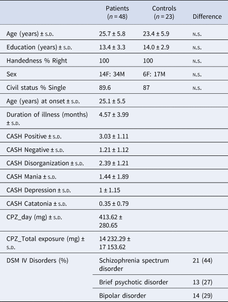
Briefly, 50 FEP consecutive admitted patients and 24 age- and sex-matched healthy controls were included. Left-handed participants were excluded to avoid the influence of laterality effects in the results. The final sample consisted of 48 FEP patients (mean age 25.7 ± 5.8, 34 males) and 23 healthy controls (mean age 23.4 ± 5.9, 17 males) (Table 1).
Demographic characteristics of the sample

CASH, the Comprehensive Assessment of Symptoms and History; CPZ, chlorpromazine equivalent doses; DSM IV, Diagnostic and Statistical Manual of Mental Disorders-IV Edition.
Written informed consent was provided for all participants and the study was approved by the Ethics Committee of the Health Navarre System.
Clinical assessments
Patients were assessed by means of the Comprehensive Assessment Symptoms and History (CASH) (Andreasen, Flaum, & Arndt, Reference Andreasen, Flaum and Arndt1992) and final DSM IV diagnosis was reached after the examination of all available information by consensus between MJC and VP. Healthy siblings and controls were evaluated by the CASH abbreviated version. The age range of FEP patients was comprised between 17 and 45 years old and exclusion criteria were the following: the absence of a lifetime substance abuse diagnosis and lack of antecedents of neurological, general medical illness, or mental retardation.
Our patients were not receiving antipsychotic treatment previously to the current treatment of their FEP. Current doses and the total exposure to antipsychotic drugs were transformed into chlorpromazine equivalents (Ho, Andreasen, Ziebell, Pierson, & Magnotta, Reference Ho, Andreasen, Ziebell, Pierson and Magnotta2011).
Motor assessments
A comprehensive battery of motor scales was used. Extrapyramidal symptoms and signs were evaluated by means of the UKU scale that includes a total score and five subscales: dystonia, rigidity, hypokinesia, dyskinesia, tremor and akathisia (Lingjaerde, Ahlfors, Bech, Dencker, & Elgen, Reference Lingjaerde, Ahlfors, Bech, Dencker and Elgen1987). Catatonic signs were assessed by means of the Bush-Francis Catatonia Rating Scale (BFCRS) (Bush, Fink, Petrides, Dowling, & Francis, Reference Bush, Fink, Petrides, Dowling and Francis1996), and NSS by means of the Neurological Evaluation Scale (NES) (Buchanan & Heinrichs, Reference Buchanan and Heinrichs1989). NES total score and scores from four neurological subscales (sensory integration, motor coordination, sequencing of complex motor acts, and ‘other’ soft signs) were included.
Motor assessments were carried out by three psychiatrists (LMI, JLI, and MR), who were specifically trained and achieved good interrater reliability scores before entering the study.
Magnetic resonance imaging protocol
Image acquisition
A 1.5 Siemens Avanto scanner with an eight-channel parallel array head coil from the Complejo Hospitalario de Navarra (Pamplona, Spain) was used for the acquisition of T1-, diffusion-, and T2*-weighted images. Patients were clinically stabilized at the time of the image acquisition, after remission of the acute symptomatology of psychosis. Clinically significant abnormal images were discarded after initial visual inspection by the neuroradiologist (TC).
Whole brain T1-weighted images were obtained with a T1-MPRAGE (magnetization-prepared rapid-acquisition gradient echo) 3D sequence (TR 1160; TE 4.24 ms; TI 600 ms; FOV 230 mm; 192 slices, 0.9 × 0.9 × 0.9 mm3 voxel; flip angle 15, matrix 256 × 256).
Diffusion-weighted images were obtained with a single-shot spin-echo echoplanar imaging sequence with the following imaging parameters: TR 4200 ms; TE 90 ms; matrix: 128 × 128; FOV 240; slice thickness 3 mm. Images were obtained with diffusion gradients applied along 30 non-collinear directions and b values of 0 and 1000 s/mm2 were used.
Finally, an axial multiecho T2* sequence was used for T2* relaxometry (TR 2000 ms; TE 6, 12, 20, 30 45, 60 ms; slice thickness 5 mm; matrix 256 × 256).
Structural image processing
The 3D axial T1-weighted acquisition sequence meets the recommendations for a successful segmentation of subcortical structures with the FreeSurfer image analysis suite. This process includes automatic Talairach transformation, non-brain tissue removal, and segmentation of white matter and deep gray matter structures. The results of each of these steps were examined and corrected when needed with the tools provided by FreeSurfer. The segmentation of each image provided masks in native space for four bilateral subcortical structures of interest: left and right putamen, GP, caudate nucleus, and accumbens. Further details about the acquisition and image analysis are detailed in Cuesta et al. (Reference Cuesta, Lecumberri, Cabada, Moreno-Izco, Ribeiro, Lopez-Ilundain and Gomez2017).
Diffusion image processing
Processing of diffusion data was performed following the TRACULA pipeline, included in FreeSurfer distributions, which in turn relies on the tools provided by the FSL suite (Smith et al., Reference Smith, Jenkinson, Woolrich, Beckmann, Behrens, Johansen-Berg and Matthews2004). First, distortions induced by eddy currents and head motion were corrected. Each image was aligned to the b0 image with a full affine transformation (mutual information cost function). A cross-modal registration with boundary-based cost function provided a rigid transformation between the b0 and structural volumes. A tensor model was fit to the diffusion data at each voxel, yielding FA and MD images. Binary masks for subcortical structures of interest were obtained after processing T1-weighted images, and thanks to the transformation between diffusion and structural spaces, which could be superimposed on FA and MD images. They were used as ROIs to compute mean FA and MD values in these deep gray matter structures.
R2* image processing
Following the protocol described in Peran et al. (Reference Peran, Cherubini, Assogna, Piras, Quattrocchi, Peppe and Sabatini2010), R2* images were computed from six T2*-weighted gradient echo volumes acquired with an echoplanar sequence at different TEs: 6, 12, 20, 30, 45, and 60 ms. The 20 ms image was chosen to find the transformation to structural space due to its higher contrast at the white matter–black matter boundary. A rigid registration with boundary-based cost function was performed between the image at 20 ms and the T1-weighted image. The other T2* volumes were realigned to the 20 ms image and least-squares fitting was applied in each voxel to obtain the parameters of the monoexponential decay function and R2* maps:

Similarly to the diffusion case, the previous transformation allowed binary masks for subcortical structures obtained from the segmentation of the structural images to be superimposed on R2* maps. They were used as ROIs to compute mean R2* values in each of these structures.
Shape analysis
Shape analysis of subcortical structures was performed with FIRST, included in the FSL suite (Patenaude, Smith, Kennedy, & Jenkinson, Reference Patenaude, Smith, Kennedy and Jenkinson2011). The shape/appearance models used in FIRST are constructed from manually segmented images provided by the Center for Morphometric Analysis (CMA), MGH, Boston. The surfaces of the segmented structures were modeled as meshes with the corresponding vertices across images in the training set. A multivariate Gaussian point distribution model was assumed for the vertices and the mean surface was computed for each structure. The principal component analysis allowed to extract the modes of variation that accounted for the variance in the distribution of the vertices. When attempting to fit the model to a new image, FIRST searches through linear combinations of these shape modes of variation for the most probable shape instance given the observed intensities in a T1-weighted image. It allows testing differences in the shape of the structures across subjects on the elements of their surface. Volumetric results have already been reported (Cuesta et al., Reference Cuesta, Lecumberri, Cabada, Moreno-Izco, Ribeiro, Lopez-Ilundain and Gomez2017); therefore, overall changes in size are disregarded in this analysis. Local atrophy or hypertrophy will refer to a surface locally curved inward or outward, respectively. Correction for multiple comparisons was performed with the Threshold-Free Cluster Enhancement method (Winkler, Ridgway, Webster, Smith, & Nichols, Reference Winkler, Ridgway, Webster, Smith and Nichols2014).
Statistical analyses
Assumptions of normality were tested by means of the Kolgomorov–Smirnov test. Normally and non-normally distributed variables were, respectively, reported as mean ± s.d. and median and interquartile range.
Data from all 48 FEP and 23 healthy controls were included in statistical analyses. Comparisons between the two groups on age and number of years of education were performed using t tests. Comparison between the two groups on gender variable was performed using the χ2 test.
Diffusion and R2* data
Multivariate analysis of variance (MANOVA), considering the values of basal ganglia structures as repeated measures, were run to compare values of FA, MD, and R2* between groups (FEP patients and controls).
A level of significance of p < 0.05 for comparative measurements was used throughout the study. Bonferroni correction was applied for multiple comparisons between FEP patients and controls (p < 0.05/8 = 0.006).
Pearson coefficient correlations between scores of motor scales in FEP patients and neuroimaging results (average FA, MD, and R2* in each subcortical structure) were examined. In order to generate greater power to counterbalance the multiple test correction penalty, we used false discovery rate (Benjamini & Hochberg, Reference Benjamini and Hochberg1995) to account for the eight correlation coefficients between the motor scores and FA, MD, and R2*MRI values for each subcortical area. The FDR-adjusted p value (or q value) was set at 0.05, which implies that 5% of significant tests will result in false positives.
Pearson correlations between motor scales and total exposure to antipsychotics in CPZ equivalents were performed. In case significant associations between motor scales and antipsychotic exposure were found, partial correlation was also calculated to account for the relationship between scores of motor scales in patients and neuroimaging results.
SPSS version 20 (Corp., 2011) was used in these analyses.
Shape data
A voxel-based analysis of group effect was performed for the surface of each subcortical structure. These analyses were repeated for patients only to test the effect of each motor assessment variable in the shape of structures. In these instances, patients were divided into two groups (mild and severe symptoms) on the criterion of their score being lower or greater than the median for the whole patient group.
In these analyses, an F statistic test is run in each surface element and a permutation method with threshold-free cluster enhancement method implemented in FSL was used for inference and correction for multiple comparisons (Winkler et al., Reference Winkler, Ridgway, Webster, Smith and Nichols2014). Statistical significance was set at p < 0.05.
Results
Sociodemographic, clinical, and motor scales
Demographic and clinical measures of FEP patient sample and healthy controls are shown in Table 1. There were no significant differences between FEP patients and controls in sociodemographic variables but FEP patients showed lower performance on all motor scales total scores and subscales regarding healthy controls except for UKU dystonia, tremor, and UKU dyskinesia subscales (online Supplementary Table S1).
MRI indices comparisons among diagnostic groups
Morphological analysis: shape analysis
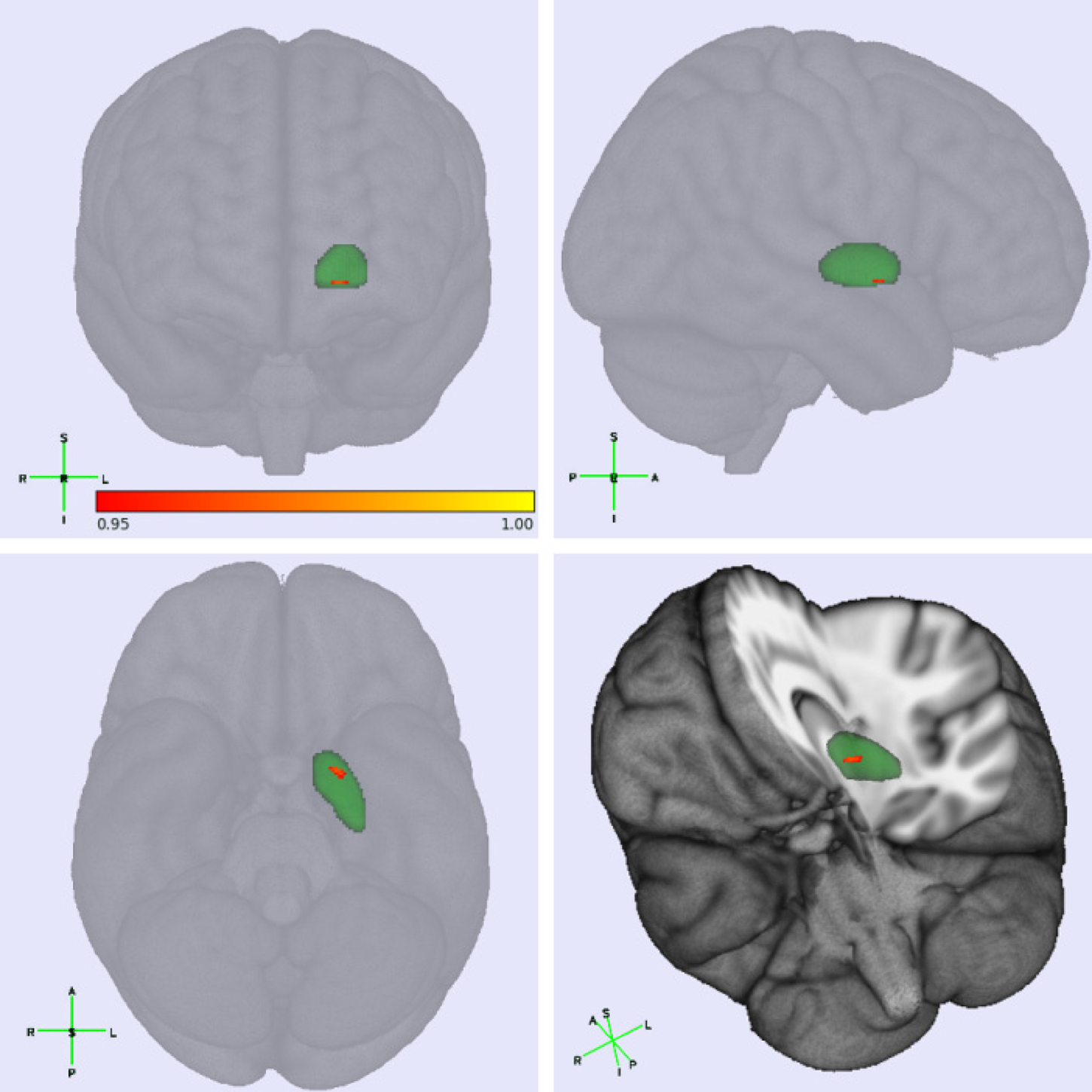
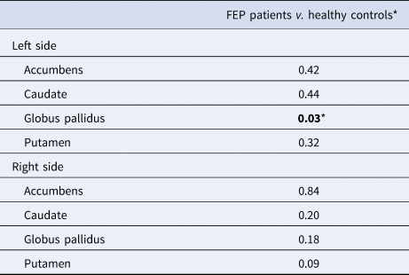
FEP patients showed small clusters of local atrophy on the caudal and anterior region of left GP regarding healthy subjects (Fig. 1), after removing global scale changes and after correcting for multiple comparisons (Table 2).
Significant shape abnormalities in first-episode psychosis patients regarding healthy subjects in left pallidum. The surface elements in grey show no significant difference between patients and controls. A grey scale is used to show the multiple comparisons corrected p value of the F statistic for the surface elements where shape is significantly different in patients and controls. Anatomical directions (anterior, posterior, left, right, superior, inferior) in each view plane are denoted by their first letter.

Statistical significance of basal ganglia shape scores between FEP patients and healthy controls (GLM model) (p value)*

* Lowest corrected p value of the F tests performed at each surface voxel. A permutation method with threshold-free cluster enhancement method implemented in FSL was used for inference and correction for multiple comparisons (statistical significance was set at p < 0.05, in bold).
Fractional anisotropy, mean diffusivity, and R2*measures
In comparison with healthy controls, FEP patients showed significantly lower FA in left and right caudate, putamen, GP, and accumbens nuclei after Bonferroni correction (F = 9.68, p = 0.003). The repeated-measures MANOVAs conducted on MD scores and R2* did not reveal a main effect for the diagnostic group (F = 1.39, p = 0.243 and F = 0.25, p = 0.617, respectively) (online Supplementary Table S2).
Associations between MRI indices and motor scales
Relation between motor scales and shape of subcortical structures
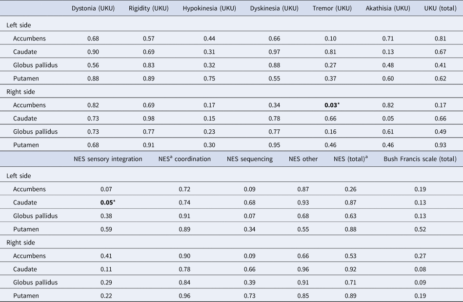
FEP patients with high UKU tremor scores showed significant small regions of hypertrophy in the shape scores of right accumbens, after correction for multiple testing comparisons as compared with patients with mild symptoms. Also, patients with high NES sensory integration scores showed significant hypertrophy in the shape scores of left caudate (Table 3). There were no significant differences in the shape scores of basal ganglia structures between mild and severe FEP groups dichotomized by other NES and UKU subscale scores (UKU total score and subscales) and by catatonia score (BFCRS total score).
Differences in basal ganglia shape scores between FEP patients dichotomized by the median of each motor assessment scale (mild and severe groups) (general lineal model, p value)*

* Lowest corrected p value of the F tests performed at each surface voxel. A permutation method with threshold-free cluster enhancement method implemented in FSL was used for inference and correction for multiple comparisons (statistical significance was set at p < 0.05, in bold).
a CPZ total exposure included as a covariate.
No differences were found between patients with severe and mild NES coordination and total scores and shape scores of basal ganglia, considering CPZ total exposure.
Pearson correlation analyses between motor scales and MRI measures
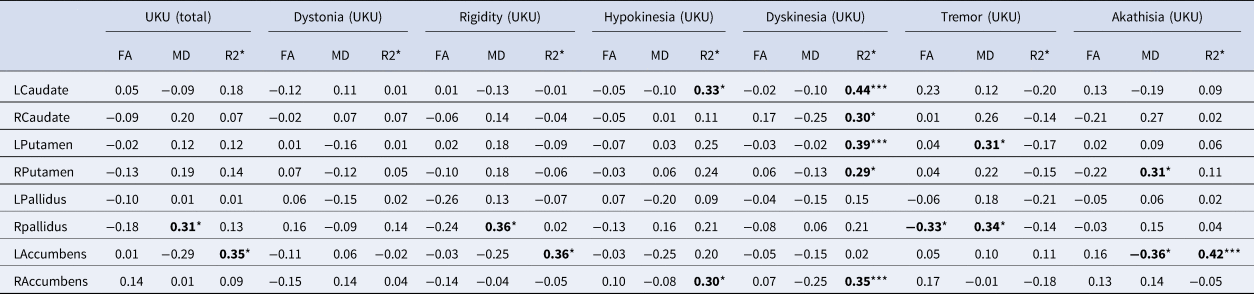
Motor scores were not significantly associated with fractional anisotropy and mean diffusivity measures, after the Benjamini–Hochberg correction for multiple testing. However, a trend toward significance (p values between 0.05 and 0.01) was found between lower left accumbens and higher right putamen MD measure and higher akathisia score, higher left putamen and right pallidus MD measures and tremor scores, and higher right pallidus MD measures and UKU total and rigidity scores. In addition, right pallidus FA measures were inversely associated with tremor scores (Table 4).
Pearson coefficient correlations between parkinsonism scores and multimodal MRI multimodal neuroimaging measures in FEP patients

FDR, false discovery rate.
In bold: *p value between p ⩽ 0.05 and >0.01;**p value ⩽ 0.01;***significant p value after Benjamini–Hochberg correction (FDR ⩽ 0.05).
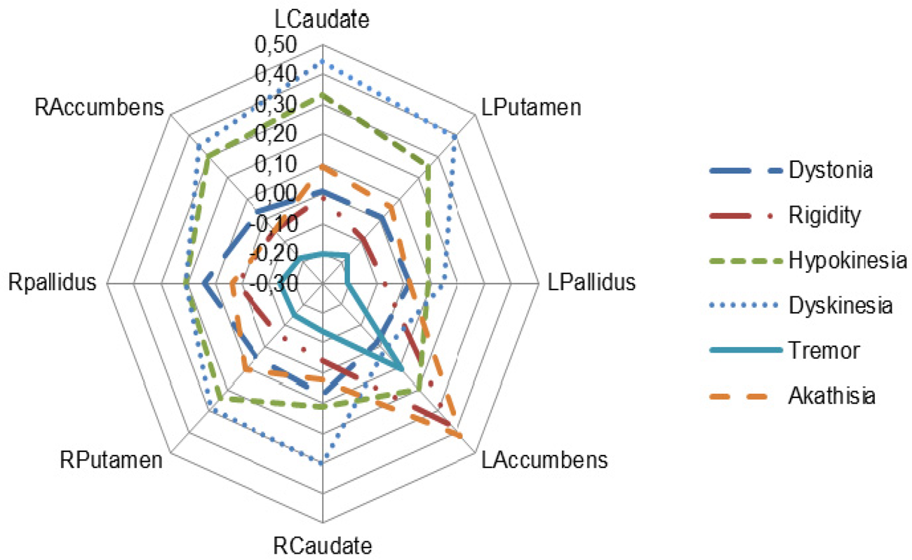
The analyses of the association between motor scores and relaxometry measures (R2* scores) and extrapyramidal scales showed that UKU dyskinesia showed significant correlations with R2* left caudate (r = 0.44, p = 0.002), with R2* left putamen (r = 0.39, p = 0.007) and with R2 right accumbens (r = 0.35, p = 0.017); and UKU akathisia with R2* left accumbens score (r = 0.42, p ⩽ 0.001). Regarding R2* scores, a trend was found toward significance (p values between 0.05 and 0.01) in the associations between right caudate and right putamen with dyskinesia scores, left caudate and right accumbens with hypokinesia, and left accumbens with rigidity and UKU total scores (Table 4 and Fig. 2).
Pearson correlations between iron loadings in basal ganglia and extra-pyramidal signs in first-episode psychosis patients. *p significant value (r ≥ 0.35) after Benjamini–Hochberg correction (FDR ⩽ 0.05); FDR, false discovery rate.

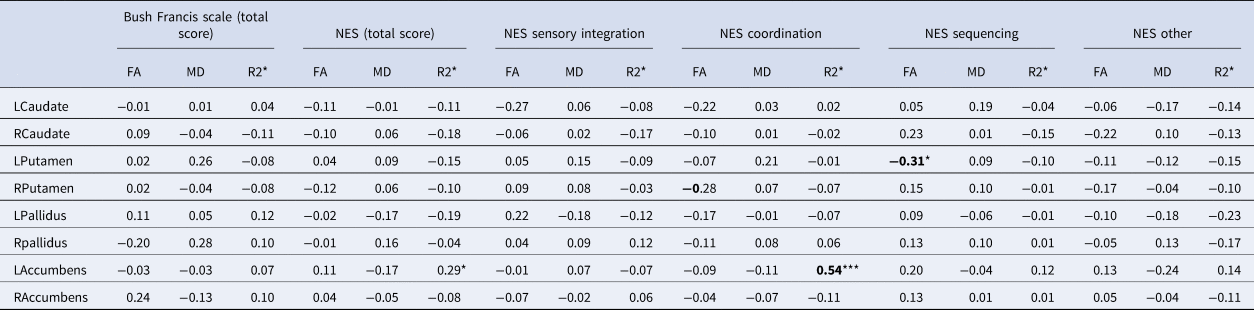
No significant correlations were found for NES and BFCRS scales regarding R2* measures except for the association between NES coordination and R2* left accumbens score (r = 0.54, p ⩽ 0.001) (Table 5).
Pearson coefficient correlations between catatonic and neurological soft signs and MRI multimodal neuroimaging measures in FEP patients

FDR, false discovery rate.
In bold: *p value between p ⩽ 0.05 and >0.01; **p value ⩽ 0.01; ***significant p value after Benjamini–Hochberg correction (FDR ⩽ 0.05).
CPZ equivalent doses of total exposure to antipsychotic drugs of FEP patients did not show significant associations with morphometric, diffusivity, and R2* measures. However, CPZ total exposure showed significant associations with NES total scores (r = 0.31, p = 0.037) and NES coordination scores (r = 0.58, p ⩽ 0.001). Therefore, partial correlations between these two NES scores and FA, MD, and R2* values were calculated, which resulted in the same significant associations as the Pearson correlations (NES coordination with left accumbens R2* values, r = 0.51, p ⩽ 0.001).
Discussion
Four main findings result from this study. First, FEP patients showed significant differences in shape and in fractional anisotropy but not in diffusivity and iron loadings of basal ganglia and accumbens nuclei regarding healthy controls subjects. In fact, FEP patients showed significant shape deformities regarding healthy controls in left GP nucleus after removing global scale changes. Second, FEP patients with high scores on the UKU tremor subscale showed significant changes in right accumbens shape, and patients with high scores on the NES sensory integration subscale showed significant changes in left caudate, regarding those FEP patients with low scores. Third, iron loading measures of mainly left-side basal ganglia in FEP patients correlated significantly with extrapyramidal abnormalities and NSS. Specifically, iron loadings in left accumbens showed positive significant associations with higher scores in NES coordination subscale and with akathisia; iron loadings in left caudate, left putamen, and right accumbens with dyskinesia scores. Fourth, we found a trend toward significance in some associations between MRI values and motor scales, which did not reach the FDR threshold for significance, but also deserve to be discussed. Specifically, right caudate and right putamen R2* measures were associated with dyskinesia scores, left caudate, and right accumbens with hypokinesia, and left accumbens with rigidity and UKU total scores. Regarding MD measures, lower left accumbens and higher right putamen were associated with higher akathisia scores, higher left putamen, and right pallidus with tremor scores, and higher right pallidus with UKU total and rigidity scores. In addition, right pallidus FA measures were inversely associated with tremor scores.
To our knowledge, this is the first study addressing the association between a comprehensive assessment of motor domains of FEP patients regarding MRI correlates including relaxometry measures of basal ganglia.
Local deformities in shape of the left pallidum nucleus might suggest that atrophic changes are already present in our FEP patients at the beginning of the illness. These findings are in agreement with the results from recent meta-analysis on subcortical and basal ganglia volumetric abnormalities of schizophrenia patients (Haijma et al., Reference Haijma, Van Haren, Cahn, Koolschijn, Hulshoff Pol and Kahn2013; Okada et al., Reference Okada, Fukunaga, Yamashita, Koshiyama, Yamamori, Ohi and Hashimoto2016; van Erp et al., Reference van Erp, Hibar, Rasmussen, Glahn, Pearlson, Andreassen and Turner2016) and bipolar disorders (Hibar et al., Reference Hibar, Westlye, van Erp, Rasmussen, Leonardo, Faskowitz and Andreassen2016) and with the previous studies examining shape deformities in schizophrenia (Hill et al., Reference Hill, Bolo, Sarvode Mothi, Lizano, Guimond, Tandon and Keshavan2017; Mamah, Alpert, Barch, Csernansky, & Wang, Reference Mamah, Alpert, Barch, Csernansky and Wang2016). Unfortunately, these meta-analyses did not examine whether grey matter abnormalities are related to MAs. No comparisons with the previous results can be performed due to the fact that no previous studies examined iron loadings in the early stages of illness of psychosis. However, a recent prospective and longitudinal approach to examine the functional interactions of the GP in a cohort of FEP patients found that abnormalities in cortical-basal ganglia circuitry, and specifically lower functional connectivity between the left internal segments of this nucleus (GPi) and a network of regions, may play a significant role in determining the outcomes in schizophrenia (Tarcijonas et al., Reference Tarcijonas, Foran, Blazer, Eack, Luna and Sarpal2019).
The basal ganglia are mainly involved in motor programming in response to external and internal inputs by enabling voluntary movements and simultaneous inhibition of competing or interfering movements (Jahanshahi, Obeso, Rothwell, & Obeso, Reference Jahanshahi, Obeso, Rothwell and Obeso2015). However, as main hubs of brain (McCutcheon, Krystal, & Howes, Reference McCutcheon, Krystal and Howes2020), they also are involved in the control of a wide range of complex non-motor behaviors, such as emotion, response inhibition, conflict, decision-making, error-detection and surprise, reward processing, language, and time processing (Eisinger, Urdaneta, Foote, Okun, & Gunduz, Reference Eisinger, Urdaneta, Foote, Okun and Gunduz2018). In this regard, some of these non-motor behaviors might be related more to the limbic loop than to the motor loop of the basal ganglia. In fact, the nucleus accumbens, ventral pallidum, and VTA seem to be more related to the reward system than the motor loop of basal ganglia (Simonyan, Reference Simonyan2019). We found that hypertrophic changes in right accumbens and left caudate were associated with higher scores in tremor UKU subscale and NES sensory integration subscale, respectively, in our FEP sample. However, we could not differentiate these portions of basal ganglia in our MRI analyses to clear distinguish the functional subdomains of basal ganglia and its counterparts in motor or limbic correlates.
Most of our associations between neuroimaging measures and abnormal movements were more focused on hyper and increasing NSS than hypokinetic abnormalities. In this regard, one can speculate that these unwarranted movements may arise from abnormal striatopallidal activity via the direct pathway of cortico-basal ganglia circuits leading to reduced output in the GP interna and disinhibition of the thalamocortical projection (Obeso, Rodriguez-Oroz, Stamelou, Bhatia, & Burn, Reference Obeso, Rodriguez-Oroz, Stamelou, Bhatia and Burn2014).
It has been reported that a reduction of certain grey matter subcortical structures is associated with higher rates of motor and sensory NSS (Dazzan et al., Reference Dazzan, Morgan, Orr, Hutchinson, Chitnis, Suckling and Murray2004). Our results concerning the association of coordination score of the NES scale and R2* score of left accumbens nucleus might be also coherent with Dazzan's et al. study. Moreover, NSS seems to be also associated with lower global sulcal index in both hemispheres and lower regional sulcal indices in left dorsolateral prefrontal and right lateral occipital cortices (Gay et al., Reference Gay, Plaze, Oppenheim, Mouchet-Mages, Gaillard, Olie and Cachia2013).
We also found reductions in FA across all basal ganglia in FEP patients regarding healthy controls. FA abnormalities are more related to deviations of tracts orientation of white but not grey matter structures that are the main component of basal ganglia. However, there are also few myelin fibers that transverse the basal ganglia and particularly the GP. In this respect, decreased FA values in GP have been reported in schizophrenia patients leading to researchers to suggest the presence of microstructural abnormalities that may be independent of antipsychotic medication (Hashimoto et al., Reference Hashimoto, Mori, Nemoto, Moriguchi, Noguchi, Nakabayashi and Ohnishi2009).
Microstructural abnormalities in left accumbens nucleus, as resulted by reduced mean diffusivity, might have a role in akathisia scores of FEP patients. On this subject, there is some evidence reporting decreased grey matter in the nucleus accumbens and worsening in cognitive performance in FEP (Dempster et al., Reference Dempster, Norman, Theberge, Densmore, Schaefer and Williamson2017). However, there are diffusivity studies reporting an increase of mean diffusivity in subcortical structures, including basal ganglia, in schizophrenia patients regarding healthy controls (Spoletini et al., Reference Spoletini, Cherubini, Banfi, Rubino, Peran, Caltagirone and Spalletta2011). Besides, there is a great variability in the literature concerning subcortical grey matter diffusivity in schizophrenia patients (Wheeler & Voineskos, Reference Wheeler and Voineskos2014) but even intersubject variability and reproducibility of diffusion tensor imaging seem to have substantial variability across the brain (Veenith et al., Reference Veenith, Carter, Grossac, Newcombe, Outtrim, Lupson and Coles2013).
The significant correlations between parkinsonian signs, but not NSS and catatonic ones, and iron loadings in left caudate and putamen, and left and right accumbens nuclei may suggest an involvement of these nuclei in the pathophysiology of MAs in early psychosis. The role of iron loadings in the brain of schizophrenia patients has been scarcely examined after the pioneer study of Casanova et al. (Reference Casanova, Comparini, Kim and Kleinman1992) in part due to the attribution of these structural abnormalities to antipsychotic drug effects (Gur et al., Reference Gur, Maany, Mozley, Swanson, Bilker and Gur1998) and to brain toxicity (Lieberman et al., Reference Lieberman, Tollefson, Charles, Zipursky, Sharma, Kahn and Group2005). However, studies from our group suggested that low serum iron levels are evidenced in a subgroup of schizophrenia patients with catatonic features and negative catatonic symptoms (Peralta et al., Reference Peralta, Cuesta, Mata, Serrano, Perez-Nievas and Natividad1999). There is some evidence reporting that a significant decrease in iron serum preceded a relapse in medication-free schizophrenia patients (Weiser, Levkowitch, Neuman, & Yehuda, Reference Weiser, Levkowitch, Neuman and Yehuda1994). These results are also in agreement with the recent studies showing a decrease in circulating iron in PD patients regarding normal subjects (Costa-Mallen et al., Reference Costa-Mallen, Gatenby, Friend, Maravilla, Hu, Cain and Anzai2017).
Iron deposits are evidenced not only in the nigrostriatal system from the early stages of PD but also they seem to be strongly associated with the severity of motor symptoms and cognitive impairment (Bunzeck et al., Reference Bunzeck, Singh-Curry, Eckart, Weiskopf, Perry, Bain and Husain2013). Iron deposits are also invoked in the pathophysiology of Alzheimer disease and other neurodegenerative diseases (Eustache, Nemmi, Saint-Aubert, Pariente, & Peran, Reference Eustache, Nemmi, Saint-Aubert, Pariente and Peran2016). These changes seem to be independent of the increase of brain deposits that are seen on aging (Pfefferbaum, Adalsteinsson, Rohlfing, & Sullivan, Reference Pfefferbaum, Adalsteinsson, Rohlfing and Sullivan2009).
Basal ganglia and other medial structures are within the most co-activated homotopic brain regions in the two hemispheres (Mancuso et al., Reference Mancuso, Costa, Nani, Manuello, Liloia, Gelmini and Cauda2019) and they closely interact with one another to coordinate the movements (Wei, Zhang, Lv, & Jing, Reference Wei, Zhang, Lv and Jing2017). However, basal ganglia abnormalities in this study were more related to the left side suggesting a dysfunctional homotopy. Clinical and neuroimaging studies may provide some explanations for our results. First, a similar asymmetry of neurological signs in never-treated patients with schizophrenia has been reported (Kamis et al., Reference Kamis, Stratton, Calvo, Padilla, Florenzano, Guerrero and de Erausquin2015). Moreover, the dominant hand is usually the most affected side (Djaldetti, Ziv, & Melamed, Reference Djaldetti, Ziv and Melamed2006) and it has reported greater contralateral striatal atrophy relative to the side of motor onset in PD (Tanner, McFarland, & Price, Reference Tanner, McFarland and Price2017). A right-sided symptom onset seems to have a less cognitive impairment, while a left-sided symptom onset appears to be associated with a slower motor progression in PD (Baumann, Held, Valko, Wienecke, & Waldvogel, Reference Baumann, Held, Valko, Wienecke and Waldvogel2014; Feis, Pelzer, Timmermann, & Tittgemeyer, Reference Feis, Pelzer, Timmermann and Tittgemeyer2015; Katzen, Levin, & Weiner, Reference Katzen, Levin and Weiner2006; Tomer, Levin, & Weiner, Reference Tomer, Levin and Weiner1993). Second, several recent studies reported reductions in resting-state functional connectivity between homotopic brain regions in schizophrenia (Guo et al., Reference Guo, Jiang, Xiao, Zhang, Zhang, Yu and Liu2014; Hoptman et al., Reference Hoptman, Zuo, D'Angelo, Mauro, Butler, Milham and Javitt2012) that may even occur early in the course of the disease and are independent of medication status (Li, Xu, Zhang, Hoptman, & Zuo, Reference Li, Xu, Zhang, Hoptman and Zuo2015).
Taken together, this model taken out from PD may imply a role for an imbalance in the process of repartitioning or redistribution of iron in FEP patients with a decrease of iron levels in circulation but accumulation in basal ganglia, and preferentially in the SN pars compacta (Costa-Mallen et al., Reference Costa-Mallen, Gatenby, Friend, Maravilla, Hu, Cain and Anzai2017). However, whether circulating iron levels are related to iron loadings in basal ganglia and other brain structures have not been examined in schizophrenia spectrum patients.
As a potent pro-oxidant, redox-active iron may be a key player in upstream mechanisms that precipitate nigrostriatal cells death in PD and this potent redox couple formed by iron and dopamine may account for neurodegeneration in PD (Hare & Double, Reference Hare and Double2016). However, the precise mechanism by which iron causes neurotoxicity in PD remains unclear (Hare & Double, Reference Hare and Double2016) though it seems to have a pathogenetic role by triggering oxidative stress, which leads to nigral cell loss and subsequently resulting motor symptoms in PD (Bunzeck et al., Reference Bunzeck, Singh-Curry, Eckart, Weiskopf, Perry, Bain and Husain2013).
Heme and non-heme are the two distinct forms of iron in the brain of vertebrates. While heme iron is bound to circulating and accumulating blood and it is essential for binding oxygen and ferrying it, non-heme iron is present in virtually all cells and it has a main role in energy-consuming processes, such as neurotransmission and myelin production and maintenance (Bulk et al., Reference Bulk, van der Weerd, Breimer, Lebedev, Webb, Goeman and Bossoni2018). Non-heme iron concentration in the brain, which is in its 90% bound to ferritin, is greater than in any organ system except the liver (see Daugherty and Raz, Reference Daugherty and Raz2015 for a review).
MR signal from brain tissue in healthy subjects is modified by paramagnetic fields created by ferritin and hemosiderin concentrations but not for other metals, such as copper and manganese, that have very low brain concentration. Thus, MRI is an in vivo method for the quantification of the differences in local magnetic susceptibility fields related to brain iron content.
Several modifiers, such as age-related, genetic and non-genetic factors, may account for the individual differences in brain iron among healthy individuals. Advanced age and neurodegeneration processes, such as Alzheimer's and Parkinson's diseases, have been involved in abnormal grey matter iron deposits. Decreased binding rate to ferritin and mutations in the human hemochromatosis protein gene have been related to the elevated iron content in the brain. Moreover, inflammatory, metabolic, and cardiovascular risk factors have been found to increase the brain iron content probably by reducing the cerebral blood flow (Daugherty & Raz, Reference Daugherty and Raz2015).
Abnormal high concentrations are typically found in the basal ganglia and have been related to motor and cognitive impairments in Parkinson's and Alzheimer's diseases (Kim & Wessling-Resnick, Reference Kim and Wessling-Resnick2014; Ward, Zucca, Duyn, Crichton, & Zecca, Reference Ward, Zucca, Duyn, Crichton and Zecca2014). A putative underlying mechanism accounting for by these disturbances may be related to the functions of iron as a cofactor for tyrosine hydroxylase and tryptophan hydroxylase, enzymes that are responsible for dopamine and serotonin synthesis, respectively. Thus, it would be argued that the MAs related to basal ganglia iron loadings in FEP patients may be related to dysfunctions on neurotransmission pathways mainly in dopamine systems in FEP patients. Future studies should address longitudinally whether this abnormal brain content in basal ganglia in FEP patients is specifically related to the acute episode or a stable feature.
MAs have been associated with cognitive impairment by our group and others. Specifically, parkinsonism and NSS demonstrated to be closely associated with cognitive impairment (Cuesta et al., Reference Cuesta, Sanchez-Torres, de Jalon, Campos, Ibanez, Moreno-Izco and Peralta2014; Molina et al., Reference Molina, Gonzalez Aleman, Florenzano, Padilla, Calvo, Guerrero and de Erausquin2016). On the contrary, much less evidence was reported regarding catatonic signs since very few studies addressed this subject and negative results have been found (Cuesta et al., Reference Cuesta, Sanchez-Torres, de Jalon, Campos, Ibanez, Moreno-Izco and Peralta2014, Reference Cuesta, Moreno-Izco, Ribeiro, Lopez-Ilundain, Lecumberri, Cabada and Peralta2018b).
In neuroimaging studies, cortical and subcortical regions, such as the basal ganglia, and the thalamus, have been involved in the neural substrates of MAs in schizophrenia (Walther & Strik, Reference Walther and Strik2012). Both structural and functional MRI studies consistently suggested a dysfunction of the ‘cerebello-thalamo-prefrontal’ brain network as an etiopathogenic model of NSS abnormalities disorders (Zhao et al., Reference Zhao, Li, Huang, Yan, Dazzan, Pantelis and Chan2014); and other MAs in schizophrenia and related psychoses (Peralta & Cuesta, Reference Peralta and Cuesta2017).
Despite MAs seem to provide a proximity to brain circuits and substrate (Walther, Reference Walther2015) and potential alternative phenotypes to current classifications of mental disorders (Cuesta et al., Reference Cuesta, Garcia de Jalon, Campos, Moreno-Izco, Lorente-Omenaca, Sanchez-Torres and Peralta2018a), MAs have been scarcely used in neurobiological research as markers or endophenotypes (Hirjak et al., Reference Hirjak, Thomann, Kubera, Wolf, Sambataro and Wolf2015). Unfortunately, at this moment, those varied evidences have not been enough to include MAs within DSM 5 classification (except for Catatonia paragraph) and only marginally within the RDoC initiative (Mittal, Bernard, & Northoff, Reference Mittal, Bernard and Northoff2017).
Limitations
Our FEP sample was made up of non-pure affective psychotic spectrum disorders such as schizophrenia spectrum disorders, brief psychotic disorders, and psychotic bipolar disorder. The rationale to include the two latter diagnostic groups was based on substantial overlap in psychopathological symptoms (Quattrone et al., Reference Quattrone, Di Forti, Gayer-Anderson, Ferraro, Jongsma, Tripoli and Reininghaus2019), neurobiology and treatment response (Kahn & Sommer, Reference Kahn and Sommer2015), and genetic liability among subtypes od psychosis (Musliner et al., Reference Musliner, Mortensen, McGrath, Suppli, Hougaard and Bybjerg-Grauholm2019).
Patients were not free of antipsychotic drugs but they did not receive previously any antipsychotic treatment and the current administration began shortly. Nonetheless, antipsychotic drugs, as potent D2 blockage agents, may account for the increase of basal ganglia volumes in patients with schizophrenia since dopamine D 2 receptors are highly expressed in these structures (Roiz-Santianez et al., Reference Roiz-Santianez, Ayesa-Arriola, Tordesillas-Gutierrez, Ortiz-Garcia de la Foz, Perez-Iglesias, Pazos and Crespo-Facorro2014) and it could not discard a potential effect of treatment in neuroimaging measures. However, morphological, anisotropy, diffusivity, and relaxometric measures of our patients were not significantly associated with chlorpromazine equivalent doses of antipsychotic drugs both in current treatment and total exposure to antipsychotic drugs.
Exposition to antipsychotic drugs has been related to smaller hippocampus, amygdala, thalamus, nucleus accumbens, and intracranial volumes but larger GP and lateral ventricle volumes in schizophrenia patients (Haijma et al., Reference Haijma, Van Haren, Cahn, Koolschijn, Hulshoff Pol and Kahn2013; van Erp et al., Reference van Erp, Hibar, Rasmussen, Glahn, Pearlson, Andreassen and Turner2016). Similarly, but a lower intensity, grey matter reductions were observed in antipsychotic naïve patients except in the caudate and thalamus that showed more pronounced volume reductions (Haijma et al., Reference Haijma, Van Haren, Cahn, Koolschijn, Hulshoff Pol and Kahn2013; Spinks et al., Reference Spinks, Nopoulos, Ward, Fuller, Magnotta and Andreasen2005). In this study, FA and MD changes were not associated with both current doses and total accumulative doses received before MRI (in CPZ equivalent doses).
Longitudinal studies of first-episode schizophrenics suggest that brain abnormalities in FEP patients may evolve during the follow-up (Shenton, Whitford, & Kubicki, Reference Shenton, Whitford and Kubicki2010). Whether iron loadings will progress or vanish over time should not be accounted for by our cross-sectional study and should be undertaken in future studies. Moreover, caution is warranted regarding the association between motor domains and iron deposits in FEP patients because there were no differences with healthy controls.
Finally, our standard magnetic resonance (1.5 Tesla) was not accurate enough to examine iron deposits in lower levels since T1- and T2-weighted, spin-echo, gradient-recalled-echo sequences, and segmented inversion-recovery ratio imaging fail to visualize in detail the anatomy of the SN.
Supplementary material
The supplementary material for this article can be found at https://doi.org/10.1017/S0033291720000343.
Acknowledgements
We appreciate very much the collaboration of patients and families in our research.
Financial support
This work was supported by the Department of Health of the Government of Navarra (grants 11/101 and 87/2014) and the Carlos III Health Institute (FEDER Funds) from the Spanish Ministry of Economy and Competitivity (grants 11/02831, 14/01621 and 16/02148), and by the Valley Baptist Legacy Foundation Center for Brain Health (NIH/D43-MH108169-01).
Conflict of interest
The authors declare that there is no conflict of interest.