Introduction
Breast milk is considered the gold standard for infant nutrition because it is essential for proper growth and development. Exclusive breastfeeding (BF) is recommended up to six months of age, as breast milk has a complex composition that makes it unique. It contains various biologically active components, such as immunoglobulins, growth hormones, oligosaccharides, and the infant’s own microbiota, which play crucial roles in intestinal homeostasis, immunity, and development. Reference Dalby and Hall1–3
In the first weeks of life, a healthy gut microbiota is characterized by limited bacterial diversity. Initially, the colonies resemble those in the mother’s gut when birth occurs by vaginal delivery (VD). In infants fed breast milk, bifidobacteria predominate. Reference Dalby and Hall1 In contrast, the gut microbiota of infants born by cesarean section (CS) is comparable to that following antibiotic ingestion, with persistent early-life disturbances characterized by reduced Bacteroides colonization and slower microbial community development. Reference Bokulich, Chung and Battaglia4 Although CS, when indicated for medical reasons, can reduce or avoid risks to mother and child, it is important to emphasize that CS rates between 10% and 15% are associated with lower maternal and infant mortality. Reference Betrán, Torloni and Zhang5,6 However, the global CS rate is 21.1%, with Brazil ranking second in the world at 55.7%, after the Dominican Republic (58.1%). In private hospitals, this rate can reach up to 90%. Reference Nakamura-Pereira, Leal and Pereira7 According to the Pan American Health Organization, the CS rate has risen from around 7% in 1990 to 21% currently, with a further increase expected over the next decade that could reach 28.5% globally by 2030, with significant regional variations. Reference Betrán, Torloni and Zhang8
Other changes associated with CS include reduced microbial diversity, delayed colonization by Bacteroides and Bifidobacterium spp., and the development of Clostridium spp. Reference Coelho, Ayres and Barreto9–Reference Rutayisire, Huang, Liu and Tao12 Although CS is necessary in certain circumstances, it has been associated with the development of immunological diseases, a phenomenon that can be explained by altered maturation of the gut microbiota. Reference Paixao, Bottomley and Pescarini13–Reference Wampach16
The infant gut microbiota develops through two key transitional phases. The first occurs immediately after birth during BF, resulting in a gut microbiota dominated mainly by Bifidobacterium. The second phase begins with the introduction of solid foods, leading to a more complex microbiota similar to that of adults, primarily composed of Bacteroides and Firmicutes. Reference Chong-Neto, Pastorino and Melo17 Clostridium difficile is widespread in the gut microbiota, present in nearly half of infants under one month old and accounting for up to 30% of the infant microbiota within the first six months of life. Reference Drall, Tun and Morales-Lizcano18 Although this bacterium is associated with diarrhea in adults, it remains unclear whether colonization with C. difficile triggers intestinal dysbiosis in infants or if pre-existing dysbiosis favors colonization by this microorganism. Reference Drall, Tun and Morales-Lizcano18 The importance of gut microbiota composition for infant health is therefore evident. Reference Selma-Royo, Calatayud Arroyo and García-Mantrana11,Reference Paixao, Bottomley and Pescarini13
BF directly affects the newborn’s microbiota and provides both short- and long-term health benefits into adulthood. Reference Lyons, Ryan, Dempsey, Ross and Stanton19 Modulating the gut microbiota through breast milk may reduce the risk of allergic disease by taking advantage of the “window of opportunity” in the first months of life to influence long-term health outcomes. Reference van den Elsen, Garssen, Burcelin and Verhasselt20,Reference Boudry, Charton and Le Huerou-Luron21 Early-life colonization with a diverse gut microbiota promotes immune tolerance by supporting the development of regulatory T cells and limiting IgE production and pro-inflammatory iNKT cell activation, thereby reducing the risk of allergic disease. Reference van den Elsen, Garssen, Burcelin and Verhasselt20 Several factors, such as delivery method and feeding type, significantly affect the composition of the infant’s gut microbiota. In recent years, infant formula consumption has increased due to positive claims on product labels and aggressive marketing that encourages caregivers to substitute formula for breast milk, leading to the misconception that it is as effective as or even better than breast milk. Reference Conway, Ritchie, Esser, Steptoe, Smith and Llewellyn22
With this in mind, the aim of this study is to provide an overview of the development of the microbial profile of the infant gut in the first months of life and to highlight the influence of factors such as mode of delivery and feeding to understand their impact on infant health.
Methods
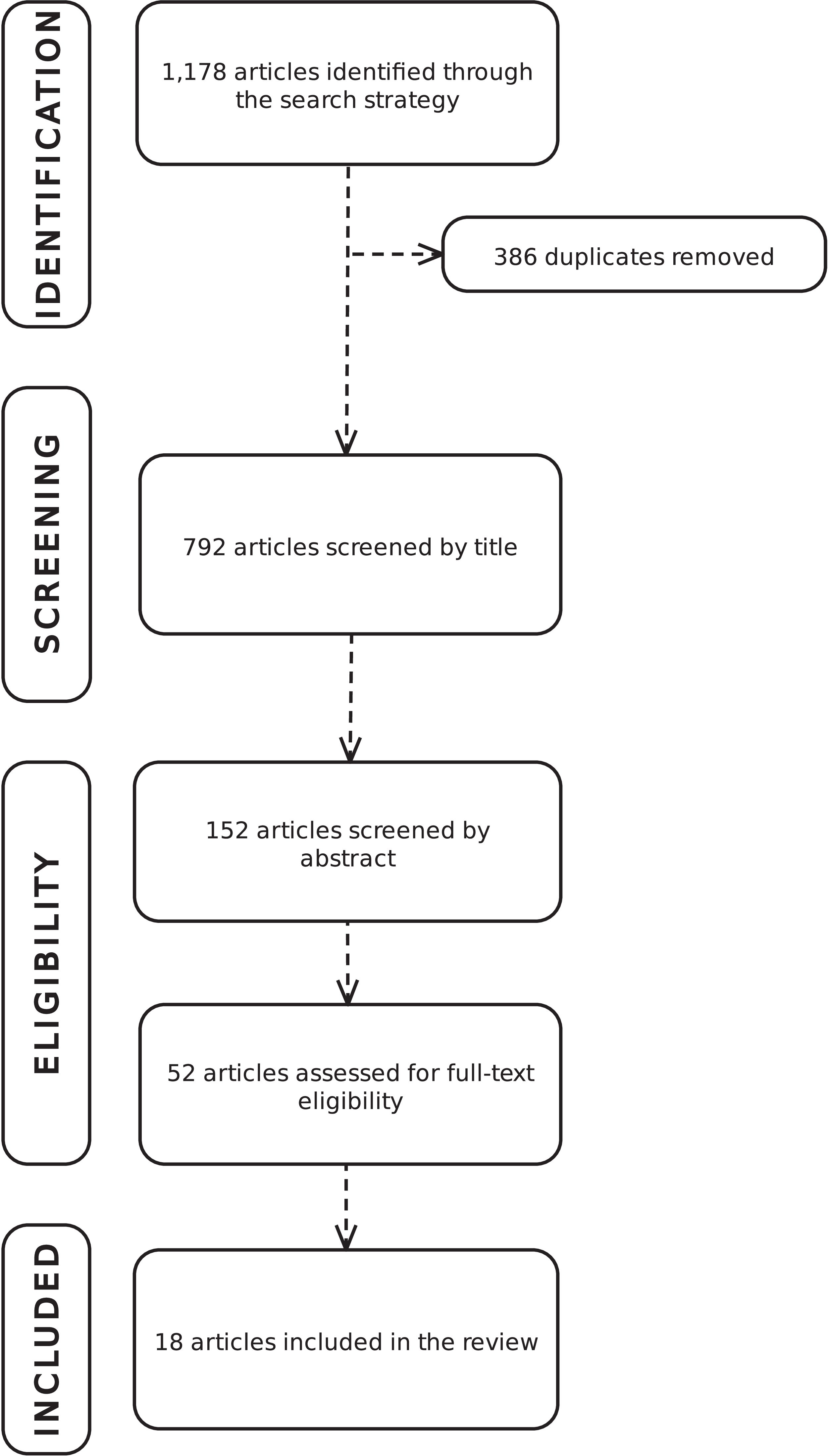
We reviewed the literature to understand the initial development of the early gut microbiota. The process followed the Preferred Reporting Items for Systematic Reviews and Meta-Analyses (PRISMA) guidelines (Figure 1). We searched the Virtual Health Library and PubMed databases using the descriptors “gut microbiota” combined with “breastfeeding” and “infant feeding” with Boolean operators (AND/OR). We applied a temporal filter for publications from the last 10 years and a language filter (Portuguese, English, and Spanish) to ensure the evidence was current.
Flowchart of the search strategy.
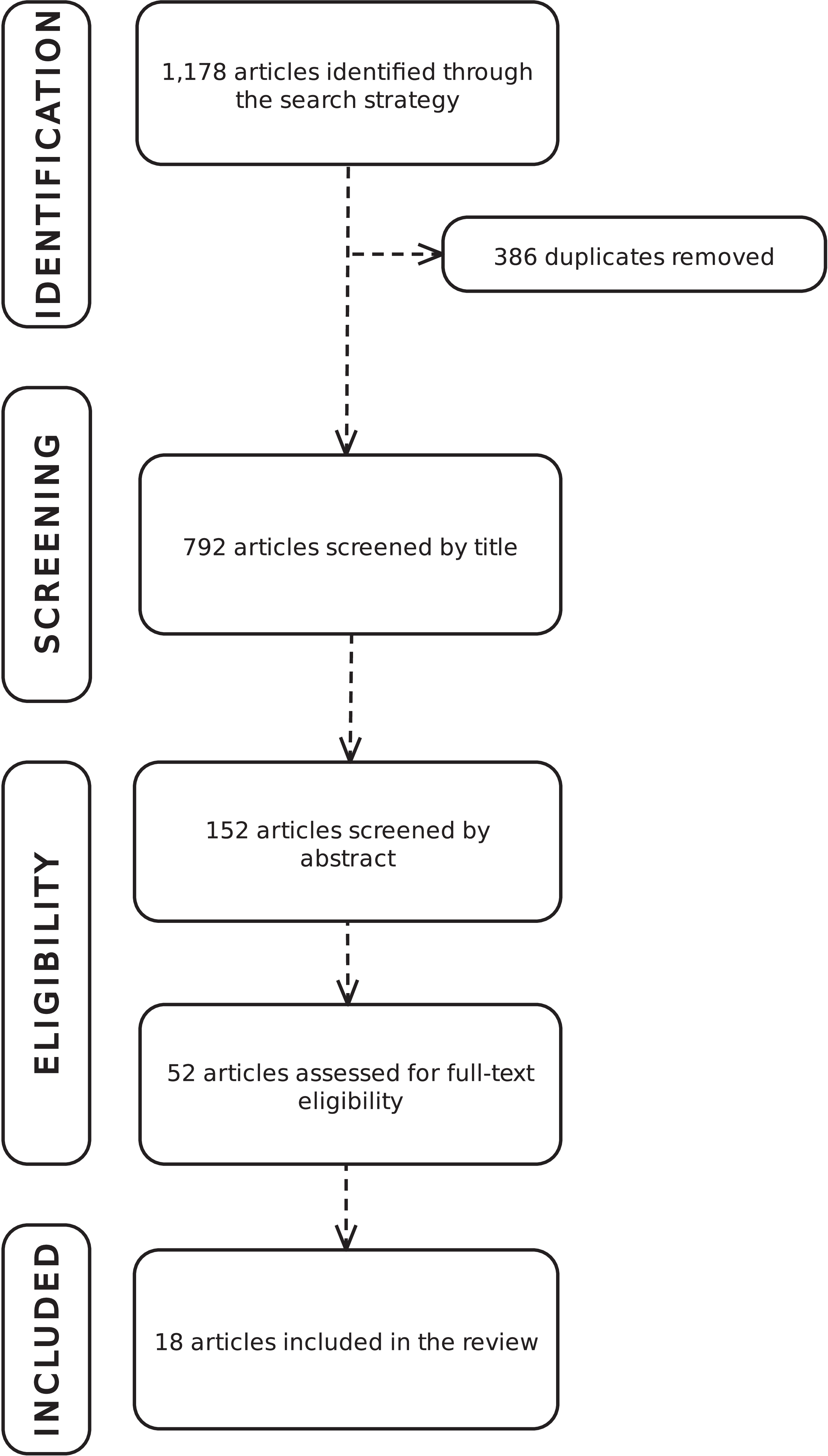
Figure 1. Long description
Flowchart illustrating the search strategy for identifying relevant articles. The process begins with 1,178 articles identified through the search strategy. After removing 386 duplicates, 792 articles are screened by title. This is followed by screening 152 articles by abstract. Next, 52 articles are assessed for full-text eligibility. Finally, 18 articles are included in the review. The flowchart visually represents the stages of identification, screening, eligibility, and inclusion, showing the progressive filtering of articles.
We then identified, selected, and analyzed relevant studies. The selection process was conducted in three rigorous steps: (1) initial screening by title, (2) review of abstracts, and (3) reading of full texts. After removing duplicates using the EndNote Web Reference Manager, we applied the exclusion criterion to studies that did not match the central theme, such as animal studies, assessments of gut microbiota as an exposure factor, and studies involving preterm infants, infants with certain diseases, or infants older than six months. Finally, 18 articles were included in the final analysis (see Figure 1).
The data extracted from the selected studies were summarized in a table and figure, including authorship, year of publication, country of origin, objectives, methodology, sample characteristics, and main findings. Data were extracted and reviewed by two authors (TBC and SP).
Results and discussion
As summarized in Table 1, the included studies were distributed across various countries, each represented to different extents. China contributed three publications, Reference Guo, Zhou and Li23–Reference Wu, Ren and Li25 as did Japan. Reference Akagawa, Tsuji and Onuma26 Italy, Reference Corvaglia, Tonti and Martini27,Reference Mazzola, Murphy and Ross28 Mongolia, Reference Ma, Li and Zhang29,Reference Ma, Li and Zhang30 the United States, Reference Bokulich, Chung and Battaglia4,Reference Wang, Li and Wu31 and the Netherlands Reference Borewicz, Fangjie Gu and Saccenti32,Reference Borewicz, Suarez-Diez and Hechler33 each contributed two articles. Spain, Reference Selma-Royo, Calatayud Arroyo and García-Mantrana11 Armenia and Georgia, Reference Lewis, Sidamonidze and Tsaturyan34 Belgium, Reference Martin, Makino and Yavuz35 the Netherlands, Reference Timmerman, Rutten and Boekhorst36 Canada, Reference Azad, Konya and Maughan37 and Thailand Reference Kongnum, Taweerodjanakarn and Hongpattarakere38 each had one publication.
Summary of the studies included in the literature review
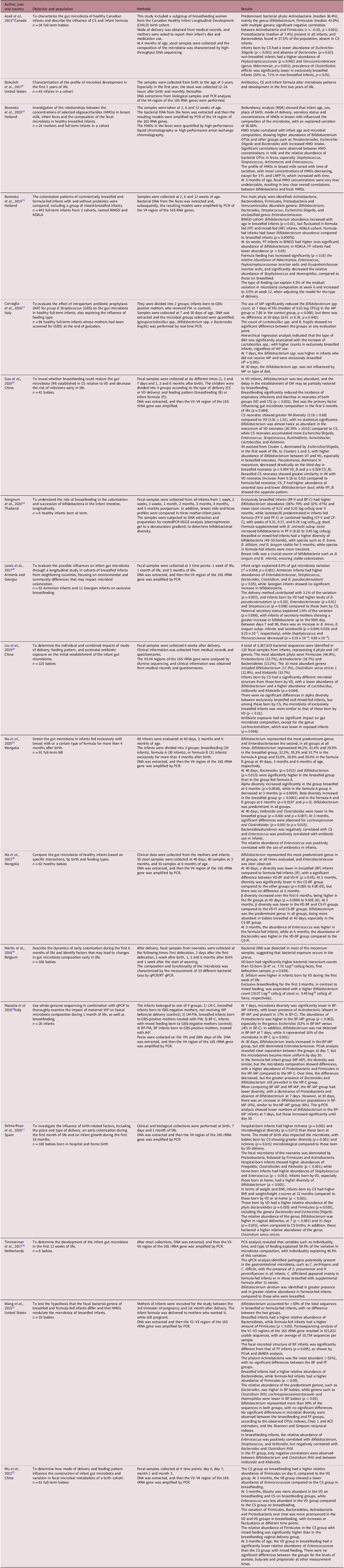
Table 1. Long description
A table summarizing studies included in a literature review, focusing on the number of publications contributed by various countries. The table has four rows and three columns, with headers for Did, N 1, and Bucket. Row 1: Did, 9; N 1, 28.221196; Bucket, bucket underscore 2. Row 2: Did, 10; N 1, 28.221196; Bucket, bucket underscore 2. Row 3: Did, 11; N 1, 28.221196; Bucket, bucket underscore 2. Row 4: Did, 12; N 1, 28.221196; Bucket, bucket underscore 2. The table highlights the distribution of studies across different countries, with China, Japan, Italy, Mongolia, the United States, and the Netherlands contributing multiple studies, while Spain, Armenia and Georgia, Belgium, Canada, and Thailand each contributed one study.
Regarding sample size, the study with the fewest participants was by Kongnum et al., Reference Kongnum, Taweerodjanakarn and Hongpattarakere38 which included only six (n = 6) full-term infants, limiting the generalizability of the results. In contrast, the study with the largest number of participants was conducted by Borewicz et al., Reference Borewicz, Suarez-Diez and Hechler33 which analyzed two different cohorts in the Netherlands – the Bingo and Koala studies – and thus had a more representative sample and greater statistical power, with 443 full-term infants.
Among the most important exposures studied, the mode of delivery stands out for its role in shaping the infant’s gut microbiota and its impact on infant health. Reference Bokulich, Chung and Battaglia4,Reference Selma-Royo, Calatayud Arroyo and García-Mantrana11,Reference Guo, Zhou and Li23–Reference Akagawa, Tsuji and Onuma26,Reference Ma, Li and Zhang30,Reference Borewicz, Fangjie Gu and Saccenti32,Reference Lewis, Sidamonidze and Tsaturyan34,Reference Martin, Makino and Yavuz35,Reference Azad, Konya and Maughan37,Reference Kongnum, Taweerodjanakarn and Hongpattarakere38 The relationship between the type of infant feeding – exclusive BF, mixed feeding, or formula feeding – has also been extensively studied. Reference Bokulich, Chung and Battaglia4,Reference Guo, Zhou and Li23–Reference Mazzola, Murphy and Ross28,Reference Ma, Li and Zhang30–Reference Kongnum, Taweerodjanakarn and Hongpattarakere38
Influential highlighted microorganisms
In addition, the selected studies identified several microorganisms relevant to the intestinal microbiota of infants. We emphasize the presence of Bifidobacterium spp., Reference Guo, Zhou and Li23,Reference Wu, Ren and Li25,Reference Ma, Li and Zhang29,Reference Borewicz, Fangjie Gu and Saccenti32,Reference Lewis, Sidamonidze and Tsaturyan34,Reference Timmerman, Rutten and Boekhorst36,Reference Azad, Konya and Maughan37,Reference Zhang, Li and Xie39 Escherichia spp., Reference Selma-Royo, Calatayud Arroyo and García-Mantrana11,Reference Guo, Zhou and Li23,Reference Mazzola, Murphy and Ross28,Reference Ma, Li and Zhang29,Reference Borewicz, Fangjie Gu and Saccenti32,Reference Borewicz, Suarez-Diez and Hechler33,Reference Azad, Konya and Maughan37 Clostridium spp., Reference Selma-Royo, Calatayud Arroyo and García-Mantrana11,Reference Liu, Qin and Song24,Reference Wu, Ren and Li25,Reference Ma, Li and Zhang29,Reference Wang, Li and Wu31,Reference Lewis, Sidamonidze and Tsaturyan34,Reference Timmerman, Rutten and Boekhorst36,Reference Azad, Konya and Maughan37 Lactobacillus spp., Reference Guo, Zhou and Li23,Reference Liu, Qin and Song24,Reference Akagawa, Tsuji and Onuma26,Reference Corvaglia, Tonti and Martini27,Reference Lewis, Sidamonidze and Tsaturyan34,Reference Kongnum, Taweerodjanakarn and Hongpattarakere38 and Bacteroides spp., Reference Selma-Royo, Calatayud Arroyo and García-Mantrana11,Reference Corvaglia, Tonti and Martini27–Reference Ma, Li and Zhang29,Reference Wang, Li and Wu31,Reference Lewis, Sidamonidze and Tsaturyan34 Additionally, representatives of Firmicutes, Reference Selma-Royo, Calatayud Arroyo and García-Mantrana11,Reference Liu, Qin and Song24,Reference Wu, Ren and Li25,Reference Mazzola, Murphy and Ross28,Reference Wang, Li and Wu31,Reference Borewicz, Suarez-Diez and Hechler33,Reference Azad, Konya and Maughan37 Staphylococcus spp., Reference Selma-Royo, Calatayud Arroyo and García-Mantrana11,Reference Borewicz, Fangjie Gu and Saccenti32–Reference Lewis, Sidamonidze and Tsaturyan34 Enterococcus, Reference Selma-Royo, Calatayud Arroyo and García-Mantrana11,Reference Guo, Zhou and Li23,Reference Wu, Ren and Li25,Reference Ma, Li and Zhang30–Reference Borewicz, Suarez-Diez and Hechler33 and Streptococcus spp. Reference Guo, Zhou and Li23,Reference Wang, Li and Wu31–Reference Lewis, Sidamonidze and Tsaturyan34,Reference Timmerman, Rutten and Boekhorst36 are repeatedly reported, reflecting the diverse microbial composition of the infant gut.
Chronology and development of the gut microbiota
Analysis of the chronological and critical development of the infant gut microbiota reveals several milestones within the first 180 days of life that provide insight into microbial succession, as distinct changes occur in the first months and different patterns emerge at each developmental stage (Figure 2). The key findings highlight the predominant bacteria during this period.
Timeline of bacterial colonization during the first six months of life. (Source: the authors).
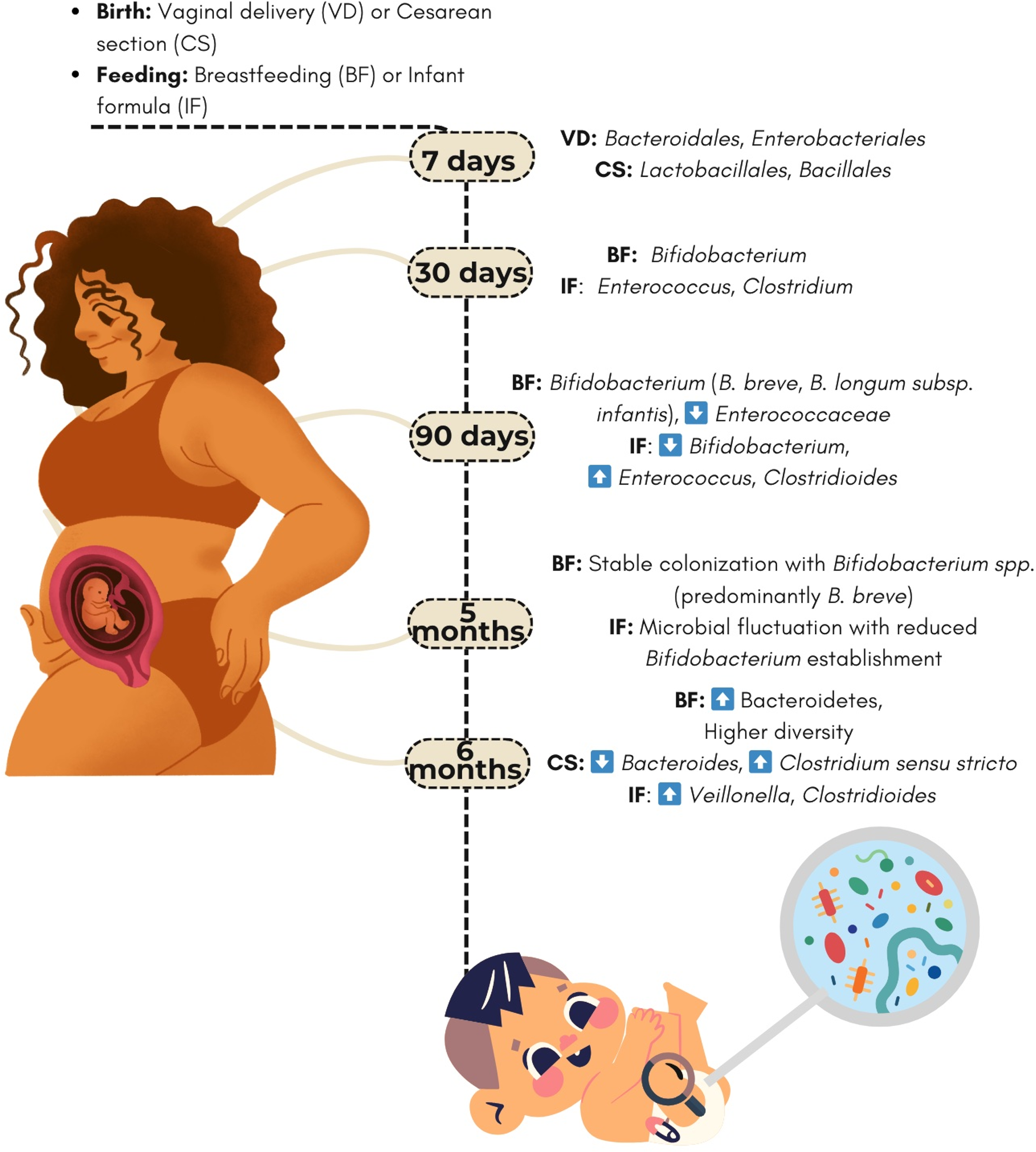
Figure 2. Long description
The diagram illustrates the timeline of bacterial colonization in infants over the first six months of life, highlighting the impact of birth and feeding methods on gut microbiota. It shows a pregnant woman on the left and a baby with a magnifying glass on the right, with a timeline in the center. The timeline is marked at 7 days, 30 days, 90 days, 5 months, and 6 months. At 7 days, vaginal delivery (VD) is associated with Bacteroidales and Enterobacteriales, while cesarean section (CS) is associated with Lactobacillales and Bacillales. At 30 days, breastfeeding (BF) is associated with Bifidobacterium, and infant formula (IF) is associated with Enterococcus and Clostridium. At 90 days, BF is associated with Bifidobacterium species such as B. breve and B. longum subsp. infantis, and an increase in Enterococcaceae, while IF is associated with Bifidobacterium, Enterococcus, and Clostridioides. At 5 months, BF leads to stable colonization with Bifidobacterium species, predominantly B. breve, while IF results in microbial fluctuation with reduced Bifidobacterium establishment. At 6 months, BF is associated with an increase in Bacteroidetes and higher diversity, while CS is associated with an increase in Bacteroides and Clostridium sensu stricto, and IF is associated with Veillonella and Clostridioides.
In the first few days – especially during the first week – vaginally delivered (VD) infants have higher levels of Bacteroidales (16% compared to 2.4% in CS) and Enterobacteriales (35% compared to 4.4%), while Lactobacillales (61%) and Bacillales (23%) predominate in infants born by CS. Reference Akagawa, Tsuji and Onuma26
After 30 days, Bifidobacterium colonization predominates in breastfed infants, accounting for up to 70 % of the microbiota, while higher proportions of Enterococcus and Clostridium are found in formula-fed infants. Reference Ma, Li and Zhang29,Reference Kongnum, Taweerodjanakarn and Hongpattarakere38
After 90 days, the gut microbiota continues to show distinct patterns based on birth type and feeding method. In breastfed infants, Bifidobacterium predominates, with increased levels of B. breve and B. longum subsp. infantis (p < 0.05) and lower levels of Enterococcaceae (p < 0.01), especially in infants born by VD. Reference Guo, Zhou and Li23,Reference Wu, Ren and Li25,Reference Lewis, Sidamonidze and Tsaturyan34 In contrast, infants fed artificial formula have lower levels of Bifidobacterium (p < 0.05) and a higher incidence of Enterococcus and Clostridioides, Reference Ma, Li and Zhang30 with these differences being more pronounced in infants born by CS. Reference Liu, Qin and Song24
Kongnum et al. (2020) Reference Kongnum, Taweerodjanakarn and Hongpattarakere38 observed that Bifidobacterium remained stable (36–70) in breastfed infants at five months of age, with species such as B. breve showing persistent colonization, in contrast to the fluctuations seen in formula-fed infants. After six months (180 days), the divergence in bacterial diversity becomes more pronounced: breastfed infants have a greater abundance of Bacteroidetes (p < 0.0001), while CS is associated with a lower abundance of Bacteroides (p = 0.035) and a higher presence of Clostridium sensu stricto. Reference Selma-Royo, Calatayud Arroyo and García-Mantrana11
According to Ma et al. (2020), differentiation of the microbiota is established by six months of age, with breastfed infants showing higher alpha diversity (p = 0.0038) and greater abundance of Bacteroides (p = 0.012), while formula-fed infants show a stronger association with Veillonella (p = 0.002) and Clostridioides (p = 0.007). Selma-Royo et al. Reference Selma-Royo, Calatayud Arroyo and García-Mantrana11 add that CS at this stage is associated with lower abundance of Bacteroides (p = 0.035) and increased Clostridium sensu stricto, highlighting the lasting influence of the type of feeding. Both studies suggest that the microbiota begins its transition to an adult-like profile at this stage, although significant differences remain depending on feeding type and mode of delivery.
Figure 2 shows the timeline of bacterial colonization during the first six months of life. According to recent literature, in breastfed infants, Bifidobacterium (especially B. breve and B. longum subsp. infantis) predominates and microbial colonization remains stable over time, whereas in formula-fed infants, greater microbial turnover occurs, with a clear presence of Enterococcus and Clostridioides.
Mode of delivery and feeding practices influence the gut microbiota
The results confirm the importance of Bifidobacterium spp. as the most abundant genus in the feces of breastfed infants, with significantly higher levels compared to formula-fed infants. Reference Ma, Li and Zhang29,Reference Chichlowski, van Diepen and Prodan40 This profile is highly beneficial for infant health, as the presence of Bifidobacterium spp. promoted by BF is associated with positive modulation of the immune system, including reduced inflammatory markers in the gut and promotion of a balanced immune response. Reference Henrick, Rodriguez and Lakshmikanth41,Reference Laursen42 According to Henrick et al., Reference Henrick, Rodriguez and Lakshmikanth41 abundant intestinal bifidobacteria in infants support immune regulation through Tregs and anti-inflammatory cytokines, while low abundance is associated with pro-inflammatory cytokines and immune activation.
The data support the importance attributed in the literature to the genera Lactobacillus and Bifidobacterium as the most widespread beneficial bacteria that play a crucial role in regulating the infant gut microbiota, exerting antibacterial effects and inhibiting colonization by pathogenic microorganisms. Reference Li, Ren and Wang43 As shown in Figure 2, the mode of delivery also influences this colonization: VD favors Bacteroidales and Enterobacteriales, while cesarean section is associated with Lactobacillales and Bacillales in the first days of life.
Studies indicate that the presence of Lactobacillus in feces varies depending on the type of infant formula provided, as many formulas are fortified with probiotics to mimic the benefits of breast milk. Reference Wang, Liu and Wang44 Additionally, probiotic lactic acid strains isolated from infant feces – Lactobacillus gasseri FWJL-4, Lactobacillus plantarum Fjias-5, and Lactobacillus rhamnosus FSJ-13 – exhibit anti-inflammatory and antioxidant effects by activating the Nrf2/HO-1 signaling pathway in macrophages. Reference Li, Pan and Sun45 Further evidence shows that Lactobacillus strains isolated from fecal samples possess relevant probiotic properties, including resistance to adverse gastrointestinal conditions and antimicrobial activity against pathogens such as Staphylococcus aureus and Escherichia coli. Reference Yadav, Mallappa and Ambatipudi46,Reference Çağal, Kıray and Kariptaş47
The scientific literature indicates differences in the presence of Firmicutes, which are found in large numbers in both exclusively breastfed and formula-fed infants. Reference Durrani, Nazli and Fatima49 Notably, formula-fed infants have a higher number of these bacteria, which may be related to the production of short-chain fatty acids (SCFAs) during the first four months of life. These SCFAs are produced through specific fermentation pathways, including acetyl-CoA and succinate fermentation to butyrate, and are significantly more abundant in formula-fed infants. SCFAs play an important role in regulating food intake and are associated with changes in body weight, highlighting their potential clinical relevance in early-life feeding behavior. This production may influence infants’ feeding responses and possibly reduce their receptivity to food. Reference Yan, Zhao and Nayar50 Additionally, the presence of Firmicutes suggests an essential role in the initial colonization of the gut, with genera such as Streptococcus and Veillonella acting as probiotics and immunomodulators, contributing to the development of the infant immune and digestive systems. These effects may be partly explained by probiotic adaptations to the intestinal environment, including changes in membrane protein and fatty acid composition, the ability to inhibit pathogen adhesion to intestinal mucus, and tolerance to bile salts, which supports microbial persistence and host-microbe interactions. Reference Li, Pan and Sun45
Regarding the genus Clostridium, studies have shown that infant formula enriched with HMOs reduces the incidence of C. difficile and makes the gut microbiome composition more similar to that of exclusively breastfed infants. Reference Bosheva, Tokodi and Krasnow51 The study by Plaza-Díaz et al. Reference Plaza-Diaz, Ruiz-Ojeda and Morales52 also suggests that Clostridium sensu stricto develops more strongly in infants fed the innovative formula studied. Holst et al. (2023) Reference Holst, Myers and Rodríguez-García53 examined HMO-supplemented infant formula and found that the relative incidence of opportunistic pathogens, including C. difficile and E. coli, was lower in the groups receiving the HMO-supplemented formula than in the control group.
The genus Akkermansia, detected in several of the studies examined, can degrade HMOs and promote syntrophy with beneficial bacteria, contributing to a healthy gut microbial network in formula-fed infants. Reference Martínez-Martínez, Martínez-Martínez and Soria-Guerra48
Regarding mode of delivery, our results show that infants born by CS have lower microbial diversity and a higher incidence of opportunistic pathogenic species. Figure 1 illustrates this difference and highlights the lower initial diversity in CS compared to VD. The absence of passage through the maternal birth canal is a key factor in these differences. Reference Coelho, Ayres and Barreto9–Reference Rutayisire, Huang, Liu and Tao12,Reference Inchingolo, Inchingolo and Palumbo54 Infants born by CS have a different gut microbiome composition, with a lower frequency of Bacteroides and Bifidobacterium and a higher presence of Klebsiella, Veillonella, and Enterococcus compared to infants born by VD. Reference Guo, Zhou and Li23,Reference Liu, Qin and Song24,Reference Liu, Ma and Zhu55
Colonization with Klebsiella, Veillonella, and Enterococcus is particularly common in infants delivered by CS or those who are formula fed. CS disrupts the natural microbial colonization of the newborn, promotes the growth of opportunistic bacteria, and potentially increases the risk of adverse disease. Reference Liu, Ma and Zhu55–Reference Shao, Forster and Tsaliki57
Given the potential negative effects of CS, formula feeding is associated with lower microbial diversity than BF in newborns delivered by CS. Reference Ma, Li and Zhang30,Reference Inchingolo, Inchingolo and Palumbo54,Reference Martín-Peláez, Cano-Ibáñez and Pinto-Gallardo58 In contrast to breast milk, infant formula may not provide the same benefits for gut colonization. Although it contains ingredients with purported benefits, these are artificial and cannot mimic the effects of their natural counterparts. Reference Inchingolo, Inchingolo and Palumbo54
Therefore, BF, especially when exclusive, plays a fundamental role in restoring the gut microbiota by promoting the growth of beneficial bacteria such as Bifidobacterium and Bacteroides. Reference Liu, Qin and Song24,Reference Henrick, Rodriguez and Lakshmikanth41,Reference Laursen42,Reference Liu, Ma and Zhu55,Reference Coker, Laue and Hoen59 This microbiota is important for protecting infants from respiratory infections and allergies. Reference Inchingolo, Inchingolo and Palumbo54,Reference Liu, Ma and Zhu55
Conclusion
The gut microbiota is a dynamic and unique ecosystem influenced by many factors, including diet, genetics, and environment. This ecosystem plays a central role in health and development during childhood and affects the risk of disease. Variability in microbial composition leads to different results across studies, highlighting the complexity of the topic and emphasizing the importance of scientific literature in expanding knowledge about this diversity.
Choosing infant formulas enriched with prebiotics or HMOs may support the development of a healthier microbiome, more similar to that of breastfed infants, and reduce the risk of colonization by potentially harmful bacteria. Despite advances in the formulation of breast milk substitutes, it is important to emphasize exclusive BF up to six months of age and continued BF up to two years or beyond, as recommended by health authorities.
In addition, promoting VD is fundamental to reducing risks for mothers and newborns, accelerating maternal recovery, and optimizing health resources. As a public health strategy, authorities should invest in health education, training multiprofessional teams, and ensuring safe, humanized conditions to increase pregnant women’s confidence and encourage the choice of this delivery method whenever possible, thus helping strengthen the baby’s immune system through the development of the intestinal microbiota.
Acknowledgments
None.
Financial support
The authors are grateful for the support of the Coordenação de Aperfeiçoamento de Pessoal de Nível Superior - Brasil (CAPES) - Financial Code 001. The authorsalsothanktheBrazilian agencies for theirsupport: CNPq – Conselho Nacional de Desenvolvimento Científico e Tecnológico, and FAPERGS - Fundação de Amparo à Pesquisa do Estado do Rio Grande do Sul – Brasil.
Competing interests
None.