Introduction
Although eosinophilic myocarditis is one of the several acute myocarditis, its nature does not differ from the ordinary acute viral myocarditis. Eosinophilic myocarditis is a rare subtype of myocarditis characterised by diffuse myocardial inflammation with infiltrating eosinophils in adults.Reference Kuchynka, Palecek and Masek 1 – Reference Rizkallah, Desautels and Malik 3 Eosinophilic myocarditis has been identified in 0.1% of cases among a cohort of patients biopsied for suspected myocarditis.Reference Maisch and Pankuweit 4 In particular, it is a rare disease in children.Reference Aslan, Fischer, Laser and Haas 5 We experienced only two cases (6%) among 30 patients, excluding neonates, in the Department of Paediatric Cardiology of our institution between 1978 and 2016.Reference Abe, Tsuda and Miyazaki 6 Failure to diagnose eosinophilic myocarditis and delay its therapy may lead to irreversible myocardial injury; however, an early diagnosis and treatment with steroid agents can lead to a favourable outcome.Reference Kuchynka, Palecek and Masek 1 , Reference Ali, Straatman and Allard 2 The spectrum of the clinical presentation in patients with eosinophilic myocarditis is diverse, and manifests with features of acute pericarditis, an acute myocardial infarction, cardiogenic shock, and cardiomyopathy.Reference Thambidorai, Korlakunta and Arouni 7 Therefore, it is difficult to diagnose eosinophilic myocarditis rapidly. We describe here the important characteristics of eosinophilic myocarditis in the view of our experience.
Patient 1
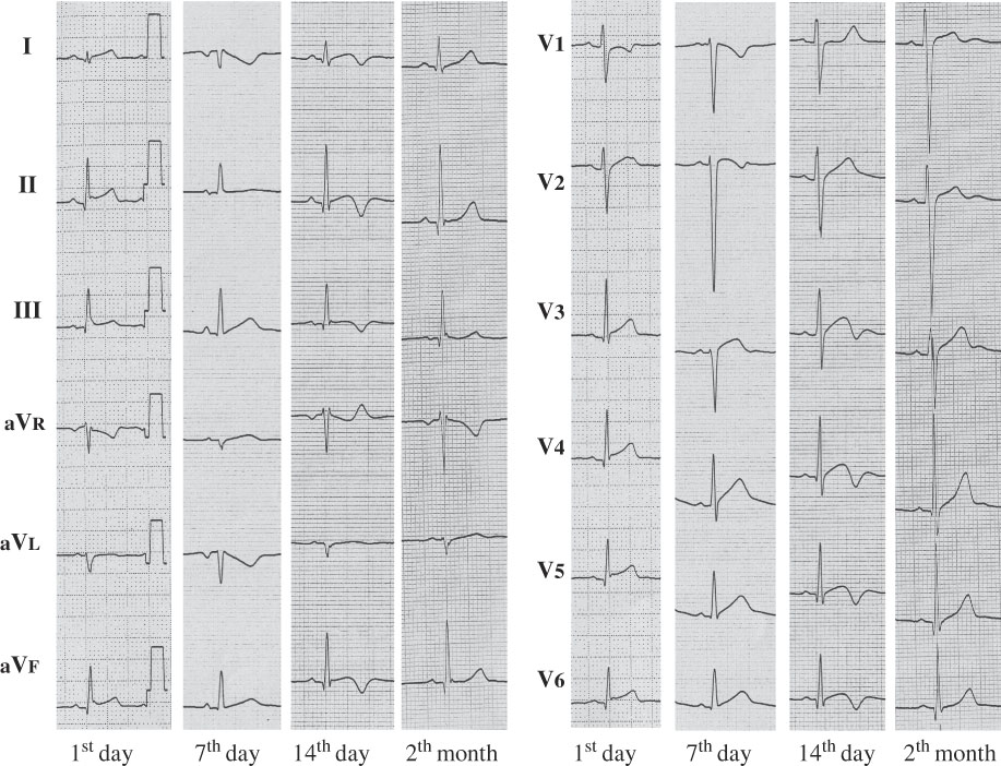
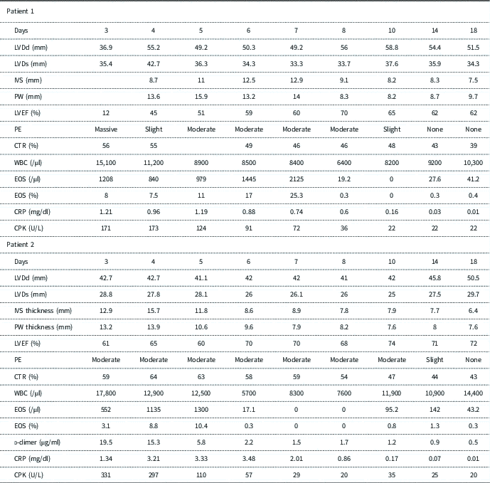
A 14-year-old boy, with a height of 168 cm and weight of 64 kg, was admitted to a nearby hospital with vomiting, dyspnoea, and chest pain. The values of his white blood cell count, C-reactive protein, and creatine phosphokinase levels were 15,600/ml, 0.825 mg/dl, and 223 U/L (30–200), respectively. His serum troponin was positive. His 12-lead electrocardiogram revealed ST-T elevation in leads II, III, aVf, and V3–6 (Fig 1). His cardiothoracic ratio was 56%. A two-dimensional echocardiogram indicated a massive pericardial effusion and decreased left ventricular ejection fraction. He was diagnosed with acute myocarditis and pericarditis. A phosphodiesterase III inhibitor was administered intravenously. He was transferred to our hospital on the third day of his illness. Dopamine was added intravenously. His heart rate and blood pressure were 120/minute and 100/70 mmHg, respectively, and his heart sounds were distant. The values of his white blood cell count, C-reactive protein, and creatine phosphokinase levels were 15,100/ml, 1.21 mg/dl, and 171 U/L (30–200), respectively (Table 1). His fibrinogen, fibrin degradation product, and d-dimer levels were 326 μg/dl, 8 μg/ml, and 4.6 μg/ml, respectively. His troponin-T and brain natriuretic peptide were 0.71 ng/ml and 445 pg/ml, respectively.
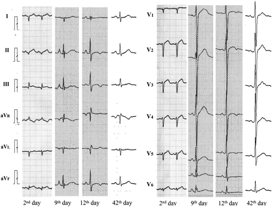
Serial changes in the 12-lead electrocardiogram during each stage in patient 1.

Changes in each parameter in acute eosinophilic myocarditis.

CPK=creatine phosphokinase; CRP=C-reactive protein; CTR=cardiothoracic ratio; EOS=eosinophil; IVS=interventricular septum; LVDd=left ventricular diastolic dimension; LVDs=left ventricular systolic dimension; PE=pericardial effusion; PW=posterior wall; WBC=white blood cell
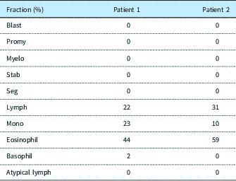
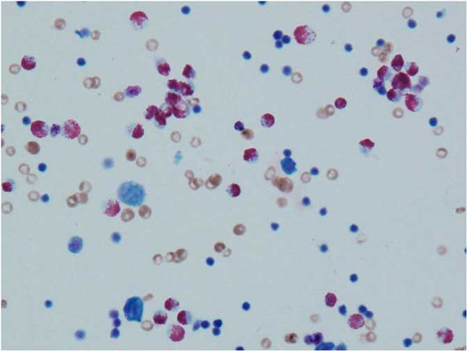
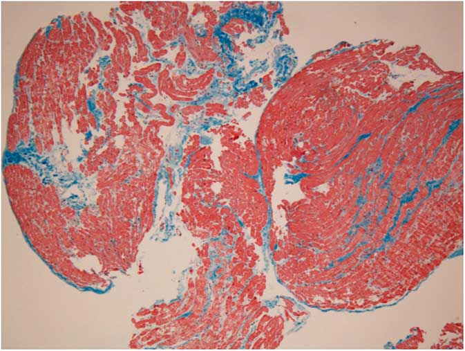
The pericardial fluid was drained, and 44% eosinophils were found in the cellular fraction of the pericardial fluid (Table 2, Fig 2). The eosinophils increased to more than 10% in the peripheral blood (Table 1). On the seventh day, he underwent a cardiac catheterisation and endomyocardial biopsy. The pathological findings showed a diffuse interstitial inflammatory process with eosinophilic degranulation. He was diagnosed with acute eosinophilic myocarditis and underwent steroid pulse therapy. The left ventricle wall thickness was remarkably decreased and his cardiac function improved after the pulse therapy with intravenous methylprednisolone for an hour at a dose of 1 g/day for 3 days (Table 1). In the second week, the inotropic support was discontinued. In the electrocardiogram, abnormal Q waves persisted until the second week, and he developed negative T waves during the third week (Fig 1). In the pathological study after 3 months of acute eosinophilic myocarditis, slight interstitial fibrosis was found (Fig 3). The steroid pulse therapy was followed by 1 mg/kg/day of oral prednisone, with a gradual tapering for 8 months. He continued to do well and had a normal echocardiographic assessment.
Cellar fraction analysis of the pericardial fluid.

Histograph of the pericardial fluid (Giemsa stain). Many eosinophils were found in the pericardial fluid.

Histograph of the endomyocardial biopsy at 3 months after the acute eosinophilic myocarditis (Maasom-trichrome stain). Slight interstitial fibrosis was detected.

Patient 2
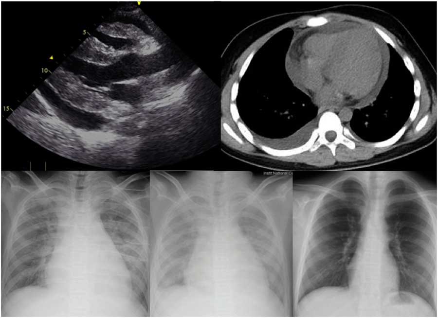
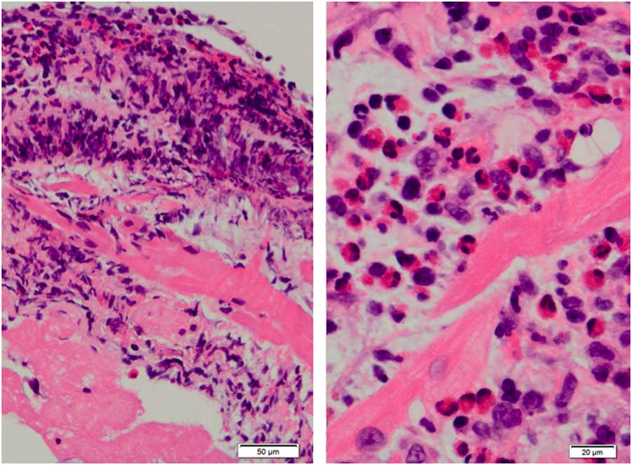
A second patient, a 13-year-old boy, with a height of 160 cm and weight of 60 kg, developed vomiting and general malaise. Cardiomegaly was detected on his chest X-ray at a nearby hospital. He had mild mental retardation and an allergic constitution. He was admitted with a fever, abdominal distension, and oedema. A 12-lead electrocardiogram revealed a low voltage and ST-T elevation in leads II, III, and aVf, and a poor r-wave progression in the chest leads (Fig 4). On the second day, a moderate pericardial effusion was found by two-dimensional echocardiography and chest computed tomography (Fig 5, upper left and upper right). After draining the pericardial effusion, wall thickening and hypokinesis of the inferoposterior wall of the left ventricle was detected. Acute myocarditis was suspected, and he was transferred to our hospital on the third day of illness. His temperature was 37.9°C, heart rate 130/minute, and blood pressure 85/50 mmHg. His heart sounds were distant on auscultation. The cardiothoracic ratio was 58% on the chest X-ray, and the pulmonary vascular area was hypolucent (Fig 5, lower left). The brain natriuretic peptide level was elevated to 1569 pg/ml. The values of the white blood cell count, C-reactive protein, creatine phosphokinase, and troponin-T levels were 17,800/μl, 1.34 mg/dl, 331 U/L, and 1.55 ng/dl, respectively (Table 2). The eosinophils were 3.1% of the white blood cell count and the count was 552/μl. The fibrinogen, fibrin degradation product, and d-dimer levels were 393 mg/dl, 22 μg/ml, and 10.0 μg/ml, respectively. Intravenous immunoglobulin, 1 g/kg/day, was slowly administered, and dopamine, dobutamine, and furosemide were given intravenously. In spite of those treatments, pulmonary congestion and the fever continued (Fig 5, lower middle). Because the SpO2 was 89% on room air, oxygen inhalation was needed. The inflammation marker did not improve. On the fifth day of illness, the eosinophils increased to 10.4% of the white blood cell count. A repeat two-dimensional echocardiogram revealed a moderate pericardial effusion (Table 2). A cardiac catheterisation and endomyocardial biopsy were performed because eosinophilic myocarditis was suspected. Steroid pulse therapy was started after the cardiac catheterisation. Coumadin and an angiotensin-converting enzyme inhibitor were added. The next day, the pathological findings showed a large quantity of eosinophils and a degree of eosinophil degranulation, confirming the diagnosis of eosinophilic myocarditis (Fig 6). His cardiac function improved by the sixth day, and the pericardial effusion regressed by the 18th day (Table 2). Eventually, he recovered after the steroid pulse therapy, followed by 1 mg/kg/day of oral prednisone, with a gradual tapering. During the second week, the inotropic support was discontinued. We investigated the pericardial fluid that was taken in the previous hospital, and found 59% eosinophils (Table 2). In his serial electrocardiogram, abnormal Q waves were observed during the second week, and negative T waves developed during the third week (Fig 4). There were no abnormal findings on his electrocardiogram and X-ray after 1 month (Figs 4 and 5, lower right). Slight interstitial fibrosis was found in the pathological study 1 month after the acute eosinophilic myocarditis, and the right ventricular ejection fraction and left ventricular ejection fraction were 53 and 67%, respectively. In the magnetic resonance findings, no late gadolinium enhancement was detected. The antithrombotic therapy was discontinued at 1 month, and the steroids were discontinued at 2 months after the onset of the acute eosinophilic myocarditis. The angiotensin-converting enzyme inhibitor was stopped 1 year after the acute eosinophilic myocarditis.
Serial changes in the 12-lead electrocardiogram during each stage in patient 2.

The findings in the two-dimensional echocardiography, CT, and chest X-ray in patient 2. (Left upper) Two-dimensional echocardiogram. Wall thickening and a moderate pericardial effusion are prominent. (Right upper) Chest CT. A moderate pericardial effusion is detected. (Lower) Serial chest X-ray; (Left) Third day of the illness; (Middle) Fifth day of the illness. Cardiomegaly and pulmonary congestion were detected. The procedures were performed in supine position. (Right) The cardiomegaly improved by 1 month after the disease. The procedure was performed in the standing position.

Histograph of the endomyocardial biopsy on the fifth day of illness (haematoxylin–eosin stain). Many eosinophils with eosinophil degranulation had infiltrated into the myocardial interstitium.

Discussion
Both our patients had eosinophilic myocarditis with more massive pericardial effusions compared with acute viral myocarditis. It is considered that the volume of the pericardial effusion in eosinophilic myocarditis is greater than that in acute myocarditis because of the complication of acute eosinophilic pericarditis.Reference Li, Gupta and Schroth 8 – Reference Mori, Morimoto and Hiramatsu 10 A moderate pericardial effusion with no evidence of cardiac tamponade, along with the progressive thickening of the left ventricle wall, suggests an infiltrative and inflammatic disorder. Further, the cellular fraction of the pericardial effusion can lead to the diagnosis of eosinophilic myocarditis. Although the diagnosis is proven by an endomyocardial biopsy with the histological detection of eosinophils, the procedure is invasive and sometimes risky. The examination of the cellular fraction of the pericardial fluid is easy if massive pericardial fluid exists. These findings can be a useful clue for the diagnosis of eosinophilic myocarditis. Unfortunately, there was no identifiable cause for the eosinophilic myocarditis in our patients.
It is also important to follow the eosinophil count in the peripheral blood samples, because the peripheral eosinophil count is not always elevated upon admission in some patients. Although the total eosinophil count on admission was 552/μl in our second patient, it increased after that. The upper normal limit of eosinophils in the peripheral blood is 3–5% with a corresponding absolute eosinophil count of 350–500/μl.Reference Mori, Morimoto and Hiramatsu 10 In the Japanese guidelines for eosinophilic myocarditis, a total eosinophil count of more than 500/μl is one of the criteria of eosinophilic myocarditis. 11 Despite inotropic support and diuretics, the pulmonary lucency remained decreased and the dyspnoea did not improve, suggesting eosinophilic pneumonia might be present. Generally, continuation of severe inflammation, including a fever, is not typical at the onset of acute viral myocarditis; however, a fever for a few days before the onset is common.Reference Abe, Tsuda and Miyazaki 6 Further, the white blood cell count in eosinophilic myocarditis is higher compared to that in acute myocarditis because of worsening inflammation. The clinical signs and changes in the peripheral blood, which differ from acute viral myocarditis, are also very important for the diagnosis of eosinophilic myocarditis.Reference Kuchynka, Palecek and Masek 1 Further, the creatine phosphokinase level is not necessarily increased to a high value during the early stage of eosinophilic myocarditis compared with that of ordinary acute myocarditis [1, −3].
Although it was well known that the changes in the electrocardiogram resemble an acute myocardial infarction, the electrocardiographic characteristics with each progressive stage of eosinophilic myocarditis are unknown.Reference Kuchynka, Palecek and Masek 1 , Reference Thambidorai, Korlakunta and Arouni 7 In the electrocardiogram in our experience, a low voltage and ST-T elevation were found during the early stage of the acute eosinophilic myocarditis. The massive pericardial effusion because of pericarditis might have been responsible for those findings. Next to develop were abnormal Q waves caused by myocardial ischaemia because of myocarditis. We conjectured that those findings indicated myocardial involvement from eosinophilic granules, which are capable of inducing tissue damage and dysfunction. In the last phase, negative T waves and ST-T abnormalities were detected during the third week of the recovery stage. Those changes resembled the changes after an acute myocardial infarction. Therefore, some patients are often misdiagnosed with an acute myocardial infarction during the first presentation because of the ST-T segment abnormalities in the electrocardiogram.Reference Thambidorai, Korlakunta and Arouni 7
There remains little consensus in the use, dose, or duration of corticosteroids in the setting of eosinophilic myocarditis.Reference Kuchynka, Palecek and Masek 1 We administered a steroid pulse therapy for 3 days, and the steroids were used for several months after the pulse therapy in our patients. In addition to inotropic support for acute heart failure, corticosteroids should be administered as soon as possible, before the necrotising phase, to minimise the myocardial involvement. Actually, the creatine phosphokinase level did not increase to a high value in either of our patients. It is important that the creatine phosphokinase level is not always increased during the early stage of eosinophilic myocarditis before myocardial necrosis occurs because of progression of the eosinophils. Further, anticoagulant therapy, such as with coumadin, is indicated during the thrombotic phase. Endomyocardial and valvular involvement occur, with the possibility of thrombus formation in the apical parts of the ventricles. Finally, angiotensin-converting enzyme inhibitors are useful for preventing post-inflammatory fibrosis. On the contrary, some cases have relapses of eosinophilic myocarditis and chronic eosinophilic myocarditis in adults, and restrictive cardiomyopathy caused by eosinophilic myocarditis are also reported. Endomyocardial fibrosis progresses and restrictive cardiomyopathy might develop. A careful follow-up of these patients after eosinophilic myocarditis is necessary.
Conclusion
Acute eosinophilic myocarditis is often complicated by a massive pericardial effusion secondary to inflammation. The analysis of the cellular fraction of the pericardial effusion is easy and useful for diagnosing eosinophilic myocarditis. With the progression of the clinical course of eosinophilic myocarditis, the serial changes, such as a low voltage with ST elevation, abnormal Q waves, and negative T waves, are detectable on the electrocardiogram.
Acknowledgements
We thank Dr. Yusuke Tanaka in Yodogawa Christian Hospital for the offer of pericardial effusion. We thank Professor Peter Olley and Dr Setsuko Olley for their kind English language consultation.
Financial Support
This study had no financial support.
Conflicts of Interest
The authors state that they have no conflict of interest.
Ethical Standards
All procedures performed in this study involving human participants were in accordance with the ethical standards of the institutional committee with the 1964 Helsinki declaration and its later amendments or comparable ethical standards.