Implications
Neonatal porcine tail docking has long-term neuroanatomical as well as morphological consequences. The observed differences in tail length and diameter at the time of slaughter may be useful as welfare surveillance tools in order to identify the docking practice of farms. The results confirm earlier findings, and show that modern docking techniques such as hot-iron cautery lead to the development of neuroanatomical changes such as the formation of neuromas, suggesting that removal of parts of the tail may have consequences in terms of long-term pain.
Introduction
In many countries, tail docking is a common practice in the production of pigs (European Food Safety Authority, 2007), which is performed in order to prevent the abnormal behaviour caudophagia (tail biting) (Hunter et al., Reference Hunter, Jones, Guise, Penny and Hoste2001). Typically, part of the tail is docked by cutting or using hot-iron cautery within the first few days of the pig’s life (reviewed by Sutherland and Tucker, Reference Sutherland and Tucker2011). The causal relationship between tail docking and the lower risk of tail biting is not known (Taylor et al., Reference Taylor, Main, Mendl and Edwards2010 and Reference Taylor, Parker, Mendl, Edwards and Main2012). Simonsen et al. (Reference Simonsen, Klinken and Bindseil1991) proposed increased sensitivity in the docked tips, decreased accessibility due to the short length, as well as lower stimulation value due to the short length, and the lack of hairs on the tail tip as possible explanations. Recently, a Danish investigation showed that the risk of tail biting decreases by increasing docking length (Thodberg et al., Reference Thodberg, Jensen and Jørgensen2010).
In other animal species, the formation of neuromas as a consequence of tail docking (lambs; French and Morgan, Reference French and Morgan1992, and dogs; Gross and Carr, Reference Gross and Carr1990) or removal of other anatomical structures (trimming of beaks in poultry; Breward and Gentle, Reference Breward and Gentle1985) has been shown. In addition, neuromas have been found after healing of shoulder ulcerations in sows (Dahl-Pedersen et al., Reference Dahl-Pedersen, Bonde, Herskin, Jensen, Kaiser and Jensen2013) and after experimental nerve transection in animal-model studies (Dorsi et al., Reference Dorsi, Chen, Murinson, Pogatzki-Zahn, Meyer and Belzberg2008) in rats. After transection of an axon, growth of fine nerve processes (sprouts) from the proximal nerve end will be initiated and – if normal nerve healing is prevented by amputation or a similar process – form a neuroma, a disorderly bundle embedded in fibrous tissue (Devor and Seltzer, Reference Devor and Seltzer1999; Burnett and Zager, Reference Burnett and Zager2004). The presence of neuromas has been linked to non-evoked pain (Devor et al., Reference Devor, Wall and Catalan1992), as well as decreased nociceptive thresholds (Wall and Gutnick, Reference Wall and Gutnick1974). In tail-docked pigs, Simonsen et al. (Reference Simonsen, Klinken and Bindseil1991) found neuromas in the healed tail tips at slaughter age, using only one docking length and docking by emasculator.
The aim of the present study was to examine the effect of hot-iron cautery tail docking on the extent of neuroma formation in the tips of pig tails, and examine whether neuroma formation was affected by the degree of tail docking.
Material and methods
Animals and housing
A total of 65 pig tails were examined. The pigs were cross-bred LYD (Landrace×Yorkshire×Duroc) and delivered and raised in the resident barn at Research Centre Foulum, University of Aarhus, Denmark. The study involved piglets from eight litters born in December 2008. The sows and litters were kept in loose-housing farrowing pens during the lactation period. All litters were weaned at 28 days of age and kept in conventional weaner pens until obtaining a BW of approximately 30 kg, after which they were mixed with pigs from another litter and kept in conventional finisher pens until slaughter at 22 weeks of age. The pigs were slaughtered at a commercial Danish slaughterhouse and the tails were removed.
Experimental design
This study involved four experimental treatments. Each treatment involved two litters, each consisting of up to 12 piglets. The treatments were as follows: intact tails (I: n=18); leaving 75% (n=17); leaving 50% (n=19); or leaving 25% (n=11) of a mean tail length on the body of the pigs, corresponding to 0, 2.9, 5.7 and 7.5 cm, respectively. These lengths were based on measures of intact pig tails on farm as part of another study (Thodberg et al., Reference Thodberg, Jensen and Jørgensen2010). The numbers in brackets indicate the number of pigs per treatment from whom tails were collected successfully at the time of slaughter, and where no lesions other than the docking had been observed during the growing period. At time of slaughter, there were 50, 59, 53 and 64% female pigs within the four treatments, respectively.
Experimental procedures
Tail docking
The piglets were tail docked between day 2 and 4 postpartum using a gas-heated apparatus (Taildocker Super Pro; Kruuse Vet., Langeskov, Denmark). As part of another study, the piglets within the docked litters were split into the following anaesthetic and analgesic treatments: three randomly chosen piglets were docked without anaesthesia or analgesia; three randomly chosen piglets were docked 15 min after being injected subcutaneously with a total of 0.3 ml of 20 mg/ml Lidocain (Lidocain; Amgros I/S, Copenhagen, Denmark) at the base of the tail (approximately 0.5 cm from the tail root); three randomly chosen piglets were docked 45 min after receiving an intramuscular injection in the neck with 0.4 mg/kg BW of the 20 mg/ml nonsteroidal anti-inflammatory drug meloxicam (Metacam®; Boehringer Ingelheim DK A/S, Copenhagen, Denmark); and three randomly chosen piglets were docked after injections with both lidocaine and meloxicam.
All experimental procedures were approved by the Danish Animal Experiments Inspectorate (permit no 2005/561-1013).
Collection and preparation of tail tissue
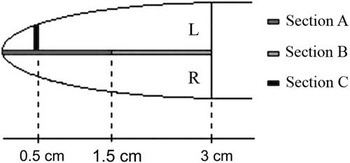
Following slaughter, tails were cut off 1 cm from the root and the length and diameter, determined 0.5 cm from the tip, were measured by a ruler. The tails were then fixed in 10% neutral buffered formalin for 5 days and then transferred to a solution of 3.3% formaldehyde and 17% formic acid for decalcification. After 11 days, when a satisfactory texture for cutting was obtained, three sections of each tail were made: a cross-section 0.5 cm from the tail tip of the left part (Section C) and two 1.5 cm longitudinal sections from the centre (Sections A and B), see Figure 1. The tissue samples were processed through graded alcohols, and xylene, and embedded in paraffin wax. Haematoxylin and eosin staining was performed on 4- to 5-μm tissue sections together with immunohistochemical visualization of nerves by S-100 staining (Dahl-Pedersen et al., Reference Dahl-Pedersen, Bonde, Herskin, Jensen, Kaiser and Jensen2013), which is staining the ensheathing Schwann cells.
Schematic drawing illustrating the sampling of tissue specimens for histology from the tails of pigs. The tail is viewed from the dorsal aspect with a right (R) and left (L) aspects. Sections A and B, both with a length of 1.5 cm, are from the midline, whereas Section C is a cross-section sampled 0.5 cm from the tail tip.

Sections – that is, three from each tail – were examined by HEJ using a Leitz-Orthoplan microscope at magnifications 4, 10 and 25×. The dimensions of structures in the sections were measured by a micrometer ocular.
Variables
The following variables were calculated: (1) mean tail length in centimeter; (2) mean tail diameter 0.5 cm from the tip in centimeter; (3) number of neuromas (aggregates of hypertrophic axons ensheathed by Schwann cells surrounded by a fibrous perineurium) per tail; (4) proportion of tails with neuromas; (5) mean diameter of neuromas in micrometer.
Statistical analysis
Statistical comparisons were carried out using SigmaPlot 12.5 (Systat Software Inc., San Jose, CA, USA). Comparison of intact v. docked tails was carried out using t-test, and in cases of non-normality or heterogeneity of variances the Mann–Whitney rank sum test was used. Comparison of the three docking lengths was carried out using the Kruskal–Wallis one-way ANOVA. For all comparisons of proportions, the Fisher exact test was used. Results are given as means±s.e. and P-values. A probability level of P<0.05 was considered statistically significant, whereas 0.05<P<0.10 was considered a tendency.
Results
Due to sporadic mortality, especially in the days after farrowing, and some cases of bitten tails throughout the life span of the pigs, the number of experimental pigs within each treatment varied.
Tail length and diameter
On the day of tail docking, the intact tails of the piglets were on average 9.2±0.9 cm (range 7.2 to 11.3 cm) and did not differ between the experimental treatments (P>0.10). After slaughter, both the length of the tails (mean tail length 30.6±0.6; 24.9±0.4; 19.8±0.6 and 8.7±0.6 cm, respectively, P<0.001) and the diameter (measured 0.5 cm from the tip of the tail: 0.5±0.03; 0.8±0.02; 1.0±0.03 and 1.4±0.04 cm, respectively, P<0.001) differed significantly between the treatments. For the tail lengths, all four treatments differed significantly from each other (P<0.05), whereas the diameters of the intact tails were significantly smaller compared with the other three treatments (P<0.05), and the diameters of the 75%-tails group were significantly smaller than the 25%-tails group (P<0.05).
Effects of tail docking and docking length
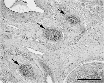
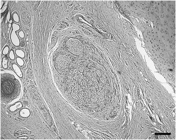
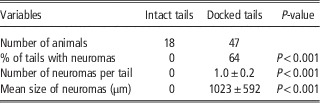
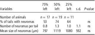
Results from the comparison between intact (n=18) and docked tails (n=47) are shown in Table 1. No neuromas were found in intact tails. No neuroanatomical differences were found between the three docking lengths, as shown in Table 2. Figures 2 and 3 show a cross-section of a normal, intact tail tip and a cross-section of a tail tip from a pig with 50% of the tail left on the body, respectively.
Cross-section of a normal, non-docked tail tip from a pig slaughtered at 22 weeks of age. The section represents Section C from the lateral part of the left side (see Figure 1). The normal nerves (→) were outlined immunohistochemically by staining the ensheathing Schwann cells by S-100. Scale bar=200 µm.

Cross-section of a tail tip from a pig slaughtered at 22 weeks of age with 50% of the tail docked. Centrally, an enormous granuloma – that is, aggregates of hypertrophic axons ensheathed by Schwann cells – is seen to be intermingled and surrounded by multiple layers of fibrous tissue. Haematoxylin and eosin. Scale Bar=200 µm.

Effects of tail docking (all docking lengths combined) at days 2 to 4 of life on the extent of neuroma formation in pigs after slaughter at 22 weeks of age

Data are presented as proportion of pigs or the mean value±s.e.
Effects of tail docking length at days 2 to 4 of life on the tail nerve pathology in pigs after slaughter at 22 weeks of age

Data are presented as proportion of pigs or the mean value, as well as the overall standard deviation and the P value
Discussion
The present study focussed on consequences of tail docking and effects of different docking lengths (removal of 25 to 75% of the tail length). Docking tails using hot-iron cautery led to the formation of neuromas, the examination of which took place when the pigs were slaughtered at 22 weeks of age. The different docking lengths were still evident in the tail length as well as diameter at slaughter. Even though the presence of neuromas have been associated with increased nociceptive sensitivity, abnormal spontaneous nervous activity and non-evoked pain in other species (Wall and Gutnick, 1974; Gross and Carr, 1990) and after other types of tissue damage (Breward and Gentle, 1985; Dahl-Pedersen et al., 2013), the existence of changes in porcine tail sensitivity after docking still need to be confirmed before the consequences of the present findings can be evaluated in terms of animal welfare.
In the present study, all piglets were docked using hot-iron cautery, by using a commercially available apparatus, which is used in modern pig production. Earlier investigations of the acute effect of tail docking in piglets have compared different docking methods. They found indications for hot-iron cautery docking being a less stressful procedure for the animals compared with docking by cutting off the tail (Sutherland et al., Reference Sutherland, Bryer, Krebs and McGlone2008 supported by results from the study by Prunier et al., Reference Prunier, Mounier and Hay2005, where hot-iron cautery was the only docking method applied). However, docking by hot-iron cautery led to slower healing (Sutherland et al., Reference Sutherland, Bryer, Krebs and McGlone2009). Until now, the only other report on neuroma formation after tail docking in pigs was based on the use of an emasculator (Simonsen et al., Reference Simonsen, Klinken and Bindseil1991). Our results confirm the findings by Simonsen et al. (Reference Simonsen, Klinken and Bindseil1991), which, however, were not quantitative as the present results, and show that modern docking techniques such as hot-iron cautery also lead to the development of neuroanatomical changes.
In the present study, we chose to leave 75, 50 or 25% of a mean tail length on the body of the pigs, due to Danish national guidelines specifying that, when docking is considered necessary, farmers are only allowed to remove up to 50% of the tails (Anonymous, 2003). Such limits in the proportion of tissue that can be removed are not specified in legislation concerning – for example, the European Union (EU Directive 91/630/EEC). Our results show that, although the piglets were docked early after birth (within 2 to 4 days postpartum), docking has long-lasting effects on the lengths and diameters of the tails. Recently, the possibilities to use data from slaughterhouses or similar animal-based measures as welfare surveillance tools have been suggested (European Food Safety Authority, 2012; Harley et al., Reference Harley, More, Boyle, O’Connell and Hanlon2012). Keeling et al. (Reference Keeling, Wallenbeck, Larsen and Holmgren2012) presented evidence that data on tail lengths obtained from Swedish slaughterhouses can be used to identify farms with tail-biting problems. Similarly, tail lengths measured at slaughterhouses may be used to classify farms according to their docking practice.
Most papers focussing on tail docking have reported the applied docking length, although the studies have been performed in countries without legal specifications on the allowed docking length. Sutherland et al. (Reference Sutherland, Davis and McGlone2011) left 2 cm of tail, whereas both Zhou et al. (Reference Zhou, Yang, Zhao, Huang, Wang, Wang, Yin, Shen, Wang and Schinckel2013) and Torrey et al. (Reference Torrey, Devillers, Lessard, Farmer and Widowski2009) left ~1/3 of the tail on the body of the piglets. Hunter et al. (Reference Hunter, Jones, Guise, Penny and Hoste2001) defined docking as any procedure, where less than 10 cm of the tail is left on the body, and defined short docking as leaving maximum 1.5 cm of the tail. Sutherland and Tucker (Reference Sutherland and Tucker2011) reviewed tail docking across farm animal species, and stated that a normal procedure within pig production would be to leave 2 cm of the tail on the piglet. However, in the majority of scientific reports, only one docking length has been used.
Within single studies of the short-term behavioural and physiological responses to tail docking in piglets, focus has mainly been on only one – and often different – docking length, which may be the source of variation underlying the lack of uniformity of the results regarding the level of acute stress or pain induced by docking (Noonan et al., Reference Noonan, Rand, Priest, Ainscow and Blackshaw1994; Prunier et al., Reference Prunier, Mounier and Hay2005; Torrey et al., Reference Torrey, Devillers, Lessard, Farmer and Widowski2009).
Neuroma formation has been shown after nerve transection in other animal species and after removal of tails (Gross and Carr, Reference Gross and Carr1990; French and Morgan, Reference French and Morgan1992), as well as other types of tissue (Breward and Gentle, Reference Breward and Gentle1985), including humans (Sehirlioglu et al., Reference Sehirlioglu, Ozturk, Yazicioglu, Tugcu, Yilmaz and Goktepe2009) and rodents as part of animal-model studies using experimental nerve transection (Dorsi et al., Reference Dorsi, Chen, Murinson, Pogatzki-Zahn, Meyer and Belzberg2008); however, previous reports have not focused on the effects of the proportion of tissue removed on neuroma formation.
On the basis of the known association between the presence of neuromas and the occurrence of non-evoked pain (Devor et al., Reference Devor, Wall and Catalan1992), as well as decreased nociceptive thresholds (Wall and Gutnick, Reference Wall and Gutnick1974), the present findings may suggest that removal of parts of the tail during docking in piglets leads to consequences in terms of long-term pain. However, not all neuromas are reported to have consequences in terms of pain (Devor, Reference Devor2013). Sandercock et al. (Reference Sandercock, Gibson, Rutherford, Donald, Lawrence, Brash, Scott and Nolan2011) did not find changes in nociceptive thresholds in piglets during 1 to 2 months after removal of half of their tails. In their study, no neuroanatomical examinations were involved, the nociceptive stimulations were directed at the tail roots and not close to the point of docking, and was carried out as early as 8 weeks after docking, all of which may be needed in order to fully examine the presence of long-term pain consequences of tail docking in piglets.
Despite the above results from Sandercock et al. (Reference Sandercock, Gibson, Rutherford, Donald, Lawrence, Brash, Scott and Nolan2011), the combination of the present findings and earlier reports on the effects of docking length on the risk of tail biting (Thodberg et al., Reference Thodberg, Jensen and Jørgensen2010) may support the suggestion of Simonsen et al. (Reference Simonsen, Klinken and Bindseil1991) that docked tails are more sensitive towards mechanical stimulation. However, at present, no studies have combined neuroanatomical and nociceptive examinations, which are warranted in order to examine long-term consequences of tail docking in terms of pain.
In conclusion, the present findings suggest that neonatal porcine tail docking has long-term neuroanatomical as well as morphological consequences. The different docking lengths led to clear differences in tail length and tail diameter. Neuromas were found throughout all docking lengths, and the extent of neuroma formation was not affected by docking length. The observed differences in tail length and diameter at the time of slaughter may be useful as welfare surveillance tools in order to identify docking practices of farms. Our results confirm earlier findings, and show that modern docking techniques such as hot-iron cautery lead to the development of neuroanatomical changes such as the formation of neuromas.
Acknowledgements
The authors thank Benita Holm, Rosa Waag, Julie S. Stausholm, Henrik Krogh Andersen and Mette Lindstrøm Bech for technical assistance, as well as the Nordic Joint Committee for Agricultural Research for financial support. Birthe Marie Damgaard is acknowledged for advice regarding the anaesthetic and analgesic treatments of the piglets during tail docking.
Conflicts of Interest
None.