Introduction
Phases with the vivianite structure type have the general structural formula M1M22(TO4)2⋅8H2O, where M1 and M2 are divalent cations (Mg, Mn, Fe, Co, Ni, Cu and Zn) in two symmetrically distinct, octahedrally coordinated sites and T is a tetrahedrally coordinated cation (P or As). To date, four phosphate minerals and seven arsenate minerals have been described with the vivianite structure. Until now, all minerals and synthetic vivianite-type phases, except barićite, have exhibited dominance of the same cation in the M1 and M2 sites, although partial ordering has been documented in some cases. The reader is referred to the detailed discussion of this subject in the descriptive paper on babánekite, Cu3(AsO4)2⋅8H2O, by Plášil et al. (Reference Plášil, Škácha, Sejkora, Škoda, Novák, Veselovský and Hloušek2017). Note that Yakubovich et al. (Reference Yakubovich, Massa, Liferovich and McCammon2001) showed Fe to be dominant in the M1 site and Mg to be dominant in the M2 site in barićite; however, the mineral has never been formally redefined to reflect this and it is still regarded as the Mg-dominant analogue of vivianite with the formula (Mg,Fe)3(PO4)2⋅8H2O. The new mineral monteneroite, Cu2+Mn2+2(AsO4)2⋅8H2O, described herein, is the first mineral with the vivianite structure to be formally defined on the basis of the dominance of different cations in the M1 and M2 sites. We are convinced that, besides monteneroite and barićite, other vivianite-structure-type phases with ordered cations exist. We intend to conduct structural investigations of other likely candidates with the purpose of formally establishing the vivianite group with cation dominance in the M1 and M2 sites considered separately.
The new mineral monteneroite is named for its type locality, the abandoned Monte Nero mine in Liguria, Italy. The new mineral and name have been approved by the Commission on New Minerals, Nomenclature and Classification of the International Mineralogical Association (IMA2020–028, Kampf et al., Reference Kampf, Plášil, Nash, Ciriotti, Castellano and Chiappino2020). One holotype specimen is deposited in the Natural History Museum of Los Angeles County, Los Angeles, California, USA, under catalogue number 67509.
Occurrence
Monteneroite was discovered by Fabrizio Castellaro on a single small specimen collected from the ‘classic’ dump at the Monte Nero mine, Rocchetta di Vara, La Spezia, Liguria, Italy (44°14′48″N, 9°45′27″E). The deposit is comprised of thin manganese stratiform ores that are located near the base of a chert sequence in the ‘Diaspri di Monte Alpe’ formation, which overlays Jurassic ophiolites of the Bracco unit. Monteneroite is a secondary mineral that crystallized from As-, Cu- and Mn-rich fluids, which circulated through a system of fractures during the final tectono–metamorphic stage of the deposit (Marescotti and Cabella, Reference Marescotti and Cabella1996). Other minerals found with monteneroite include braunite, copper, cuprite, rhodochrosite and strashimirite. The Monte Nero mine is also the type locality for coralloite (Callegari et al., Reference Callegari, Boiocchi, Ciriotti and Balestra2012) and castellaroite (Kampf et al., Reference Kampf, Cámara, Ciriotti, Nash, Balestra and Chiappino2016). More information on the deposit can be found in Callegari et al. (Reference Callegari, Boiocchi, Ciriotti and Balestra2012) and references therein.
Physical and optical properties
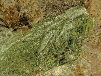
Monteneroite occurs as light green, thick blades up to ~2.5 mm long (Fig. 1). Blades are elongated on [001] and flattened on {010} and exhibit the forms {010}, {110}, {101} and {30 $\bar{1}$} (Fig. 2). Note that it was impractical to measure the forms; the forms listed are considered the best fit to the general shape exhibited by the crystal in Fig. 1. No twinning was observed. The streak is white. Crystals are transparent with vitreous lustre. Monteneroite does not fluoresce under long- or shortwave ultraviolet light. Scratch tests indicated a Mohs hardness of ~2. The mineral is somewhat sectile and crystals exhibit two cleavages, {010} perfect and {001} fair, and irregular stepped fracture. The density measured by flotation in a mixture of methylene iodide and toluene is 2.97(2) g cm–3. The calculated density is 2.983 g cm–3 for the empirical formula and 2.988 g cm–3 for the ideal formula. Monteneroite crystals dissolve rapidly in dilute HCl at room temperature.
Intergrown blades of monteneroite on the holotype specimen; field of view: 1.8 mm across (Natural History Museum of Los Angeles County, catalogue number 67509).

Crystal drawing of monteneroite; clinographic projection in nonstandard orientation.

Monteneroite is optically biaxial (+), with the indices of refraction α = 1.604(2), β = 1.637(2) and γ = 1.688(2), determined in white light. The 2V is 80(1)° measured directly on a spindle stage; the calculated 2V is 79.8°. The dispersion is r < v slight and the optical orientation is X = b, Z ^ c = 52° in the obtuse angle β. No pleochroism was observed. The Gladstone–Dale compatibility index, 1 – (K p/K c), is –0.003 for the empirical formula, in the range of superior compatibility (Mandarino, Reference Mandarino2007).
Raman spectroscopy
The Raman spectrum was obtained using a micro/macro Jobin Yvon LabRam HRVIS, equipped with a motorised x–y stage and an Olympus microscope. The back-scattered Raman signal was collected with a 50× objective and the spectrum was obtained from a randomly oriented crystal. The 632.8 nm line of a He–Ne laser was used as excitation; laser power was controlled by means of a series of density filters. The minimum lateral and depth resolution was set to a few μm. The system was calibrated using the 520.6 cm–1 Raman band of silicon. The spectrum was collected with multiple acquisitions and processed using LabSpec 5 software. The spectrum from 4000 to 100 cm–1 is shown in Fig. 3.
Raman spectrum of monteneroite.

The Raman spectrum of monteneroite is dominated by the stretching and bending vibrations of AsO4 tetrahedra and O–H stretching vibrations. The very broad band of relatively low intensity in the range ~3600 to ~2900 cm–1 is attributed to the stretching O–H vibrations of molecular H2O. According to the empirical correlation of Libowitzky (Reference Libowitzky1999), the H⋅⋅⋅OA lengths of the corresponding hydrogen bonds are in the range 2.7 to 1.7 Å, which is in line with the structure analysis. There are barely perceptible bands at ~1658 and ~1608 cm–1, which can be ascribed to the ν2 (δ) H–O–H deformation vibrations of molecular H2O. The overlapping composite band of highest intensity, composed of bands at 898, 880, 866 (100% rel. int.) and 812 cm–1, is attributed to overlapping ν3 antisymmetric and ν1 symmetric As–O vibrations of the AsO4 tetrahedron. The bands of low intensity at ~468, 449, 435 and 428 cm–1 are related to the ν4 (δ) AsO4 vibrations. At least two of the overlapping bands of medium–weak intensity at 435 and 428 cm–1 are related to the ν2 (δ) AsO4 bending vibrations. The composite band at ~380 and 370 cm–1 may be related to the stretching vibrations of the M1 and M2 octahedra. There are numerous low-energy overlapping bands (348, 340, 317, 304, 270, 259, 240, 226, 192, 146 and 112 cm–1) that are related to various M–Ox stretching vibrations, deformations and phonons.
Chemical composition
Analyses of monteneroite (7 points on 5 crystal fragments) were done at the University of Utah on a Cameca SX–50 electron microprobe with four wavelength dispersive spectrometers using Probe for EPMA software. Analytical conditions were 15 kV accelerating voltage, 20 nA beam current and beam diameter of 10 μm. Raw X-ray intensities were corrected for matrix effects with a ϕρ(z) algorithm (Pouchou and Pichoir, Reference Pouchou, Pichoir, Heinrich and Newbury1991). There was significant beam damage during the analyses. Super-exponential time-dependent X-ray intensity corrections were applied for significant ‘grow in’ on all three elements. Insufficient material is available for a direct determination of H2O, so it was calculated based on the structure. The high analytical total is probably due to H2O loss under vacuum or during the analyses. Analytical data are given in Table 1.
Electron microprobe analytical data (in wt.%) for monteneroite.

* Based on the structure; S.D. – standard deviation.
The empirical formula (based on 16 O atoms per formula unit) is Cu2+0.88Mn2+2.11As2.00O16H15.99 or formulated in accord with the structure, (Cu2+0.88Mn2+0.11)Σ0.99Mn2+2.00(As1.00O4)2⋅8H2O. The simplified formula is (Cu2+,Mn2+)Mn2+2(AsO4)2⋅8H2O. The ideal formula is Cu2+Mn2+2(AsO4)2⋅8H2O, which requires CuO 13.36, MnO 23.83, As2O5 38.60, H2O 24.21, total 100 wt.%.
X-ray crystallography and structure determination
Powder X-ray diffraction data were obtained on a Rigaku R-Axis Rapid II curved imaging plate microdiffractometer utilising monochromatised MoKα radiation. Observed d values and intensities were derived by profile fitting using JADE Pro software (Materials Data, Inc.). The observed powder data fit well with those calculated from the structure, also using JADE Pro (Table 2). The unit-cell parameters refined from the powder data using JADE Pro with whole-pattern fitting are: a = 10.301(8), b = 13.667(8), c = 4.824(8) Å, β = 106.076(19)° and V = 652.6(13) Å3.
Powder X-ray data (d in Å) for monteneroite. Only calculated lines with I ≥ 1.5 are listed.

The strongest lines are given in bold
Single-crystal X-ray studies were carried out using the same diffractometer and radiation noted above. The Rigaku CrystalClear software package was used for processing the structure data, including the application of an empirical multi-scan absorption correction using ABSCOR (Higashi, Reference Higashi2001). The structure was solved using SHELXT (Sheldrick, Reference Sheldrick2015a). Refinement proceeded by full-matrix least-squares on F 2 using SHELXL–2016 (Sheldrick, Reference Sheldrick2015b). The occupancies of the two octahedrally coordinated cation sites were refined. The M2 site was shown to be fully occupied by Mn, while the M1 site was refined to a joint occupancy of Cu0.69Mn0.31. All non-hydrogen sites were refined with anisotropic displacement parameters. Difference-Fourier syntheses located all H atom positions, which were then refined with soft restraints of 0.82(3) Å on the O–H distances and 1.30(3) Å on the H–H distances, and with the U eq of each H set to ×1.2 that of its donor O atom. Data collection and refinement details are given in Table 3, atom coordinates and displacement parameters in Table 4, selected bond distances in Table 5 and a bond-valence analysis in Table 6. The crystallographic information files have been deposited with the Principal Editor of Mineralogical Magazine and are available as Supplementary material (see below).
Data collection and structure refinement details for monteneroite.

R int = Σ|F o2–F o2(mean)|/Σ[F o2]. GoF = S = {Σ[w(F o2–F c2)2]/(n–p)}½. R 1 = Σ||F o|–|F c||/Σ|F o|. wR 2 = {Σ[w(F o2–F c2)2]/Σ[w(F o2)2]}½; w = 1/[σ2(F o2) + (aP)2 + bP] where a is 0.123, b is 3.000 and P is [2F c2 + Max(F o2,0)]/3.
Atom positions and displacement parameters (Å2) for monteneroite.

*Occupancy Cu0.69Mn0.31(5)
Selected interatomic distances (Å), angles (°) and distortion measures of polyhedra in monteneroite.

ECoN – effective coordination number defined by Hoppe (Reference Hoppe1979); Δ – distortion index defined after Baur (Reference Baur1974); D – donor; A – acceptor.
Bond-valences analysis for monteneroite. Values are in valence units (vu).

Multiplicities indicated by ×↓→. The bond valences for the Cu site are based on its refined joint occupancy. Bond-valence parameters are from Gagné and Hawthorne (Reference Gagné and Hawthorne2015). Hydrogen-bond strengths are based on O–O distances according to the relation of Ferraris and Ivaldi (Reference Ferraris and Ivaldi1988).
Description of the structure
Monteneroite is isostructural with other minerals with vivianite-type structures. Vivianite-type structures consist of MlO2(H2O)4 octahedra and M22O6(H2O)4 edge-sharing octahedral dimers that are linked via XO4 tetrahedra (where X = P or As) and hydrogen bonds to form layers parallel to {010} (Fig. 4). Adjacent layers are linked by hydrogen bonds only (Fig. 5). The coordinations of the M1 and M2 sites, while both octahedral, are significantly different. Several investigations for Mg-bearing vivianite-structure-type phases have shown partial ordering of cations between the M1 and M2 sites and, as noted above, barićite was shown by Yakubovich et al. (Reference Yakubovich, Massa, Liferovich and McCammon2001) to have Fe dominant in the M1 site and Mg dominant in the M2 site; however, for those phases in which the principal cations are Co, Zn, Ni or Cu, site ordering has not been shown conclusively, possibly because of the similarities in scattering powers of these cations (see Plášil et al., Reference Plášil, Škácha, Sejkora, Škoda, Novák, Veselovský and Hloušek2017, and references therein).
Layer composed of CuO2(H2O)4 octahedra, Mn2O6(H2O)4 octahedral dimers (two octahedra superimposed) and AsO4 tetrahedra in the structure of monteneroite. Hydrogen bonds are shown as thin black lines. The view is down [010] and the unit cell outline is shown with dashed lines.

Stacking of polyhedral layers in the structure of monteneroite. Hydrogen bonds are shown as thin black lines. The view is slightly canted along [001].

The coordination geometries of the M1 and M2 sites in the structure of monteneroite differ more markedly than those in other minerals with vivianite-type structures. While the average bond lengths of the sites are similar, 2.142 and 2.181 Å, respectively, the M1 site is much more strongly distorted (see Table 5). The M1 site exhibits a compressed Jahn–Teller distortion with short apical bonds of 1.981(7) Å and long equatorial bonds of 2.223(7) Å. In contrast, the M2 site has a much narrower range of bond lengths (2.168–2.201 Å). Comparisons of the differences in the coordinations for the M1 and M2 sites are provided by the effective coordination number (ECoN) defined by Hoppe (Reference Hoppe1979) and the distortion index (Δ) after Baur (Reference Baur1974). The ECoN values for M1 and M2 are 5.079 and 5.991, respectively; the Δ values are 0.050 and 0.006, respectively (see Table 5).
Besides the strong ordering of Cu and Mn indicated by the refinement, the ordering is also supported by bond-valence summations (BVS) at these sites. The M1 site has a BVS of 1.76 valence units (vu) with occupancy only by Cu2+ and 2.38 vu with only Mn2+. Based on the refined M1 occupancy (Cu2+0.69Mn2+0.31), the BVS is 1.94 vu. The M2 site has a BVS of 2.08 vu with only Mn2+, but a BVS of 1.49 vu with only Cu2+.
Monteneroite is an ordered intermediate between manganohörnesite (Gabrielson, Reference Gabrielson1954), Mn2+3(AsO4)2⋅8H2O and babánekite (Plášil et al., Reference Plášil, Škácha, Sejkora, Škoda, Novák, Veselovský and Hloušek2017), Cu2+3(AsO4)2⋅8H2O. Selected data for arsenate minerals with the vivianite structure are compared in Table 7. The vivianite group has not yet been formally approved by the CNMNC; however, a vivianite-group proposal is in preparation.
Arsenate minerals with the vivianite structure.

M 2+ indicates both octahedral cation sites in the structure.
The unit-cell parameters for hörnesite are from Jambor and Dutrizac (Reference Jambor and Dutrizac1995).
The M 2+ measured value for köttigite and the unit-cell parameters for parasymplesite are from Sturman (Reference Sturman1976).
The strongest powder X-ray diffraction lines for köttigite are calculated from the crystal structure of Hill (Reference Hill1979).
The optical properties for babánekite, erythrite, annabergite, hörnesite, manganohörnesite and parasymplesite are from Anthony et al. (Reference Anthony, Bideaux, Bladh and Nichols1990).
Supplementary material
To view supplementary material for this article, please visit https://doi.org/10.1180/mgm.2020.76
Acknowledgements
Anonymous reviewers are thanked for their constructive comments. A portion of this study was funded by the John Jago Trelawney Endowment to the Mineral Sciences Department of the Natural History Museum of Los Angeles County and by the by the Czech Science Foundation (GACR 17-09161S) to J.P.