Case report
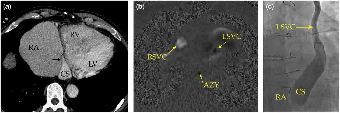
A 57-year-old man with congenitally corrected transposition of the great arteries and previous ventricular septal defect patch repair presented to our hospital for cardiac resynchronisation therapy. A CT scan was requested by the cardiologist to define the cardiac venous anatomy. Electrocardiogram-gated contrast-enhanced CT angiogram demonstrated a small left superior caval vein, and a dilated mildly tortuous coronary sinus with a dominant left posterolateral marginal vein. There was a bridging innominate vein connecting the left and right superior caval vein. The terminal coronary sinus tapers close to the right atrial wall. However, no clear patent ostium or contrast jet was identified to confirm patency (Fig 1a). Therefore, it was determined that this patient had coronary sinus ostial atresia or severe stenosis with cardiac venous drainage via the small calibre left-sided superior caval vein, draining cranially to the left innominate vein. A previous phase contrast MRI demonstrated that the flow within left superior caval vein was cranial, in the same direction as the azygous vein, and in the opposite direction as the right superior caval vein and abdominal aorta (Fig 1b). There was no evidence of unroofing of the coronary sinus or left superior caval vein to left atrial connection.
(a) Axial view of CT scan with contrast injection into coronary sinus tributaries. Lack of opacification of right atrium due to coronary sinus ostial atresia. (b) Phase contrast MRI demonstrating cranial flow of left superior vena cava. (c) Contrast injection into left superior vena cava in catherization lab failed to opacify the right atrium. RA=right atrium, RV=right ventricle, LV=left ventricle, cs=coronary sinus, rSVC=right superior vena cava, lSVC=left superior vena cava, av=azygous vein.

The ostial atresia was later confirmed at cardiac catheterisation. A catheter was inserted via the left subclavian vein to the left superior caval vein and contrast was injected. Contrast opacified the coronary sinus but failed to opacify the right atrium (Fig 1c). CT is important to identify these congenital anomalies of the coronary sinus before cardiac resynchronisation therapy. In this case, the findings on CT changed the course of management.
Discussion
Congenitally corrected transposition of the great arteries is an rare condition occurring in 1/33,000 live births and characterised by discordant atrio-ventricular and ventriculo-arterial connections.Reference Wallis, Debich-Spicer and Anderson 1 In this condition, the atrio-ventricular conduction system is abnormal, and patients typically experience atrio-ventricular block and other arrhythmias, with increasing incidence in life.Reference Wallis, Debich-Spicer and Anderson 1 In addition, the subaortic morphological right ventricle is unable to fully accommodate afterload of the systemic circulation, leading to eventual heart failure.Reference Wallis, Debich-Spicer and Anderson 1 The complications of heart block and heart failure often require percutaneous cardiac interventions such as cardiovascular resynchronisation therapy, pacemaker placement, or radiofrequency ablation.Reference Bottega, Kapa and Edwards 2 Cardiovascular resynchronisation therapy requires access to the cardiac veins draining the subpulmonic left ventricle, as well as the subaortic morphologic right ventricle, via the coronary sinus.
Coronary sinus ostial atresia is a rare condition often associated with persistent left-sided superior caval vein.Reference Lim, Baskaran, Ho, Teo and Ching 3 In roughly 40% of the cases, blood from the coronary sinus flows into the left superior caval vein, and then cranially to the left innominate or other veins in the area, before flowing caudally within the right superior caval vein.Reference Lim, Baskaran, Ho, Teo and Ching 3 Other possibilities of drainage include directly to the left atrium via an unroofed coronary sinus or to the right atrium through collateral venous pathways.Reference Lim, Baskaran, Ho, Teo and Ching 3 A review of literature in 2016 revealed 99 reported cases of coronary sinus ostial atresia.Reference Song, Ren and Chen 4 They report associations with cardiac malformations such as ventricular septal defect, atrial septal defect, transposition of the great arteries, hypoplastic left/right ventricle, double-outlet right ventricle, and other valvular anomalies.Reference Song, Ren and Chen 4
A number of patients with congenitally corrected transposition of the great arteries have been reported to have coronary sinus anomalies. In a study of Mayo Clinic pathology database, 7 of 51 (14%) patients with this condition had abnormal coronary sinus anatomy, with two (4%) cases having complete ostial atresia and the rest having other anomalies.Reference Bottega, Kapa and Edwards 2 Out of the two atretic cases, one had the coronary sinus connecting with the left atrium via unroofing.Reference Bottega, Kapa and Edwards 2
This case highlights the importance of imaging coronary venous anatomy before cardiac resynchronisation therapy in patients with congenitally corrected transposition of the great arteries, owing to the high prevalence of coronary sinus anomalies in these patients. We recommend that all patients with congenitally corrected transposition of the great arteries undergo an electrocardiogram-gated contrast-enhanced CT angiogram before cardiac resynchronisation therapy, pacemaker placement, or radiofrequency ablation. Because it was not known at the time of the CT angiogram that coronary sinus ostial atresia was present, a right-arm injection was performed as per our institutional protocol for CT angiograms. However, injection of contrast via the left arm would have more clearly demonstrated the coronary sinus ostial atresia.
Acknowledgements
None.
Financial Support
This research received no specific grant from any funding agency or from commercial or not-for-profit sectors.
Conflicts of Interest
None.
Ethical Standards
The case report was written according to ethical standards.