



Introduction
Reproduction is the most important process in the life cycle of species, which involves physiological and morphological modifications of the gonads (Mazzoni et al., Reference Mazzoni, Bombardelli and Quagio-Grassiotto2020; Tenugu et al., Reference Tenugu, Pranoty, Mamta and Senthilkumaran2021; Cole and Parenti, Reference Cole and Parenti2022; Hagihara et al., Reference Hagihara, Wakiya, Maeda and Kimura2022). In most teleost fish, the ovaries are paired organs, which are found in the abdominal cavity and can be two types: cystovarian, showing continuity with the oviduct, and gymnovarian, where the oocytes are released directly into the abdominal cavity for subsequent spawning in the external environment (Redding and Patiño, Reference Redding, Patiño and Evans1993; Mylniczenko, Reference Mylniczenko, Lewbart and Harms2021).
In the oogenesis, oogonia undergo successive mitotic divisions and subsequently enter meiosis, giving rise to oocytes at different stages of growth, which finalize this process by releasing mature oocytes into the environment (Viana et al., Reference Viana, Gonçalves, Ferreira, Mendes and Rocha2018; Mazzoni et al., Reference Mazzoni, Bombardelli and Quagio-Grassiotto2020). In this context, the oocytes development mode can be classified in synchronous and asynchronous. In synchronous development, it can be total or in groups. In total, oocyte development occurs without the presence of reserve oocytes. The maturation of two or more oocyte populations at different growth stages takes place in a group, in this case there are reserve oocytes and small oocytes groups that are in synchronous development. In asynchronous development, there are many oocyte populations in different stages of maturation, in which the oocytes that reach the mature stage are released (Wallace and Selman, Reference Wallace and Selman1981; Murua and Saborido-Rey, Reference Murua and Saborido-Rey2003). This information is important to attribute the type of spawning that the animal performs during the reproductive cycle.
In teleost fish, during the process of gonadal maturation, it is possible to analyze the color and morphology of the ovary and the number and size of oocytes. These factors may indicate reproductive characteristics in species, such as ovarian maturation stages (Qadri et al., Reference Qadri, Shah, Balkhi, Bhat, Bhat, Najar, Asmi, Farooq and Aalia2019; Wali et al., Reference Wali, Shah, Bhat and Mohd2022), fecundity (Mendes et al., Reference Mendes, Lee, Viana, Rocha and Ferreira2018; Zhukova et al., Reference Zhukova, Maznikova and Orlov2018), parental care (Viana et al., Reference Viana, Gonçalves, Ferreira, Mendes and Rocha2018) and period and types of spawning (Shimizu et al., Reference Shimizu, Ijiri, Izumi, Gen, Kurogi, Hashimoto, Tanaka, Jinbo, Saito and Chow2021). Such aspects are essential to establish criteria for the survival of species into the environment. In the last years, Amazonian region has been showing anthropic influence, such as the implementation of hydroelectric dams that promote hydrological changes in rivers, this situation can compromise the life of various aquatic species, especially endemic species (Winemiller et al., Reference Winemiller, McIntyre, Castello, Fluet-Chouinard, Giarrizzo, Nam, Baird, Darwall, Lujan, Harrison and Saenz2016).
In addition to the impact of environmental changes, several species are captured for the international ornamental fish market, in which the family Loricariidae has great commercial importance in the international ornamental fish market due to its peculiar shapes, colours and behaviour patterns (Lujan et al., Reference Lujan, Winemiller and Armbruster2012; Sousa de et al., Reference Sousa de, Lucanus, Arroyo-Mora and Kalacska2021). Despite its commercial importance all King Tiger Pleco specimens come from artisanal fisheries (Raghavan et al., Reference Raghavan, Ali, Philip and Dahanukar2018; Sousa de et al., Reference Sousa de, Lucanus, Arroyo-Mora and Kalacska2021), which can lead to overfishing in natural populations.
Hypancistrus seideli (Sousa et al., Reference Sousa, Sousa, Ribeiro, Sabaj, Zuanon and Py-Daniel2025)., formerly known as L333, King Tiger Pleco, or Acari-Pão, is endemic to the Xingu River, part of the Amazon Basin. It is one of the species widely exported to the ornamental fish market (Cardoso et al., Reference Cardoso, Carvalho, Benathar, Serrão, Nagamachi, Pieczarka, De Sousa, Ready and Noronha2016; Reis et al., Reference Reis, Alves, Abe, da Costa Sousa, Paixão, Palheta, de Melo, Fujimoto, Luz and Takata2021, Reference Reis, Oliveira, da Silva Viana, Abe, Takata, de Sousa and da Rocha2022), being sold for prices ranging from US$90 to US$100 (Live Aquaria, 2023). Despite its economic value and international demand, the commercialization of H. seideli . still relies exclusively on wild-caught individuals, as no captive breeding protocol has been developed for the species (Reis et al., 2020). In this context, the present study provides the first detailed morphological description of the ovarian structure and oogenesis up to final oocyte maturation in H. seideli, addressing a critical knowledge gap in the reproductive biology of the species. By providing essential biological data, our findings contribute not only to the development of reproductive management protocols for captive breeding but also to conservation strategies aimed at reducing pressure on wild populations subjected to intense exploitation.
Materials and methods
Study area and collecting sample
A total of 60 female of H. seideli were collected in the Xingu River basin between Vitória do Xingu (2°53′5.11″S, 52°0′45.24″W) and Porto de Moz (1°45′6.29″S, 52°14′14.53″W), in the period of 2018 September. After they were transported to the cell ultrastructure laboratory (Federal University of Pará). The specimens were anesthetized with benzocaine hydrochloride (0.1 g L−1) and euthanized with sodium pentobarbital solution (60–100 mg/kg−) according to the guidelines of the National Council for the Control of Animal Experimentation (CONCEA) and were performed in accordance with approved guidelines, FIPERj No. 002/2019.
The animals were measured in terms of total mass in grams with accuracy of 0.1 g (TM – g) and total length in centimeters (TL – cm) with accuracy of 0.1 cm. The gonads were removed through a ventral incision and classified into four stages: immature, maturing, mature and spawning. The classification of stages development was adapted from the Mendes et al. (Reference Mendes, Oliveira, Montag, Andrade, Giarrizzo, Rocha, Auxiliadora and Ferreira2021) and oocyte development Viana et al. (Reference Viana, Gonçalves, Ferreira, Mendes and Rocha2018).
Light microscopy and scanning Electron Microscopy (SEM)
Fragments of H. seideli ovaries were fixed in Bouin’s solution for 24 h. The samples were then dehydrated in increasing concentrations of ethanol, cleared in xylene, and infiltrated and embedded in paraffin. Sections 5 μm thick were cut and stained with hematoxylin/eosin (HE) and methylene blue. Stained sections were analyzed, and photomicrographs were obtained using a Nikon Eclipse Ci-S H550S optical photomicroscope, equipped with a digital camera (Nikon DS-Ri1).
For SEM analysis, ovarian fragments were fixed in Karnovsky’s solution for 3 h at 4°C. Samples were post-fixed in a 1% osmium tetroxide solution buffered with sodium cacodylate (0.1 M, pH 7.3) for 2 h at room temperature, dehydrated in a graded ethanol series, and subjected to critical point drying using CO2 (CPD 030 Baltec). Specimens were mounted on stubs, coated with gold, and analyzed using an LEO 1430 scanning electron microscope (LEO-ZEISS, Cambridge, UK).
Morphometry and statistical analysis
Samples from 100 oogonia and 100 oocytes of each type (I, II, III and IV) were analyzed. Only cells that contained a nucleus were measured. Serial sections were cut and the slides were evaluated under a photomicroscope with the software NIS-elements BR (4.00.07-bit) and measurements were made 40X magnification.
For the analysis of the follicular complex thickness, four females were used for each maturation stage (immature, maturing, mature, and post-spawning), and measurements of the total thickness — including the theca, follicular cells, and zona radiata, when present — were performed on 100 oocytes of each type (I, II, III, and IV), totaling 400 oocytes analyzed. It is important to note that not all oocytes exhibited a fully developed follicular complex, and measurements were conducted based only on the structures present in each oocyte type.
The data were tested through the Kruskal-Wallis non-parametric test, followed by pairwise comparisons with Bonferroni-adjusted p-values, adopting a significance level of 5% (α).
Results
Ovarian structure
Females of H. seideli, which exhibited an average length of 11.69 ± 0.63cm and an average total mass of 25.3 ± 3.13g. The ovaries are paired organs located in the abdominal cavity, in a cystovarian tubular format, united caudally forming a single duct (gonoduct) that extends to the urogenital pore. Microscopically, the ovary is covered by the tunica albuginea, which emits septa into the organ, forming the ovigerous lamellae, which support the germ cells (Figure 1A). The ovarian parenchyma is formed by loose connective tissue, collagen, reticular fibers, fibroblasts, nerves and blood vessels (Figure 1B). It shows a random distribution of oocytes in different maturation stages, with asynchronous oocyte development (Figures 1B–D).
(A) Lateral, ventral view of H. seideli Anatomy of the mature gonad showing oocytes of different types and the ovigerous lamellae inside the gonad. (B–D) Ovarian ultrastructure. (B) Internal view of the ovigerous lamellae with different oocytes (C) Type I and II oocytes. (D) Type III and IV oocytes. (E) Mean diameters (µm) of cell types in H. seideli I: type I oocyte, II: type II oocyte, III: type III oocyte, IV: type IV oocyte.

Morphology of oogonia and oocytes of Hypancistrus seideli
During gonadal maturation, the germ cells showed different diameters, it was possible to observe oogonia and oocytes at different development stages (Figure 1E). Oogonia were characterized by having a voluminous nucleus and a central or peripheral nucleolus, and these cells were organized in groups, forming nests of oogonia with a mean cell diameter of 2.30 ± 0.63 μm (Figure 2A).
Female germ cells of H. seideli (A) Nest of oogonia. (B) Type I oocyte. (C) Type II oocyte. (D) Oocyte type III. (E) Type IV oocyte. (F,G) Type IV oocyte ultrastructure. (F) Oocyte IV. (G–J) Formation of the layers that form the follicular complex. (L) Schematic drawing of the formation of the follicular complex. I: type I oocyte, II: type II oocyte, III: type III oocyte, IV: type IV oocyte, arrow: theca layer, arrowhead: follicular cell, asterisk: zona radiata.
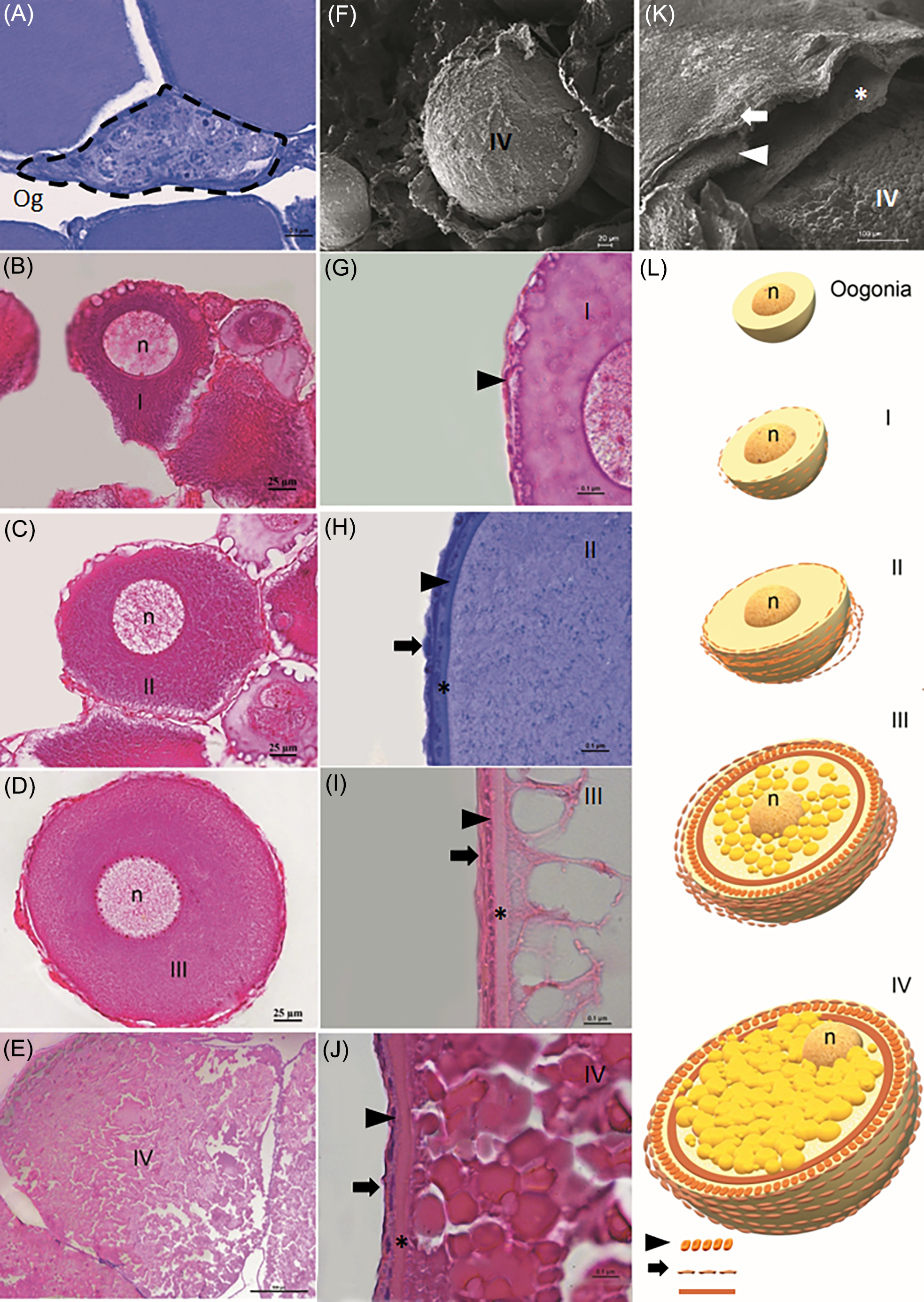
Type I oocytes showed an increase in cytoplasm volume, basophilic eccentric nucleus, with nucleoli arranged centrally or along the periphery, varying between one or more per nucleus (Figure 2B), with a mean cell diameter of 5.09 ± 17.24 μm (Figure 2G). These cells exhibited an enveloping structure formed by a homogeneous layer in development and flattened follicular cells (Figure 2G)
Type II oocytes showed the nucleus and cytoplasm equal in size, and they were less basophilic and had fewer nucleoli located at the periphery of the nucleus compared to type I oocytes. In these cells, we observed cortical alveoli on the periphery of the cytoplasm, zona radiata close to the plasma membrane, follicular cells with an even flatter appearance and theca layer in development, it had an average cell diameter of 143.44 ± 17.79 μm (Figure 2H)
Type III oocytes were characterized by having a central nucleus and several nucleoli at the periphery. The acidophilic cytoplasm was occupied by yolk globules. The cortical alveoli were located close to the plasma membrane. At this stage, it was possible to observe the zona radiata and the follicular cell and theca layers (Figure 2I). These cells had a mean diameter of 250 ± 35.36 μm (Figure 2D).
Type IV oocytes were the largest germline cells with a mean diameter of 2391.47 ± 504.63 μm. These cells showed acidophilic cytoplasm with a high presence of cortical alveoli close to the plasma membrane. The nucleus was not visible because of its migration to the cell periphery (Figure 2E). The complete development of the layers around the oocyte was observed, being from outside to inside the theca layer, follicular cells and zona radiata, which together formed the follicular complex (Figure 2K).
Development of the follicular complex
Starting with the type I oocyte, it was possible to observe a basal membrane covering this cell type (Figure 2G), thus the follicular complex in formation presents a median of 3.09 μm (IQR = 1.86).
The type II oocyte had an outermost layer consisting of flattened cells, called the theca layer, with the presence of follicular cells just below, also with a flattened shape, while the zona radiata was very thin and difficult to visualize (Figure 2H) For the type II oocyte, the median thickness of the follicular complex was 4.05 μm (IQR = 1.86).
In the type III oocyte, the three layers were already fully formed, where the follicular cells still had a flattened shape but now having greater thickness, while the zona radiata also had greater thickness compared to the previous phase (Figure 2I). In type III oocytes, a substantial increase in the follicular complex was observed, with a median thickness of 16.8 μm (IQR = 6.81).
In the type IV oocyte, the three layers were fully formed, i.e., the theca, follicular cells and zona radiata (Figure 2K), and they had reached their greatest thickness, mainly the zona radiata (Figure 2J). Finally, type IV oocytes exhibited the greatest development of the follicular complex, with a median thickness of 35.4 μm (IQR = 6.11). The development of the follicular complex is represented in Figure 2L.
Comparisons between the different oocyte types revealed statistically significant differences in the observed thicknesses. The thickness of type I was significantly lower than that of type II (p < 0.05), as well as compared to types III and IV, which showed substantially higher medians (p < 0.05).
These results indicate a progressive and statistically significant increase in thickness as the oocytes advance through the developmental stages, from type I to type IV.
Stages of gonadal maturation
The stages of gonadal maturation were identified on the basis of histological analysis of the ovary and the types of germ cells during oogenesis (Figure 3), indicating four stages of gonadal development: immature; maturing; mature and spawned.
Ovary of H. seideli. in three stages. (A) Macroscopically immature. (B) Surface of the immature ovary under SEM. (C) Gonad in immature stage showing oocytes I and II. (D) Ovary under maturation macroscopically. (E) Surface of ovary in maturation under SEM. (F) Gonad in maturation showing oocytes II and III. (G) Mature ovary showing oocytes and vascularization. (H) Surface of mature ovary under SEM. (I) Gonad in mature stage showing type III and IV oocytes.
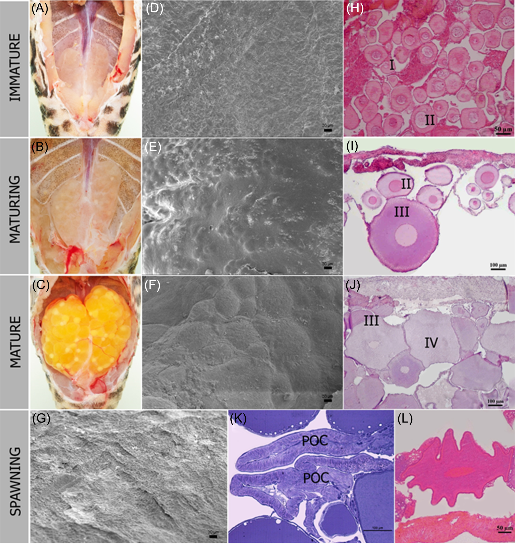
Macroscopically, the immature ovary occupied one-third of the abdominal cavity, was opaque (Figure 3A) and had a smooth surface wall (Figure 3D). Histologically, the ovarian parenchyma had numerous type I and II oocytes, where the cells were grouped and inserted in the ovigerous lamellae (Figure 3H).
The maturing ovary occupied half of the abdominal cavity and was still opaque (Figure 3B), and the wall had a rough appearance resulting in the increase of oocytes’ volume (Figure 3E). At this stage, numerous type II and III oocytes were observed distributed in the ovigerous lamellae (Figure 3I).
The mature stage was characterized by a voluminous ovary that occupied a large part of the abdominal cavity, was intensely vascularized and showed the presence of yellowish oocytes (Figure 3C). The organ wall surface exhibited intense irregularity due to the presence of large oocytes inside the organ (Figure 3F). At this stage, lamellae that presented all oocyte types were found, but type IV oocytes were observed in greater numbers in the gonad (Figure 3J).
In the spawned stage, the ovary had a flaccid appearance (Figure 3G). Several post-ovulatory complexes formed by the theca and follicular cell layers were also found (Figure 3K), along with oocytes in the process of cellular atresia (Figure 3L).
Discussion
Macroscopically, the ovaries of H. seideli. are paired organs and have an arrangement classified as cystovarian. This pattern is found for most teleosts (Mylniczenko, Reference Mylniczenko, Lewbart and Harms2021), being similar to those observed in Siluriformes: catfish such as Horabagrus brachysoma (Chandran and Prasad, Reference Chandran and Prasad2018) and other loricarids such as Loricariichthys platymetopon (Cardim et al., Reference Cardim, de Brito Pereira, de Santana and Dei Tos2019) and Hypancistrus zebra (Viana et al., Reference Viana, Gonçalves, Ferreira, Mendes and Rocha2018). In addition to other species belonging to the order Osteoglossiformes such as Brevimyrus niger, Gnathonemus petersii and Mormyrus rume (Dymek et al., Reference Dymek, Piprek, Boroń, Kirschbaum and Pecio2022) and also Gymnotus sylvius (Gymnotiformes) (França et al., Reference França, Grier and Quagio-Grassiotto2010). Nevertheless, the cystovarian ovary can not be considered a phylogenetic feature, since it appears in basal species as representatives of the order Osteoglossiformes and in recent orders, observed Gymnotiformes.
During gonadal maturation, the ovaries undergo morphological changes and color pattern (Duarte et al., Reference Duarte, Araújo, Sales and Bazzoli2007; Gomes et al., Reference Gomes, Araújo, do Nascimento and Sales2015). In H. seideli. presented the shape of the lobular ovary and the color pattern that varied in the initial whitish stage and in the final intense yellow stage. These results were similar to those described for the loricariidae family (Rodrigues-Filho et al., Reference Rodrigues-Filho, Honji, Mello, Borella, Hilsdorf and Moreira2017; Dien et al., Reference Dien, Pavlov, Ganzha and Bush2022). These aspects demonstrate that these characteristics are highly conserved among the species of this family. However, the color change is related to the oocyte’s maturation, which start to produce and accumulate yolk in the cytoplasm. At the same time there is an increase in the branching of blood vessels, ensuring the nutrition and development of the oocytes themselves.
The growth of oocytes provides a significant increase in ovaries (Cook-Hildreth et al., Reference Cook-Hildreth, Bonner and Huffman2016). In the maturing stage, the ovaries can generate batches of oocytes at the same developmental stage as in Hypostomus affinis (Duarte and Araújo, Reference Duarte and Araújo2002), produce two batches with distinct phases commonly seen in Hypostomus auroguttatus (Gomes et al., Reference Gomes, Araújo, do Nascimento and Sales2015), and produce different batches of oocytes with different sizes and characteristics, as is the case with H. seideli. Therefore, we suggest that the species under study presents oocyte development of the asynchronous type, which enables the continuous production of offspring. Species with asynchronous ovaries normally have multiple spawning, considering that there is an apparent recruitment of germ cells in different periods, as observed in Millerichthys robustus (Domínguez-Castanedo and Uribe, Reference Domínguez-Castanedo and Uribe2019), Acanthopagrus berda (Shilta et al., Reference Shilta, Suresh Babu, Asokan, Vinod, Joseph and Joseph2022) and Hypancistrus zebra (Roman, Reference Roman2011) belonging to the same genus as H. seideli. Therefore, we suggest that multiple spawning occurs in the species under study.
One of the criteria to determine an animal’s reproductive investment is to evaluate the oocyte diameter. In most loricarids, oocytes have a large cell volume (Perini et al., Reference Perini, Sato, Rizzo and Bazzoli2010, Melo et al., Reference Melo, Nunes, Moreira, Weber, Bazzoli and Rizzo2023). In H. seideli., we found the IV type oocytes mean diameter of 2391.47 μm, larger compared to other species of the same family, such as 487.81 μm in Hypancistrus zebra (Viana et al., Reference Viana, Gonçalves, Ferreira, Mendes and Rocha2018), 1360.80 μm in Rhinelepis aspera (Perini et al., Reference Perini, Sato, Rizzo and Bazzoli2010). The oocytes mean size is not necessarily correlated with the female’s body length (Jonsson and Jonsson, Reference Jonsson and Jonsson2014; Samarin et al., Reference Samarin, Policar and Lahnsteiner2015; Súarez et al., Reference Súarez, Silva and Viana2017). However, recent studies indicate that the width of the abdomen of the female H. seideli. strongly influences the ability to contain bulky eggs (Reis et al., Reference Reis, Oliveira, da Silva Viana, Abe, Takata, de Sousa and da Rocha2022). In addition, the large volume of eggs may be related to reproductive strategies and slower embryonic development, a common characteristic in species that have parental care, such as Loricariichthys platymetopon (2320 μm), Hypostomus ternetzi (4360 μm) and Megalancistrus aculeatus (4290 μm) (Suzuki et al., Reference Suzuki, Agostinho and Winemiller2000). These species belong to the family Loricariidae, which have sedentary behavior, parental care, large and fewer oocytes, and slower larval development (Melo et al., Reference Melo, Arantes, Sato, dos Santos, Rizzo and Bazzoli2011; Jakobsen et al., Reference Jakobsen, Fogarty, Megrey and Moksness2016).
The follicular complex consists of the basement membrane, theca layer, follicular cells, zona radiata and mature oocyte (Grier, Reference Grier2012). In H. seideli. the appearance of these layers was observed from type II oocyte and completely formed in type IV, with greater thickness and distinct characteristics such as theca squamous layer, cuboidal follicular cells and thick zona radiata. In other loricarids, the morphology of the layers is similar, but the appearance of these layers has been reported from type I oocytes (Viana et al., Reference Viana, Gonçalves, Ferreira, Mendes and Rocha2018). H. seideli. is endemic, exhibits parental care behavior, and inhabits a region with many rapids. These characteristics reflect the morphological aspects of oocytes III and IV, specifically in the follicular complex, which is closely linked to different reproductive strategies, as seen in Auchenipterichthys longimanus, Ageneiosus ucayalensis, Hypophthalmus marginatus, Baryancistrus xanthellus, Panaqolus tankei and Peckoltia oligospila (Viana et al., Reference Viana, Ferreira, Mendes, Silva, Gonçalves and Rocha2023).
Description of the development of gonads and oocytes in H. seideli. is necessary, as they support reproductive biology and contribute to aquaculture through the development of protocols for induced reproduction. Since the species is in high demand in the ornamental fish market, it is endemic, and its habitat undergoes environmental changes due to the implementation of a hydroelectric plant in the Xingu River region.
Acknowledgements
The authors thank the National Council for Scientific and Technological Development (CNPq) for the financial support to Rossineide Martins da Rocha (303140/2022-4) and Maria Auxiliadora Pantoja Ferreira (308895/2022-3).
Author contributions
Iann Leonardo Pinheiro Monteiro and Ivana Kerly da Silva Viana: Conceived and designed the experiments; Performed the experiments; Wrote the paper. Maria Auxiliadora P. Ferreira; Rossineide M. Rocha: Conceived and designed the experiments; Contributed reagents, materials, analysis tools or data; Wrote the paper. Ryuller Gama Abreu: Performed the experiments; Analyzed and interpreted the data; Wrote the paper. Leandro Melo de Sousa: Analyzed and interpreted the data; Wrote the paper.
Competing interests
The authors have no conflict of interest to declare.



