Introduction
The family Simuliidae consists of 2129 nominal species, of which 22% are distributed among 24 species groups in the subgenus Simulium Latreille (Adler and Crosskey Reference Adler and Crosskey2011). About 20 nominal species in the subgenus were discovered by analyzing the giant polytene chromosomes in the larval silk glands, and another 50 cytoforms have been recognized, many probably representing valid species yet to be formally described (Adler and Crosskey Reference Adler and Crosskey2011). The pervasive nature of cytotaxonomy in the study of the Simuliidae also is demonstrated by the nearly 600 papers that have dealt to some degree with the chromosomes of about one-quarter of the world's nominal species (Adler et al. Reference Adler, Cheke and Post2010). The taxonomic structure of the family is built on more than 250 years of morphological work and over half a century of chromosomal study. Future studies of the systematics of the family will need to build on this foundation.
Within the subgenus Simulium, the S. malyschevi Dorogostaisky, Rubtsov, and Vlasenko and S. reptans (L.) species groups account for 39 and 17 formally described species, respectively (Adler and Crosskey Reference Adler and Crosskey2011). Historically the S. malyschevi group has been recognized by a distally narrowed male ventral plate, the presence of a small subbasal tooth on the female claw, 6–16 pupal gill filaments, a shoe-shaped cocoon, and a postgenal cleft reaching the hypostomal groove, whereas the S. reptans group has been defined by an abruptly narrowed ventral plate at midlength, the absence (sometimes presence) of a subbasal tooth on the claw, 6–12 gill filaments, a slipper-shaped or slightly shoe-shaped cocoon, and a postgenal cleft typically not reaching the hypostomal groove (Rubtsov Reference Rubtsov1956, Reference Rubtsov1962Reference Rubtsov [“Rubzow”] and Lindner1963). With the discovery of additional species and an increased appreciation for intraspecific variation, the taxonomic limits of the two groups have become ill-defined, making assignment of some species problematic; whether or not the groups are independently monophyletic is questionable.
As an opportunity to investigate relationships in the subgenus Simulium, we examined an enigmatic and little-known species, Simulium (Simulium) acrotrichum Rubtsov, previously recorded from Kazakhstan, Mongolia, Siberia, and Uzbekistan (Adler and Crosskey Reference Adler and Crosskey2011). Here we redescribe the male and properly associated female, pupa, and larva, and map the polytene chromosomes to gain insights into the evolutionary relationships. We relied on published studies and our unpublished data on the cytotaxonomy of the subgenera Boophthora Enderlein, Psilozia Enderlein, and Simulium to understand the relationships. Our analysis of S. acrotrichum is based on material from Mongolia. The only previous report of this species from Mongolia is based on three females (Rubtsov Reference Rubtsov1971).
Taxonomic history of S. acrotrichum
Simulium acrotrichum was described from a male, designated the holotype, collected on 25 May 1940 from the Fergana Valley of Uzbekistan; a few larval features were illustrated at the time of the original description, although the larval association was considered tentative (Rubtsov Reference Rubtsov1956). Previously the species had been lumped with Simulium flavidum Rubtsov (Rubtsov Reference Rubtsov1951). Rubtsov (Reference Rubtsov [“Rubzow”] and Lindner1963) repeated the figures of his original (1956) description and again questioned his earlier larval association. Rubtsov and Violovich (Reference Rubtsov and Violovich1965) redescribed the male and illustrated its genitalia, and described and illustrated the female and pupa, based on material from Tuva (southern Siberia), but did not mention the status of the earlier larval association. Patrusheva (Reference Patrusheva1982) repeated the illustrations of Rubtsov and Violovich (Reference Rubtsov and Violovich1965), and Konurbaev (Reference Konurbaev1984) included the male in an identification key for a limited number of species in Central Asia. Yankovsky (Reference Yankovsky2003) presented the original figures of the male and larva (illustrated by Rubtsov Reference Rubtsov1956), and stated that the female and pupa were unknown. Thus, all taxonomic treatments other than those of Rubtsov (Reference Rubtsov1956) and Rubtsov and Violovich (Reference Rubtsov and Violovich1965) simply recycled the presentation in these two papers.
Complicating the taxonomy of S. acrotrichum is the morphological similarity to S. flavidum Rubtsov, originally described from the Dyushambinka River of Tadzhikistan (Rubtsov Reference Rubtsov1947) and subsequently reported by Rubtsov (Reference Rubtsov1956) from the type locality of S. acrotrichum in Uzbekistan. Rubtsov (Reference Rubtsov1956) in his keys to males indicated that the two species could be distinguished on the basis of the number of teeth on the posterior margin of the ventral plate, 3 in S. acrotrichum versus 4 in S. flavidum. We relied largely on this character and the shape of the ventral plate, both of which were based on the type of S. acrotrichum, to establish the identity of our material because subsequent illustrations and descriptions of both species (e.g., Rubtsov and Violovich Reference Rubtsov and Violovich1965) from areas other than the type localities cloud the distinction. Nonetheless, variation in the number of teeth on the ventral plate of our material confounds the identification.
Despite their apparent morphological similarities, the two species have, at times, been placed in different taxonomic groups. Rubtsov (Reference Rubtsov1956) originally put S. acrotrichum, along with S. flavidum, in the S. reptans species group. Rubtsov and Violovich (Reference Rubtsov and Violovich1965) later placed S. acrotrichum in the genus Gnus Rubtsov (now equivalent to the S. malyschevi species group), while maintaining S. flavidum in the genus Simulium. Takaoka and Saito (Reference Takaoka and Saito2007) established a new species group of Simulium, the S. argentipes Edwards group, in which they included S. flavidum.
Materials and methods
Simulium acrotrichum was collected from the Tuul River at Songino Camp, about 20km southwest of the center of Ulaanbaatar, Mongolia (47°50′57′′N, 106°40′11′′E), on 25 July 2004. The river at this site was at an elevation of 1235m, about 36 m wide, stony, silted, and 24 °C. We fixed 49 larvae and 6 pupae (1 pharate male) in 1:3 acetic ethanol, and 3 larvae, 35 pupae (5 pharate females, 7 pharate males), and 6 pupal exuviae in 95% ethanol. Simulium decimatum Dorogostaisky, Rubtsov, and Vlasenko (66 larvae, 1 pupa) and S. malyschevi (17 larvae) were also collected at this site.
Two pharate adults of each gender were dissected from pupae, dried with hexamethyldisilazane (Brown Reference Brown1993), mounted on Minuten pins, and associated with their pupal exuviae and cocoons in glycerin vials. Because the adults were pharate, specific colors of insufficiently tanned and sclerotized areas are not reported, nor are characters of incompletely formed structures such as wings. The heads and terminalia of selected pharate adults were cleared by boiling in 85% lactic acid and then transferred to glycerin in a depression slide for illustration using a drawing tube on an Olympus BH-2 compound microscope. Dissected structures were placed in microvials with glycerin and pinned beneath the adults. Adults were deposited in the Clemson University Arthropod Collection, South Carolina (CUAC). Morphological terminology follows that of Adler et al. (Reference Adler, Currie and Wood2004).
Polytene chromosomes from the larval silk glands of 45 larvae (25 females, 20 males) of sufficient size for analysis (penultimate to ultimate instars) were prepared using the Feulgen technique of Rothfels and Dunbar (Reference Rothfels and Dunbar1953). The chromosomes and one gonad (for gender identification; Adler et al. Reference Adler, Currie and Wood2004) were dissected from the abdomen of each larva, transferred to a drop of 50% acetic acid on a microscope slide, and flattened with thumb pressure applied to a cover slip. The stained abdomens were repatriated with the anterior portions of the body and deposited, with pupae, pupal exuviae, and unstained larvae, in 80% ethanol in the CUAC. Photographic negatives of all arms of the polytene complement also were deposited in the CUAC.
A standard chromosomal reference map does not exist for the S. malyschevi or S. reptans groups, although within the S. malyschevi group, maps have been presented for the Nearctic S. arcticum complex (Shields and Procunier Reference Shields and Procunier1982; Procunier Reference Procunier1984; Adler et al. Reference Adler, Currie and Wood2004) and scattered species in the Palearctic Region (Chubareva and Petrova Reference Chubareva and Petrova2008), without addressing commonalities among other species. We therefore used a more inclusive framework to compare the banding sequence of S. acrotrichum, selecting the Simulium subgeneric standard maps of Rothfels et al. (Reference Rothfels, Feraday and Kaneps1978) for the IS, IL, IIL, and IIIS arms and those of Adler and Kachvorian (Reference Adler and Kachvorian2001) and Tangkawanit et al. (Reference Tangkawanit, Kuvangkadilok, Baimai and Adler2009) for the IIS and IIIL arms. A more universal subgeneric standard map for IIIL was established by Adler and Kachvorian (Reference Adler and Kachvorian2001), whose figure 6 shows the sequence and numbering for the S. noelleri species group standard; the subgeneric standard for IIIL is obtained through their inversions IIIL-1 and IIIL-2. Actual numbering of the IIIL subgeneric standard map was first used by Tangkawanit et al. (Reference Tangkawanit, Kuvangkadilok, Baimai and Adler2009), based on the recommended subgeneric standard sequence of Adler and Kachvorian (Reference Adler and Kachvorian2001); we use the standard numbering of Tangkawanit et al. (Reference Tangkawanit, Kuvangkadilok, Baimai and Adler2009).
Inversion breakpoints, relative to the Simulium subgeneric standard map, are indicated on our composite photomaps of S. acrotrichum by brackets — solid if the inversion is fixed or an autosomal polymorphism, dotted if X-linked, and dashed if Y-linked. Only maps of those arms that differ from the standard by one or more fixed rearrangements are presented. Inversions are named by chromosome arm and sequential numbers. Fixed inversions are italicized in the text and underlined on the maps, whereas all other inversions are given in roman type. Intact chromosomal fragments of complex inversions, relative to the subgeneric standard, are labeled with lower-case letters on our maps, which, when ordered alphabetically, yield the standard sequence. Additional chromosomal terminology and mapping conventions follow those of Rothfels et al. (Reference Rothfels, Feraday and Kaneps1978) and Adler et al. (Reference Adler, Currie and Wood2004).
Simulium (Simulium) acrotrichum Rubtsov
[Simulium flavidum, Rubtsov Reference Rubtsov1951: 826–830 (misidentification in part; male: Fig. 78A, B, E, G, I, V; larva: Fig. 76A, B, V, Zh)].
Simulium acrotrichum Rubtsov Reference Rubtsov1956: 823–824 (original description: male, larva).
Simulium acrotrichum, Rubtsov Reference Rubtsov [“Rubzow”] and Lindner1963: 582 (male, larva).
Simulium acrotrichum, Violovich Reference Rubtsov and Violovich1965, 834 (Tuva record).
Gnus acrotrichum, Rubtsov and ViolovichReference Rubtsov and Violovich1965: 55–58 (female, male, pupa).
Gnus acrotrichum, Gornostaeva et al. Reference Gornostaeva, Balkarova and Gachegova1969: 715 (Siberian records).
Simulium acrotrichum, Rubtsov Reference Rubtsov1971: 49 (Mongolian records).
Gnus acrotrichum, Patrusheva Reference Patrusheva and Zolotarenko1976: 316 (Siberian records).
Gnus acrotrichum, Rubtsov and ShakirzyanovaReference Rubtsov and Shakirzyanova1976: 29, 32 (Kazakhstan records).
Gnus acrotrichum, Patrusheva Reference Patrusheva1982: 250–253 (female, male, pupa).
Simulium (Simulium) acrotrichum, KonurbaevReference Konurbaev1984: 106 (male).
Gnus acrotrichum, Yankovsky Reference Yankovsky2003: 437, 446 (male, larva).
Morphological redescription
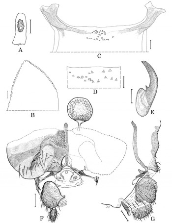
Female. Scutum 0.81–0.94mm long. Head. Slightly narrower than thorax. Frontal ratio 1.27–1.29:1.00:1.02–1.04; frons to head ratio 1.00:3.67–4.04. Maxillary palpus with proportional lengths of palpomeres III, IV, and V 1.00:1.03–1.05:2.28–2.31; sensory vesicle (Fig. 1A) ellipsoidal, with rugged surface, 0.36–0.43 times as long as palpomere III, with medium-sized opening submedially. Maxillary lacinia with 10–15 inner and 16 or 17 outer teeth. Mandible (Fig. 1B) with 26 inner and 14 outer teeth. Cibarium (Figs. 1C, 1D) centrally with 17–22 minute, pointed, and rounded tubercles. Thorax. Scutum dark brown, densely covered with golden recumbent pile; in anterior view with 2 small widely spaced pollinose spots anteriorly. Scutellum pale brown, with golden hairs. Postscutellum and katepisternum brown, shiny, bare. Pleural membrane bare. Legs. Pale, except with dark pigment distributed as follows: on foreleg: apex of tibia, entire basitarsus and tarsus; on midleg: extreme apex of basitarsus, apical three-quarters of tarsomere I, and all of remaining tarsomeres; on hindleg: apex of tibia, apex of basitarsus, apical half of tarsomere I, and all of remaining tarsomeres. Calcipala slightly shorter than its basal width, 0.40 times as wide as greatest width of basitarsus. Pedisulcus distinct. Claws (Fig. 1E) each with small subbasal tooth. Genitalia. Sternite VIII (Fig. 1F) well sclerotized, bare medially but with 23–29 long hairs on each side. Hypogynial valves (ovipositor lobes) (Fig. 1F) in ventral view truncate posteriorly, with posteromedial area broadly rounded; ventral surface produced and rather bulbous (Fig. 1G); inner margins slightly convave, with 8–10 hairs on each side and densely covered with microtrichia. Genital fork (Figs. 1F, 1G) with each arm bearing well-sclerotized, rounded, anterodorsally directed apodeme. Anal lobe (paraproct) in ventral view (Fig. 1F) large, rounded, well defined except for weakly sclerotized area on mesal margin; in lateral view (Fig. 1G) broadly rounded, slightly wider than long, with 14–16 long dark hairs; in anterior view with 7–9 sensilla on anteromedial surface. Cercus in lateral view (Fig. 1G) rounded posteriorly, about twice as wide as long. Spermatheca (Fig. 1F) faintly reticulate, subspherical, well sclerotized except at junction with duct.
Female of Simulium acrotrichum from Mongolia. A, Maxillary palpomere III with sensory vesicle (left side); B, mandible (apex only); C, cibarium; D, central portion of cibarium; E, claw; F and G, terminalia (F, ventral view; G, lateral view): sternite 8, hypogynial valves, genital fork, anal lobes, cerci, and spermatheca. Scale bars: 0.01mm for B–E; 0.05mm for A, F, and G.

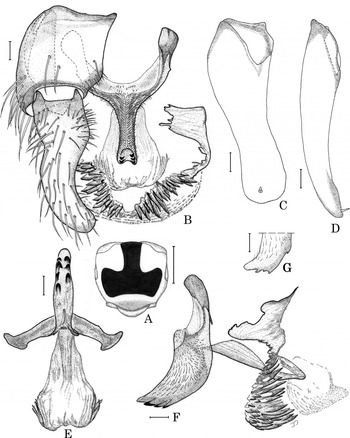
Male. Scutum 0.64–0.90mm long. Head. About as wide as thorax. Upper eye with 17–19 vertical columns and 17–19 horizontal rows of large facets. Antenna with first flagellomere 1.68–1.73 times as long as second. Maxillary palpus with proportional lengths of palpomeres III, IV, and V 1.00:1.18–1.32:2.61–2.79; palpomere III widened apically; sensory vesicle ellipsoidal, 0.18–0.21 times as long as palpomere III, with medium-sized opening apically or laterally. Thorax. Scutum velvety black, densely covered with golden recumbent pile and outlined by wide, shiny, steel-grey border anterolaterally, laterally, and posteriorly; anterolateral areas somewhat bluish in certain light (Fig. 2A). Scutellum light brown, with erect golden hairs. Postscutellum dark brown, shiny, bare. Pleural membrane bare. Legs. Calcipala slightly shorter than its basal width, 0.29 times as wide as greatest width of basitarsus. Pedisulcus distinct. Genitalia. Gonocoxite in ventral view (Fig. 2B) subquadrate. Gonostylus in ventral view (Fig. 2B) elongate, longer than gonocoxite, approximately 3.0 times longer than its basal width, slightly tapered apically, with subapical spine; in dorsal view (Fig. 2C) slightly sinuous, somewhat broadened near basal one-quarter, narrowed to middle, and nearly parallel-sided toward apex; in medial view (Fig. 2D) somewhat broad in basal one-quarter, smoothly tapered toward apex. Ventral plate and basal arms in ventral view (Fig. 2B) roughly Y-shaped; in terminal view (Fig. 2E) lip narrow with lateral margins parallel, and posterior margin with 2–4 teeth per side; in lateral view (Figs. 2F, 2G) gently curved, margins nearly parallel, with 2–4 teeth posteriorly in addition to apical point; body of ventral plate with short fine setae. Median sclerite (Fig. 2E) well sclerotized, widened toward apex, rounded posterolaterally, with posterior margin slightly concave. Paramere (Fig. 2B, F) ragged, with numerous long and short spines apically. Dorsal plate not apparent. Abdominal segment X bare. Cercus rounded, with 10–12 hairs.
Male of Simulium acrotrichum from Mongolia. A, Scutum (dorsal); B, genitalia: gonocoxite, gonostylus (omitted from left side), ventral plate, median sclerite, paramere, and parameral spines (ventral view). C and D, gonostylus, right side (C, dorsal view; D, medial view); E, ventral plate and median sclerite (terminal); F, ventral plate, median sclerite, paramere, and parameral spines (left lateral view); G, ventral plate (apex of body), showing variation on the posterior margin (left lateral view). Scale bars: 0.5mm for A; 0.02mm for B–G.

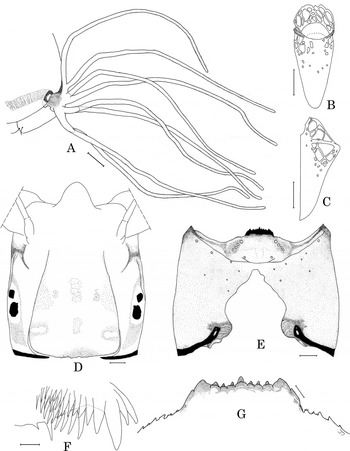
Pupa. Body length 3.0–3.7mm. Head. Frons densely covered with small rounded tubercles, and with 3 pairs of simple trichomes (0.07–0.09mm long). Thorax. Integument densely covered with small rounded tubercles dorsally on anterior two-thirds, and with relatively smaller, sparser tubercles elsewhere; each side with about 8 simple, bifurcated or trifurcated trichomes (0.07–0.14mm long). Gill (Fig. 3A) about 28%–42% as long as pupal body, with 8 slender filaments in 4 pairs; dorsalmost filament shorter than other filaments; all filaments arising from short petioles, except 2nd pair from dorsum typically sessile; filaments, except petioles, with annular ridges and furrows; base densely covered with minute tubercles dorsally. Abdomen. Tergite I with 1 simple, slender, short seta on each side; tergite II with 3–5 simple, slender, short setae and 3 simple, short, variably sized brown spinose setae on each side; tergites III and IV each with 4 dark recurved hooks on each side; tergite VIII with short row of dark spinecombs on each side; tergite IX with 1 pair of minute dark bluntly rounded triangular terminal spines. Sternite IV with 2 simple, slender, short, pale setae on each side; sternites V–VII with 0–3 simple pale setae and 1 pair of dark recurved hooks per side. Cocoon (Figs. 3B, 3C). Length 3.7–4.7mm, width 1.4–1.9mm. Shoe-shaped, with variously sized openings anterolaterally; floor present on posterior half.
Larva and pupa of Simulium acrotrichum from Mongolia. A, Gill filaments (right side); B and C, cocoon (B, dorsal view; C, lateral view); D and E, larval head capsule (D, dorsal view; E, ventral view); F, larval mandible (apex only); G, larval hypostoma (anterior margin). Scale bars: 0.01mm for F and G; 0.05mm for D and E; 0.1mm for A; 1.0mm for B and C.

Larva (final instar in Carnoy's fixative). Body 4.9–5.5mm long, slightly expanded at abdominal segment V. Body pale, faintly grayish in females, faintly rusty brown in males. Head capsule pale yellowish, sometimes suffused with brown pigment laterally and ventrally; headspots absent or faintly negative (Fig. 3D); eyebrow faintly brown or absent. Antenna slightly longer than stem of labral fan; length ratios of articles (proximal to distal) 1.0:1.24–1.35:0.78–0.85. Labral fan with 32–42 primary rays. Mandible (Fig. 3F) without supernumerary serrations. Hypostoma (Figs. 3E, 3G) with 9 teeth, of which median and lateral teeth longest; lateral serrations well developed; 3–5 hypostomal bristles per side. Postgenal cleft (Fig. 3E) widest at midpoint, narrowed anteriorly, nearly or sometimes reaching hypostoma. Subesophageal ganglion typically unpigmented, or sometimes partially ensheathed with faint grayish pigment. Gill histoblast with 8 slender filaments in 4 pairs. Abdominal setae pale, unbranched, sparse. Male gonads ensheathed with pale, rusty brown pigment. Rectal papilla of 3 lobes, each with 3–6 fingerlike secondary lobules ventrally. Anal sclerite X-shaped, with anterior arms 0.66 times as long as posterior arms. Abdominal segment IX without ventral tubercles. Posterior proleg with 14 or 15 hooks in each of 78–82 rows.
Chromosomal description
Fixed rearrangements
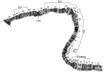
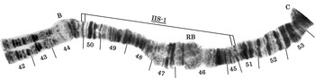
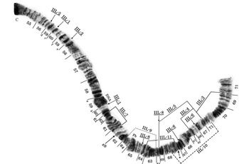
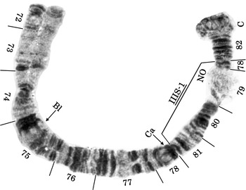
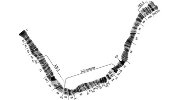
The banding sequences of all 45 larvae prepared for analysis were read in their entirety. Simulium acrotrichum is highly rearranged, differing from the subgeneric standard by nearly 30 fixed rearrangements distributed as follows: about 9 in IL (IL-1, IL-2, IL-3, IL-4, IL-complex; Fig. 4), 1 in IIS (IIS-1; Fig. 5), 8 in IIL (IIL-1, IIL-2, IIL-3, IIL-4, IIL-5, IIL-6, IIL-7, IIL-8; Fig. 6), 1 in IIIS (IIIS-1; Fig. 7), and about 9 in IIIL (IIIL-2, IIIL-3, IIIL-complex; Fig. 8), plus a nucleolar organizer transposed, without an inversion, from section 87 in IIIL of the subgeneric standard to section 79 in IIIS (Fig. 7). IS is the only arm that retains the subgeneric standard sequence. The number of inversions required to derive the scrambled IL-complex sequence from the subgeneric standard is five, based on the section numbers in Figure 4, which represent one of several hypotheses for reconciling bands with the standard. IIL is resolved with the fewest steps, but also might include one or more mimic inversions; a band or two on either side of the 64/69 interface are not convincingly assigned to a section (Fig. 6). IIIL-complex partially overlaps IIIL-3; the possible band identities of IIIL-complex are shown in Fig. 8, representing perhaps seven inversions. To test the hypotheses of band homologues and determine the actual inversions involved in IL-complex, the distal half of IIL, and IIIL-complex, intermediate steps need to be discovered, if they exist, in other species.
Photocomposite of IL of the female larva of Simulium acrotrichum from Mongolia, showing fixed inversions IL-1, IL-2, IL-3, IL-4, and IL-complex; section numbers reflect the standard banding sequence of Rothfels et al. (Reference Rothfels, Feraday and Kaneps1978) (C, centromere; 2°NO, location of the secondary nucleolar organizer in one male. The letters “a”–“l” for fragments in IL-complex represent one hypothesis concerning the order of bands required to generate the standard sequence).

Photocomposite of IIS of the female larva of Simulium acrotrichum from Mongolia, showing fixed inversion IIS-1; section numbers reflect the standard banding sequence of Adler and Kachvorian (Reference Adler and Kachvorian2001) for subgenus Simulium (B, bulge; C, centromere; RB, ring of Balbiani).

Photocomposite of IIL of the female larva of Simulium acrotrichum from Mongolia, showing fixed inversions IIL-1, IIL-2, IIL-3, IIL-4, IIL-5, IIL-6, IIL-7, and IIL-8; X-linked IIL-9; and breakpoints of Y-linked IIL-10 and autosomal IIL-11; section numbers reflect the standard banding sequence of Rothfels et al. (Reference Rothfels, Feraday and Kaneps1978) for subgenus Simulium (C, centromere; DNA, DNA puff; gB, gray band; Pb, parabalbiani; the arrow with asterisk indicates the location of the heteroband in three females; the letters “a”–“l” for fragments of section 59 indicate the sequence of bands, proximal to distal, on the standard map).

Photocomposite of IIIS of the female larva of Simulium acrotrichum from Mongolia, showing fixed inversion IIIS-1; section numbers reflect the standard banding sequence of Rothfels et al. (Reference Rothfels, Feraday and Kaneps1978) for subgenus Simulium (Bl, blister; C, centromere; Ca, capsule; NO, nucleolar organizer).

Photocomposite of IIIL of the female larva of Simulium acrotrichum from Mongolia, showing fixed inversions IIIL-2, IIIL-3, and IIIL-complex; section numbers reflect the standard banding sequence of Adler and Kachvorian (Reference Adler and Kachvorian2001) and Tangkawanit et al. (Reference Tangkawanit, Kuvangkadilok, Baimai and Adler2009) for subgenus Simulium. The dotted right side of the bracket for IIIL-3 indicates that the distal portion of the inversion is buried in IIIL-complex (C, centromere; CS, cup and saucer; letters “a”–“r” for fragments in IIIL-3 and IIIL-complex represent one hypothesis for the order of bands required to generate the standard sequence).

Sex chromosomes. The sex chromosomes were differentiated, with IIL-9 and IIL-10 absolutely linked to the X and Y chromosomes, respectively (Fig. 6). Thus, all females were IIL-9 inverted homozygotes (i.e., 9/9) and completely lacked the IIL-10 inversion (i.e., st/st), whereas all males were double heterozygotes in trans configuration (i.e., 9/st, st/10). In other words, X1=IIL-9 and Y1=IIL-10 such that all females were X1X1 and all males were X1Y1. Although the short region between the near-tandem IIL-9 and IIL-10 inversions provides the possibility for crossover types to occur, our sample had none, suggesting that the sex chromosomes are completely coadapted, at least in this population.
Floating inversions. Polymorphisms were infrequent, occurring in only 7.8% of the 90 chromosomal homologues. They included a mid-arm heterozygous inversion (breakpoints at sections 7/8 and 10/11) in IS of one male, a secondary nucleolar organizer expressed heterozygously in section 25 of IL in one male (Fig. 4), a thick heteroband carried heterozygously in section 68 of IIL in three females (Fig. 6), IIL-11 heterozygous in one female (Fig. 6), and heterozygous expression of the primary nucleolar organizer in one male.
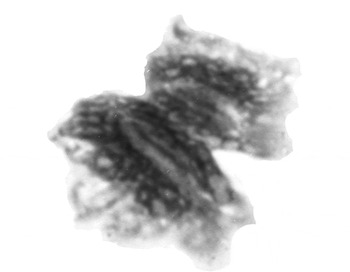
B chromosomes. A high proportion (73.3%) of larvae had supernumerary (B) chromosomes (Fig. 9); 19 of 25 females and 14 of 20 males had B chromosomes, but they were not significantly more frequent in either sex (χ2=0.205, df =1, P=0.651). The Bs in S. acrotrichum are small metacentrics heavily endowed with heterochromatin in the polytene complement, and with about six discernible bands.
B chromosome of the female larva of Simulium acrotrichum from Mongolia.

Discussion
A comparison of the chromosomal banding sequence of S. acrotrichum with the map for S. flavidum presented by Chubareva and Petrova (Reference Chubareva and Petrova2008), based on material from Kyrgyzstan, suggests a number of similarities, although some critical portions of the latter map are unreadable. The subterminal IL-complex inversion of S. acrotrichum appears homologous with the sequence in S. flavidum; IL-1, but not IL-2, appears to be shared; and IIS-1, IIL-1, IIL-2, IIL-3, IIIS-1, and IIIL-2 are shared. The nucleolar organizers of the two species are in opposite arms, however, remaining in the standard position in S. flavidum and providing a useful means of distinguishing the two species. The position of the nucleolar organizer in IIIS of S. acrotrichum is unique among all studied members of the subgenus Simulium; its position in the base of IIIS in the S. tuberosum group is only superficially similar. Although it is tempting to interpret the entire IIL sequence of S. flavidum as equivalent to that of S. acrotrichum, including perhaps the same X- and Y-linked inversions producing what appears to be a double heterozygote in the photograph of Chubareva and Petrova (Reference Chubareva and Petrova2008), the map of S. flavidum simply does not provide sufficient detail. Other fixed rearrangements of S. acrotrichum cannot be compared with the unreadable portions of the map of S. flavidum. The two species, nonetheless, are sister species, no other studied species having IL-complex or IIL-3 inversions. Lacking specimens of S. flavidum, we are unable to provide diagnostic morphological characters to distinguish this species from S. acrotrichum, although the confusion in the literature attests to their similarity. Two alternative hypotheses, however, should be considered regarding the relationship of S. acrotrichum and S. flavidum, ideally by analyzing freshly collected material from the respective type localities: (i) S. acrotrichum in our study, S. flavidum in that of Chubareva and Petrova (Reference Chubareva and Petrova2008), or both, are misidentified; (ii) S. acrotrichum and S. flavidum are conspecific, the putative morphological differences and any chromosomal differences (e.g., differential location of the nucleolar organizer) representing intraspecific variation.
The banding sequence of S. acrotrichum carries all inversions shared among the S. jenningsi, S. malyschevi, and S. reptans groups. All studied members of these three groups share IIL-1, IIL-2, IIIS-1, and IIIL-2. These inversions are absent in other species groups that we have examined in the subgenus Simulium, viz. the S. bezzii, S. nobile, S. noelleri, S. ornatum, S. parnassum, S. petersoni, S. tuberosum, S. variegatum, and S. venustum groups.
A persistent limitation of cytotaxonomy has been the inability to determine the directionality of rearrangements (Rothfels Reference Rothfels1979). This inability stems from the difficulty of rooting a phylogeny by resolving banding sequences relative to an outgroup; typically, the greater the phylogenetic distance between the outgroup and the taxon under study, the more jumbled are the banding patterns. Using an outgroup defined on chromosomal criteria for the first time in the Simuliidae, we establish the polarity of occurrence of the four inversions shared among the S. jenningsi, S. malyschevi, and S. reptans groups. Both of our selected outgroups—Simulium (Psilozia) vittatum Zetterstedt (based on maps of Pasternak Reference Pasternak1964) and Simulium (Boophthora) erythrocephalum (De Geer)—lack these inversions. IIL-1, IIL-2, IIIS-1, and IIIL-2, therefore, are derived in the S. jenningsi, S. malyschevi, and S. reptans groups, providing evidence that these groups form a chromosomally well defined, monophyletic cluster. With S. acrotrichum unequivocally a member of this clade, and as the sister species of S. flavidum, the current placement of S. flavidum in the S. argentipes group becomes untenable.
IL-1 of S. acrotrichum is shared minimally with the S. arcticum complex, S. defoliarti Stone and Peterson, S. subvariegatum Rubtsov, and S. colombaschense (Scopoli). IIS-1 is shared at least with S. decimatum and S. murmanum Enderlein from Mongolia, S. reptans, and S. colombaschense. The monophyly of the S. malyschevi group+S. reptans group can be corroborated morphologically by the uniquely shaped, large anal lobe, which in lateral view is subquadrate and anteriorly rounded (Moulton and Adler Reference Moulton and Adler1995). Based on both chromosomal and morphological criteria, S. acrotrichum is, therefore, a member of this lineage.
We have found IIIL-3 in S. acrotrichum, the two examined members of the S. reptans group (i.e., S. reptans and S. colombaschense), in all members of the S. arcticum complex, and in the map of Chubareva and Petrova (Reference Chubareva and Petrova2008) for S. flavidum. It is lacking, however, in the S. jenningsi group, S. decimatum, and S. subvariegatum. Therefore, IIIL-3 probably defines a nested group in the malyschevi + reptans clade, although it does not correspond to the currently recognized malyschevi and reptans groups. Though it is tempting to speculate that these two groups, as currently delimited (Adler and Crosskey Reference Adler and Crosskey2011), are not monophyletic, the banding patterns of more members of the groups need to be resolved.
More broadly, S. acrotrichum illustrates the problems inherent in species recognition and phylogenetic inference of black flies. Morphology and polytene chromosomes provide rich character sources for the Simuliidae, and the now-routine use of molecular tools affords additional opportunities to reveal and classify the biodiversity of black flies. Although an integrated approach offers the best promise for resolving these problems, little progress toward full integration has been realized (Adler et al. Reference Adler, Cheke and Post2010). Of more than 600 papers on the chromosomes of black flies, just over 10% have included at least a modicum of morphological information, while of more than 120 papers on the molecular aspects of black flies, fewer than 8% have included chromosomal information. Only about four papers have integrated morphology, chromosomes, and DNA.
The large number of species names for black flies in the Palearctic Region, including more than 360 authored by Rubtsov, is based on morphology, which ipso facto must provide the starting point for sorting out the limits and relationships of the species. The task, however, is often fettered by incomplete descriptions, nonstandardized orientations for illustrations of structures (e.g., ventral plates), missing or inaccessible types and other specimens, misassociated life stages, or a lack of properly fixed material, forcing a return to a type locality often vaguely identified or since obliterated. The task of integrating the past 250 years of taxonomic information with current and future information is perhaps the greatest challenge facing today's simuliid systematists.
Acknowledgements
We dedicate this paper to the coordinators and artists of the Manual of Nearctic Diptera, especially our colleague and friend D. Monty Wood and the Manual's principal artist Ralph Idema, in recognition of the high standards of scholarship that they set. We thank Zagas Banzragch (Hydrobiological Section of the Institute of Meteorology and Hydrology, Ulaanbataar, Mongolia) for gracious field assistance to P.H.A. We are grateful to D. Currie (Royal Ontario Museum) and two anonymous reviewers for comments on an earlier draft of the manuscript. This work was funded in part by National Science Foundation award DEB-0206674 (Survey and Inventory of the Aquatic Macroinvertebrates of the Selenge River Drainage Basin, Mongolia) to J. Gelhaus, B. Hayford, and J. Morse, and by National Science Foundation award DEB-0841636 (Discovery and Prediction of Hidden Biodiversity in Black Flies (Diptera: Simuliidae)) to P.H.A. under the American Recovery and Reinvestment Act of 2009. This is Technical Contribution No. 5904 of the Clemson University Experiment Station, and is based on work supported in part by the National Institute of Food and Agriculture, United States Department of Agriculture, under project number SC-1700276.