Introduction
Children with functionally univentricular hearts are commonly managed with staged surgical palliation culminating in a total cavopulmonary connection (Fontan circulation), although staging strategies may vary and not all patients undergo all classical stages. The bidirectional superior cavopulmonary anastomosis, first described by Glenn, represents a critical intermediate stage that reduces ventricular volume load while improving systemic oxygenation. Reference Glenn1 Despite improved survival following bidirectional superior cavopulmonary anastomosis, long-term morbidity remains significant, particularly due to complications such as pulmonary arteriovenous malformations, progressive cyanosis, and reduced exercise tolerance. Reference Kavarana, Jones, Stroud, Bradley, Ikonomidis and Mukherjee2–Reference Duncan and Desai4
The original Glenn procedure described an end-to-end anastomosis between the superior vena cava and the right pulmonary artery. The modern bidirectional superior cavopulmonary anastomosis is a modification that allows blood flow to both lungs and has become a standard stage in single-ventricle palliation. Pulmonary arteriovenous malformations are a well-recognised complication following superior cavopulmonary connections and are an important cause of late hypoxaemia. Early studies demonstrated that intrapulmonary right-to-left shunting frequently develops after bidirectional superior cavopulmonary anastomosis, even in the absence of anatomic obstruction. Reference Srivastava, Preminger and Lock3,Reference McFaul, Tajik, Mair, Danielson and Seward5 The bidirectional Glenn procedure is a well-established surgical strategy with acceptable early mortality and good intermediate- and long-term outcomes, and it plays an important role in ventricular unloading and preparation for Fontan circulation. However, despite these favourable outcomes, late complications such as pulmonary arteriovenous malformations and progressive cyanosis remain important clinical concerns. The pathogenesis of pulmonary arteriovenous malformations is multifactorial; however, two dominant mechanisms have been proposed: loss of pulsatile pulmonary blood flow and exclusion of hepatic venous effluent from the pulmonary circulation. Reference Vettukattil, Slavik and Lamb6–Reference Uemura, Yagihara, Kawashima, Okada, Kamiya and Anderson8 Experimental and clinical observations strongly support the role of a hepatic-derived angiogenic inhibitor, the absence of which promotes abnormal pulmonary vascular proliferation and arteriovenous shunting. Reference Vettukattil, Slavik and Lamb6,Reference Gossage and Kanj7
The management of antegrade pulmonary blood flow at the time of bidirectional superior cavopulmonary anastomosis remains controversial. Preservation of antegrade pulmonary blood flow has been advocated to maintain pulmonary artery growth, improve systemic oxygen saturation, and preserve pulsatility within the pulmonary circulation. Reference Allgood, Alejos, Drinkwater, Laks and Williams9,Reference Chen, Tulloh, Caputo, Stoica, Kia and Parry10 Several studies have demonstrated higher post-operative oxygen saturations and larger pulmonary artery dimensions in patients with preserved antegrade pulmonary blood flow. Reference Chen, Tulloh, Caputo, Stoica, Kia and Parry10–Reference Baek, Park and Choi12 However, other studies have shown that additional pulmonary blood flow may result in persistent ventricular volume loading, higher central venous pressures, prolonged pleural effusions, and no clear improvement in Fontan outcomes. Maintaining antegrade flow also imposes an additional volume load on the single ventricle and has been associated with prolonged pleural effusions, higher central venous pressures, and potentially inferior long-term outcomes. Reference Frommelt, Frommelt and Berger13,Reference Schreiber, Cleuziou, Cornelsen, Hörer, Eicken and Lange14
Conversely, interruption of antegrade pulmonary blood flow at the time of bidirectional superior cavopulmonary anastomosis effectively unloads the ventricle and may facilitate subsequent Fontan completion by maintaining favourable pulmonary artery pressures. Reference Schreiber, Cleuziou, Cornelsen, Hörer, Eicken and Lange14,Reference Mahle, Wernovsky, Bridges, Linton and Paridon15 However, concerns remain that complete elimination of antegrade flow may accelerate the development of pulmonary arteriovenous malformations by eliminating pulsatile flow and limiting hepatic factor delivery to the pulmonary vasculature. Reference Duncan and Desai4,Reference Vettukattil, Slavik and Lamb6 Despite these theoretical considerations, existing literature presents conflicting conclusions regarding the relationship between antegrade pulmonary blood flow status and pulmonary arteriovenous malformation formation. While some studies suggest that preservation of antegrade pulmonary blood flow mitigates intrapulmonary shunting, others report no protective effect and even worse long-term outcomes. Reference Baek, Park and Choi12–Reference Zahr, Kirshbom and Kopf16
Importantly, although several studies have examined oxygenation, pulmonary artery growth, and survival following bidirectional superior cavopulmonary anastomosis with or without antegrade pulmonary blood flow, direct comparative data evaluating the incidence of pulmonary arteriovenous malformations in relation to antegrade pulmonary blood flow status remain limited, particularly from low- and middle-income settings. In regions where delayed presentation, variable follow-up, and resource constraints are common, understanding the impact of antegrade pulmonary blood flow on long-term pulmonary vascular outcomes is especially relevant.
The present study was therefore designed to evaluate the effect of antegrade pulmonary blood flow on the development of pulmonary arteriovenous malformations following bidirectional superior cavopulmonary anastomosis. We aimed to compare the incidence of pulmonary arteriovenous malformations, oxygen saturation, and haemodynamic suitability for Fontan completion between patients with preserved versus interrupted antegrade pulmonary blood flow, thereby addressing an important gap in current literature and informing surgical decision-making in the management of univentricular physiology. Given the ongoing controversy and limited data directly comparing pulmonary arteriovenous malformation incidence in patients with preserved versus interrupted antegrade pulmonary blood flow, this study aimed to evaluate the impact of antegrade pulmonary blood flow on pulmonary arteriovenous malformation development, oxygen saturation, and Fontan suitability.
Methods
This retrospective observational study was conducted in the Department of Cardiothoracic and Vascular Surgery at the All-India Institute of Medical Sciences, New Delhi, after approval from the Institutional Ethics Committee (Ref. No. IECPG-472/24.08.2023, dated 02.09.2023). Children with functionally univentricular physiology who had undergone a bidirectional superior cavopulmonary anastomosis before 4 years of age were included in order to create a relatively homogeneous cohort and to reflect the typical timing of staged palliation in our patient population. Patients who underwent bidirectional superior cavopulmonary anastomosis after 4 years of age, those with pre-existing pulmonary arteriovenous malformations, pulmonary atresia, or incomplete medical or imaging records were excluded. Patients who had previously undergone pulmonary artery banding as part of staged palliation were not excluded, provided they met inclusion criteria and had complete follow-up data. Based on operative records, patients were categorised into two groups: those in whom antegrade pulmonary blood flow was interrupted at the time of bidirectional superior cavopulmonary anastomosis and those in whom antegrade pulmonary blood flow was intentionally preserved. The decision to preserve or interrupt antegrade pulmonary blood flow was made intraoperatively based on haemodynamic and anatomical considerations, including pulmonary artery pressure, ventricular function, atrioventricular valve regurgitation, pulmonary artery size, and the overall clinical condition of the patient, as determined by the operating surgeon.
Demographic data, clinical characteristics, surgical details, and follow-up parameters were obtained from institutional databases and medical records. Clinical assessment at follow-up included evaluation of symptoms, resting oxygen saturation measured by pulse oximetry, and echocardiographic assessment of ventricular function and valvular competence. Imaging evaluation for pulmonary arteriovenous malformations was performed using CT pulmonary angiography and/or cardiac catheterisation, as deemed appropriate by the treating paediatric cardiologist. CT angiography was performed using a dual-source 256-slice scanner with weight-adjusted contrast dosing and bolus tracking to ensure optimal pulmonary arterial opacification. Pulmonary arteriovenous malformations were identified based on early pulmonary venous opacification and abnormal vascular channels and were categorised as absent, present but non-significant, or significant based on radiological extent and haemodynamic relevance.
The primary outcome of the study was the incidence of pulmonary arteriovenous malformations at follow-up. Secondary outcomes included resting oxygen saturation, pulmonary artery dimensions, mean pulmonary artery pressures, and overall suitability for Fontan completion. Continuous variables were expressed as mean ± standard deviation and categorical variables as frequencies and percentages. Comparisons between groups were performed using the independent samples t-test for continuous variables and the chi-square test or Fisher’s exact test for categorical variables, as appropriate. Statistical analysis was carried out using SPSS version 18.0 (SPSS Inc., Chicago, Illinois, USA), and a p-value of less than 0.05 was considered statistically significant.
Results
A total of 60 patients who had undergone bidirectional superior cavopulmonary anastomosis were included in the analysis, with 30 patients in each group based on the status of antegrade pulmonary blood flow. In the antegrade pulmonary blood flow-interrupted group, 22 patients (73.3%) were male and 8 (26.7%) were female, while in the antegrade pulmonary blood flow-preserved group, 18 patients (60%) were male and 12 (40%) were female. The age distribution at the time of follow-up was comparable between groups, with the majority of patients being younger than 15 years. The baseline demographic and clinical characteristics were broadly comparable between the two groups, except for baseline oxygen saturation, which was significantly lower in the antegrade pulmonary blood flow-interrupted group.
Clinical presentation at follow-up
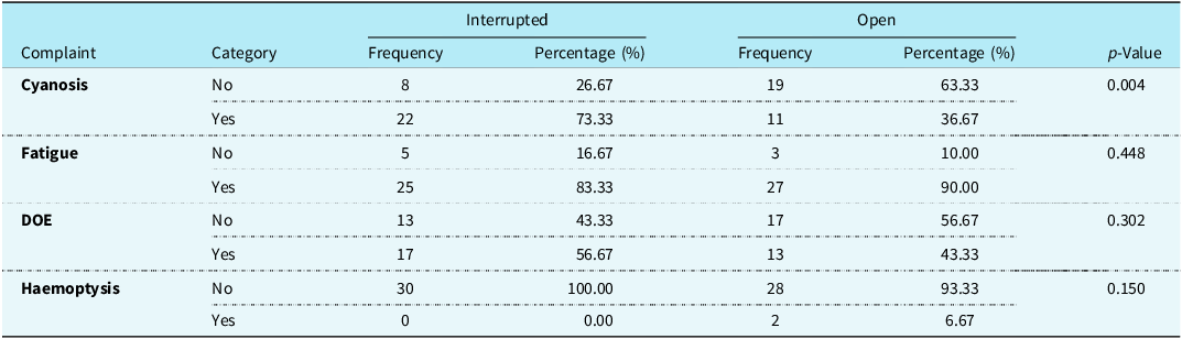
At the time of follow-up, cyanosis was significantly more common in patients with interrupted antegrade pulmonary blood flow. Twenty-two patients (73.3%) in the interrupted group reported cyanosis compared with 11 patients (36.7%) in the preserved group (p = 0.004). Fatigue and dyspnoea on exertion were frequent in both groups but did not differ significantly. Haemoptysis was observed only in the antegrade pulmonary blood flow-preserved group in two patients (6.7%), although this difference was not statistically significant. The distribution of presenting symptoms is detailed in Table 1. The higher incidence of cyanosis in the antegrade pulmonary blood flow-interrupted group is likely related to lower baseline oxygen saturation and underlying haemodynamic differences in patients in whom antegrade pulmonary blood flow was interrupted, rather than the effect of antegrade pulmonary blood flow interruption alone, and may reflect selection bias inherent to the retrospective study design.
Table showing the symptom frequency between the two groups

Table 1 Long description
The table presents the frequency and percentage of various symptoms in two groups of patients: those with interrupted antegrade pulmonary blood flow and those with open antegrade pulmonary blood flow. The table has four rows and six columns, including headers for complaint categories, frequency, and percentage for both interrupted and open groups, along with a p-value column. The complaints listed are cyanosis, fatigue, dyspnoea on exertion (DOE), and haemoptysis. For cyanosis, 22 patients (73.3%) in the interrupted group reported it compared to 11 patients (36.7%) in the open group, with a significant p-value of 0.004. Fatigue was reported by 25 patients (83.3%) in the interrupted group and 27 patients (90%) in the open group, with a p-value of 0.448. Dyspnoea on exertion was reported by 17 patients (56.7%) in the interrupted group and 13 patients (43.3%) in the open group, with a p-value of 0.302. Haemoptysis was observed in 0 patients in the interrupted group and 2 patients (6.7%) in the open group, with a p-value of 0.150. The table highlights that cyanosis is significantly more common in the interrupted group, while other symptoms do not show significant differences between the groups.
Pulmonary arteriovenous malformations
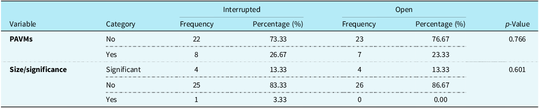
Pulmonary arteriovenous malformations were detected in 8 patients (26.7%) in the antegrade pulmonary blood flow-interrupted group and in 7 patients (23.3%) in the antegrade pulmonary blood flow-preserved group. This difference was not statistically significant (p = 0.766). Among these, clinically significant pulmonary arteriovenous malformations were identified in 4 patients (13.3%) in each group. The overall distribution and severity of pulmonary arteriovenous malformations are summarised in Table 2.
Table showing the incidence of PAVMs between the two groups

Table 2 Long description
The table presents data on the incidence of pulmonary arteriovenous malformations (PAVMs) in two groups: Interrupted and Open. It includes variables such as PAVMs presence and size/significance, with frequencies and percentages for each category. The Interrupted group has 22 patients without PAVMs (73.33%) and 8 with PAVMs (26.67%), while the Open group has 23 patients without PAVMs (76.67%) and 7 with PAVMs (23.33%). For size/significance, 4 patients in each group have significant PAVMs (13.33%), and 25 patients in the Interrupted group and 26 in the Open group have non-significant PAVMs (83.33% and 86.67%, respectively). One patient in the Interrupted group has significant size/significance (3.33%), with no such cases in the Open group. The p-values for these comparisons are 0.766 for PAVMs and 0.601 for size/significance.
PAVM = pulmonary arteriovenous malformation.
Echocardiographic and functional assessment
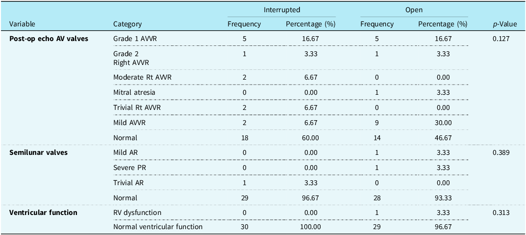
Post-operative echocardiographic evaluation demonstrated no significant differences between groups with respect to atrioventricular valve regurgitation, semilunar valve regurgitation, or ventricular function. The majority of patients in both groups had preserved ventricular function. Mild to moderate atrioventricular valve regurgitation was seen in a small proportion of patients without intergroup statistical significance. These findings are detailed in Table 3.
Showing the functional variables like atrioventricular valve status, semilunar valve status, and ventricular function between the two groups

Table 3 Long description
The table presents a comparison of functional variables between two groups, focusing on atrioventricular valve status, semilunar valve status, and ventricular function. It consists of three main categories: Post-op echo AV valves, Semilunar valves, and Ventricular function, each with various subcategories. The table has five columns: Variable, Category, Interrupted Frequency, Interrupted Percentage, Open Frequency, Open Percentage, and p-Value. Notable trends include a higher percentage of normal ventricular function in both groups, with 100 percentage in the Interrupted group and 96.67 percentage in the Open group. Mild AVVR is observed in 6.67 percentage of the Interrupted group and 30 percentage of the Open group. The p-Values indicate no significant differences between the groups for the variables measured.
AVVR = atrioventricular valve regurgitation; Rt = right; AR = aortic regurgitation; PR = pulmonary regurgitation; RV = right ventricle.
Oxygen saturation
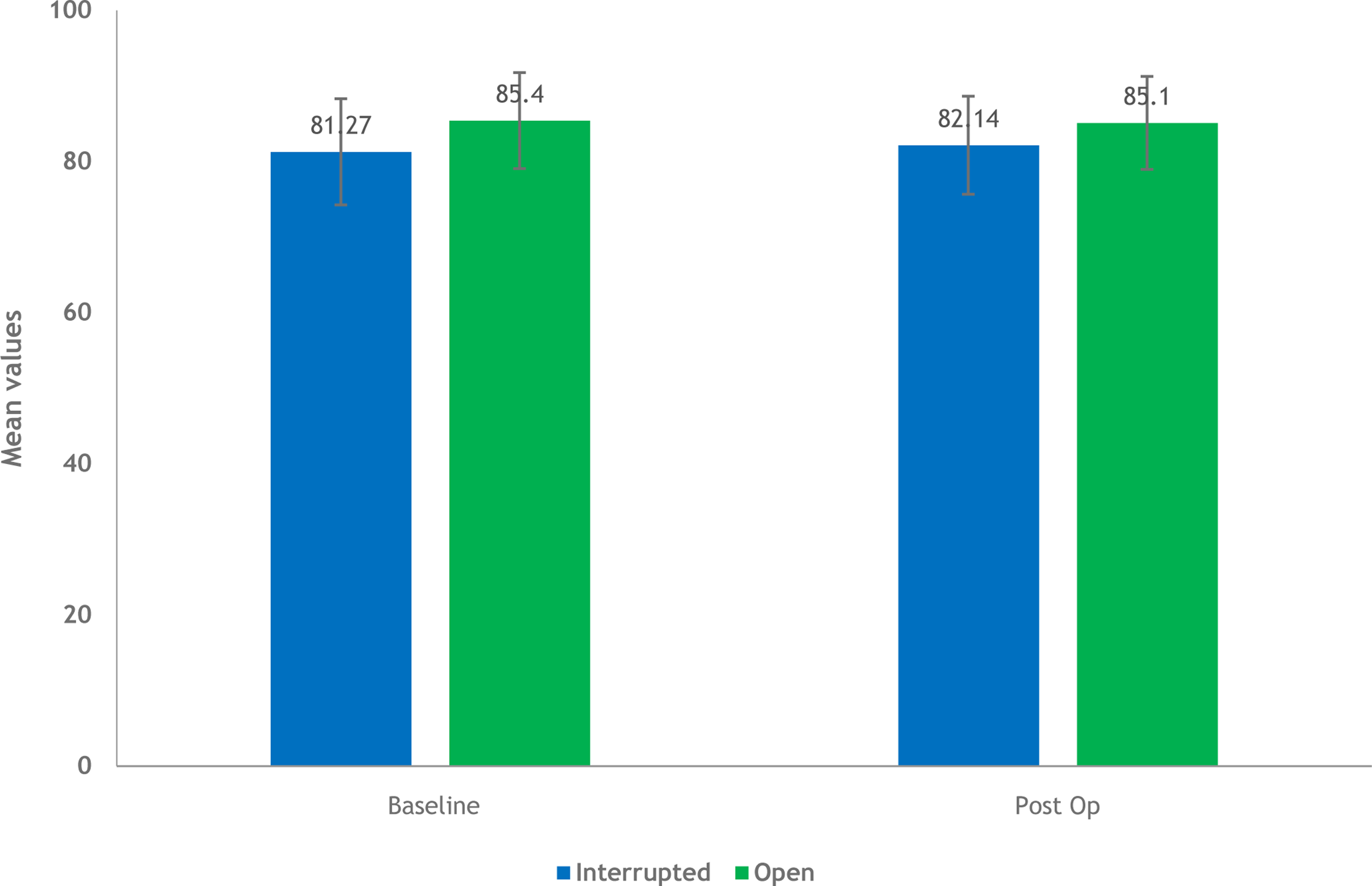
Baseline pre-Glenn oxygen saturation was significantly lower in the antegrade pulmonary blood flow-interrupted group compared with the antegrade pulmonary blood flow-preserved group (81.27 ± 7.03% versus 85.40 ± 6.35%; p = 0.020). At follow-up, oxygen saturation remained lower in the antegrade pulmonary blood flow-interrupted group (82.14 ± 6.48%) compared to the preserved group (85.10 ± 6.16%), although this difference did not reach statistical significance (p = 0.077). Trends in oxygen saturation are illustrated in Figure 1. The difference in baseline oxygen saturation between groups likely reflects underlying haemodynamic differences that influenced the intraoperative decision to interrupt antegrade pulmonary blood flow. The relatively modest increase in oxygen saturation following bidirectional superior cavopulmonary anastomosis in both groups may be related to the older age at bidirectional superior cavopulmonary anastomosis and long-standing cyanosis prior to surgery in this cohort. Importantly, although oxygen saturation tended to be higher in the antegrade pulmonary blood flow-preserved group, this did not translate into a reduction in pulmonary arteriovenous malformation incidence.
Bar chart showing the baseline saturations pre-Glenn and at follow-up.

Figure 1 Long description
The bar chart compares mean saturation values for two groups, interrupted and open, at two stages: baseline and post-operation. The x-axis represents the stages, with two categories: Baseline and Post Op. The y-axis represents mean values, ranging from 0 to 100. There are four vertical bars in total, two for each stage. At the baseline stage, the interrupted group has a mean value of 81.27, represented by a blue bar, while the open group has a mean value of 85.4, represented by a green bar. At the post-operation stage, the interrupted group has a mean value of 82.14, represented by a blue bar, and the open group has a mean value of 85.1, represented by a green bar. Each bar includes error bars indicating variability in the data. The color scheme uses blue for the interrupted group and green for the open group. All values are approximated.
Pulmonary artery dimensions and haemodynamics
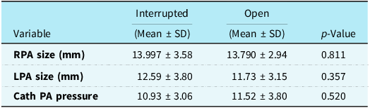
Assessment of pulmonary artery dimensions revealed no significant difference between the two groups. Mean right pulmonary artery diameter was 13.99 ± 3.58 mm in the interrupted group and 13.79 ± 2.94 mm in the preserved group (p = 0.811). Mean left pulmonary artery diameter was 12.59 ± 3.80 mm and 11.73 ± 3.15 mm, respectively (p = 0.357). Mean pulmonary artery pressures were also comparable between groups (10.93 ± 3.06 mmHg versus 11.52 ± 3.80 mmHg; p = 0.520). These findings suggest that interruption of antegrade pulmonary blood flow did not adversely affect pulmonary artery growth or haemodynamic suitability for Fontan completion in this cohort. These parameters, which reflect suitability for Fontan completion, are summarised in Table 4.
Showing the mean right pulmonary artery (RPA) and left pulmonary artery (LPA) sizes and pulmonary artery mean pressures between the two groups—showing Fontan feasibility

Table 4 Long description
The table presents a comparison of mean right pulmonary artery (RPA) size, left pulmonary artery (LPA) size, and pulmonary artery (PA) pressure between two groups: interrupted and open. The table has four rows and three columns. The columns are labeled Variable, Interrupted (Mean ± SD), Open (Mean ± SD), and p-Value. The rows list RPA size in millimeters, LPA size in millimeters, and catheter PA pressure in millimeters of mercury. For RPA size, the interrupted group has a mean of 13.997 millimeters with a standard deviation of 3.58, while the open group has a mean of 13.790 millimeters with a standard deviation of 2.94, and a p-value of 0.811. For LPA size, the interrupted group has a mean of 12.59 millimeters with a standard deviation of 3.80, while the open group has a mean of 11.73 millimeters with a standard deviation of 3.15, and a p-value of 0.357. For catheter PA pressure, the interrupted group has a mean of 10.93 millimeters of mercury with a standard deviation of 3.06, while the open group has a mean of 11.52 millimeters of mercury with a standard deviation of 3.80, and a p-value of 0.520. The data indicates no significant differences between the two groups in terms of RPA size, LPA size, and PA pressure.
Cath = catheterisation; PA = pulmonary artery.
Discussion
The present study evaluated the influence of antegrade pulmonary blood flow on the development of pulmonary arteriovenous malformations, systemic oxygenation, and haemodynamic suitability for Fontan completion following bidirectional Glenn surgery. The principal finding of this study is that preservation of antegrade pulmonary blood flow did not significantly reduce the incidence of pulmonary arteriovenous malformations when compared with interruption of antegrade pulmonary blood flow. Although patients with preserved antegrade pulmonary blood flow demonstrated higher oxygen saturations at follow-up, this did not translate into a measurable reduction in pulmonary arteriovenous malformation burden, supporting the concept that factors other than pulsatile pulmonary blood flow are primarily responsible for pulmonary arteriovenous malformation formation after cavopulmonary connection. Reference Chen, Tulloh, Caputo, Stoica, Kia and Parry10,Reference Baek, Park and Choi12,Reference Schreiber, Cleuziou, Cornelsen, Hörer, Eicken and Lange14 However, the management of additional pulmonary blood flow at the time of bidirectional superior cavopulmonary anastomosis remains controversial, with studies demonstrating both potential benefits and disadvantages of preserving antegrade flow. Reference Zahr, Kirshbom and Kopf16–Reference Alghamdi19
Pulmonary arteriovenous malformations are a well-recognised late complication following bidirectional superior cavopulmonary anastomosis and are an important contributor to progressive cyanosis and exercise intolerance. Reference Kavarana, Jones, Stroud, Bradley, Ikonomidis and Mukherjee2,Reference Duncan and Desai4,Reference McFaul, Tajik, Mair, Danielson and Seward5 The prevailing hypothesis attributes pulmonary arteriovenous malformation development to exclusion of hepatic venous effluent from the pulmonary circulation, resulting in loss of hepatic-derived angiogenesis inhibitors and subsequent abnormal pulmonary vascular remodelling. Reference Srivastava, Preminger and Lock3,Reference Vettukattil, Slavik and Lamb6,Reference Gossage and Kanj7 This mechanism has been supported by both experimental and clinical observations, including resolution of pulmonary arteriovenous malformations following redirection of hepatic venous blood to the pulmonary arteries. Reference Srivastava, Preminger and Lock3,Reference Vettukattil, Slavik and Lamb6 The findings of the present study are consistent with this paradigm, as pulmonary arteriovenous malformation incidence remained similar in both groups despite differences in pulmonary blood flow patterns, reinforcing the concept that hepatic factor deprivation rather than flow pulsatility is the dominant driver of pulmonary arteriovenous malformation development. Reference Srivastava, Preminger and Lock3,Reference Vettukattil, Slavik and Lamb6,Reference Gossage and Kanj7
Patients in whom antegrade pulmonary blood flow was preserved demonstrated higher resting oxygen saturations both at baseline and at follow-up. This is an expected physiological consequence of increased pulmonary blood flow and has been reported consistently in previous studies. Reference Allgood, Alejos, Drinkwater, Laks and Williams9–Reference Baek, Park and Choi12 However, improved oxygenation did not correlate with a lower prevalence of pulmonary arteriovenous malformations, suggesting that higher saturations reflect increased pulmonary perfusion rather than improved pulmonary vascular integrity. This distinction is clinically important, as reliance on oxygen saturation alone may obscure ongoing pulmonary vascular pathology. Reference Kavarana, Jones, Stroud, Bradley, Ikonomidis and Mukherjee2,Reference Duncan and Desai4,Reference Vettukattil, Slavik and Lamb6 While improved saturations may offer symptomatic benefit in the intermediate term, they do not appear to confer protection against long-term complications related to abnormal pulmonary vascular development. Reference Chen, Tulloh, Caputo, Stoica, Kia and Parry10,Reference Baek, Park and Choi12,Reference Schreiber, Cleuziou, Cornelsen, Hörer, Eicken and Lange14
The modest and statistically non-significant increment in oxygen saturation observed in the antegrade pulmonary blood flow-preserved group at follow-up should not be considered problematic. Prior studies have demonstrated variability in the magnitude and durability of oxygenation benefit associated with antegrade pulmonary blood flow, influenced by patient selection, timing of bidirectional superior cavopulmonary anastomosis, and evolving pulmonary vascular physiology. Reference Chen, Tulloh, Caputo, Stoica, Kia and Parry10,Reference Frommelt, Frommelt and Berger13 Moreover, additional pulmonary blood flow after bidirectional superior cavopulmonary anastomosis has been associated with increased ventricular volume load, prolonged pleural effusions, and higher central venous pressures in some reports, potentially offsetting the perceived benefit of higher saturations. Reference Frommelt, Frommelt and Berger13,Reference Schreiber, Cleuziou, Cornelsen, Hörer, Eicken and Lange14 Recent studies have also shown that although additional pulmonary blood flow may improve oxygenation in the short term, its effect on long-term Fontan outcomes and pulmonary vascular development remains uncertain, and individualised decision-making is generally recommended. Reference Chen, Yuan and Liu18–Reference Lee, Amabile, Geirsson, Gruber and Kopf21 Accordingly, the lack of a large saturation increase in our cohort does not contradict the short-term physiological effects of antegrade pulmonary blood flow but highlights that any oxygenation advantage is limited and does not equate to protection from pulmonary arteriovenous malformation formation. Reference Chen, Tulloh, Caputo, Stoica, Kia and Parry10,Reference Baek, Park and Choi12,Reference Schreiber, Cleuziou, Cornelsen, Hörer, Eicken and Lange14
An important observation in this study is the comparable pulmonary artery dimensions and pulmonary artery pressures between the two groups. Patients with interrupted antegrade pulmonary blood flow did not demonstrate inferior pulmonary artery growth or unfavourable haemodynamics, indicating that elimination of antegrade flow does not compromise pulmonary vascular development or Fontan candidacy. In fact, mean pulmonary artery pressures tended to be lower in the antegrade pulmonary blood flow-interrupted group, a finding that may be advantageous in the context of Fontan physiology, where elevated pulmonary vascular resistance is a key determinant of long-term failure. Reference Mainwaring, Lamberti, Uzark, Spicer, Cocalis and Moore11,Reference Frommelt, Frommelt and Berger13 These findings align with prior reports suggesting that ventricular unloading achieved by eliminating antegrade pulmonary blood flow may offset the theoretical benefits of increased pulmonary blood flow. Long-term follow-up studies of cavopulmonary connections have similarly demonstrated acceptable survival and functional outcomes with different sources of pulmonary blood flow, further supporting an individualised approach. Reference Zahr, Kirshbom and Kopf16,Reference Steele, Zahr, Kirshbom, Kopf and Karimi17,Reference Delikatnyi, Sekelyk, Maksymenko and Kurkevych20
With regard to age at bidirectional superior cavopulmonary anastomosis, all patients in the present study were younger than 15 years at follow-up, reflecting a cohort in which Glenn procedures were performed later than current international practice, where bidirectional superior cavopulmonary anastomosis is typically undertaken in early infancy. Reference Uemura, Yagihara, Kawashima, Okada, Kamiya and Anderson8,Reference Chen, Tulloh, Caputo, Stoica, Kia and Parry10 Delayed presentation and staged palliation are common in low- and middle-income settings due to referral patterns, socio-economic factors, and limited access to specialised paediatric cardiac care. Although timing of bidirectional superior cavopulmonary anastomosis may influence pulmonary vascular adaptation, both antegrade pulmonary blood flow groups in our cohort were demographically and haemodynamically comparable, supporting the validity of intergroup comparisons. Importantly, the persistence of similar pulmonary arteriovenous malformation rates across both groups despite differences in antegrade pulmonary blood flow status further supports the hypothesis that hepatic venous exclusion, rather than age at bidirectional superior cavopulmonary anastomosis or the presence of pulsatile flow, is the principal determinant of pulmonary arteriovenous malformation development. Reference Srivastava, Preminger and Lock3,Reference Vettukattil, Slavik and Lamb6,Reference Gossage and Kanj7 The higher incidence of cyanosis observed in the antegrade pulmonary blood flow-interrupted group should be interpreted in the context of lower baseline oxygen saturation and underlying haemodynamic differences rather than as a direct effect of antegrade pulmonary blood flow interruption, reflecting the retrospective and non-randomised nature of the study.
The clinical implications of these findings are particularly relevant in resource-limited settings, where timing of staged palliation and long-term follow-up may be variable. Preservation of antegrade pulmonary blood flow may offer short-term improvement in oxygenation but at the potential cost of increased ventricular volume load. Reference Frommelt, Frommelt and Berger13,Reference Schreiber, Cleuziou, Cornelsen, Hörer, Eicken and Lange14 Conversely, interruption of antegrade pulmonary blood flow appears to provide a haemodynamically favourable profile without increasing the risk of pulmonary arteriovenous malformation formation. Reference Allgood, Alejos, Drinkwater, Laks and Williams9,Reference Schreiber, Cleuziou, Cornelsen, Hörer, Eicken and Lange14 These observations support an individualised surgical strategy rather than a uniform approach, with consideration of ventricular function, pulmonary artery pressures, and clinical status when deciding on antegrade pulmonary blood flow management. Reference Kavarana, Jones, Stroud, Bradley, Ikonomidis and Mukherjee2,Reference Srivastava, Preminger and Lock3,Reference Vettukattil, Slavik and Lamb6,Reference Mahle, Wernovsky, Bridges, Linton and Paridon15
Several limitations must be acknowledged. The retrospective design introduces potential selection bias, particularly as the decision to preserve or interrupt antegrade pulmonary blood flow was surgeon-dependent. The sample size, while comparable to other single-centre studies, may be underpowered to detect small differences in pulmonary arteriovenous malformation severity. Quantitative assessment of shunt fraction and exercise capacity was not available, and follow-up duration, although sufficient to detect early pulmonary arteriovenous malformation formation, may not capture late-onset changes. Additionally, the absence of biochemical or molecular markers of hepatic factor activity limits mechanistic interpretation.
Despite these limitations, this study contributes meaningful data to an area where evidence remains limited and conflicting. By directly comparing pulmonary arteriovenous malformation incidence between antegrade pulmonary blood flow-preserved and antegrade pulmonary blood flow-interrupted cohorts using imaging-based assessment, the study provides clinically relevant insights into the long-standing debate regarding optimal Glenn physiology. Reference Chen, Tulloh, Caputo, Stoica, Kia and Parry10,Reference Baek, Park and Choi12–Reference Schreiber, Cleuziou, Cornelsen, Hörer, Eicken and Lange14 The findings reinforce the concept that prevention of pulmonary arteriovenous malformations is primarily related to restoration of hepatic venous flow rather than maintenance of antegrade pulmonary flow.
Conclusion
The preservation of antegrade pulmonary blood flow after bidirectional Glenn was not associated with a reduction in pulmonary arteriovenous malformation incidence in this study, although it was associated with slightly higher systemic oxygen saturation. Interruption of antegrade pulmonary blood flow did not adversely affect pulmonary artery growth or haemodynamic suitability for Fontan completion. These findings suggest that the development of pulmonary arteriovenous malformations is more closely related to the absence of hepatic venous effluent than to the presence or absence of antegrade pulmonary blood flow. However, given the retrospective design and limited sample size, the decision to preserve or interrupt antegrade pulmonary blood flow should be individualised based on ventricular function, pulmonary artery pressure, atrioventricular valve competence, and overall haemodynamic considerations rather than applied as a uniform strategy.