Primary cardiac tumours, particularly vascular tumours, are rare in children. Our understanding of their natural history and optimal management is limited to a small number of cases described in the literature. We present a diagnostically challenging case of a ventricular mass in which echocardiography, cardiac magnetic resonance imaging, and pathological specimens obtained via left heart catheterisation led to the diagnosis of an intramuscular vascular malformation of the heart.
Presentation
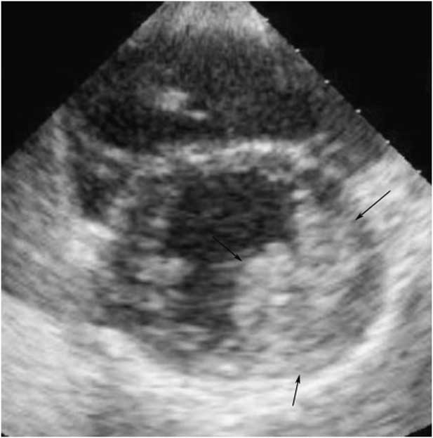
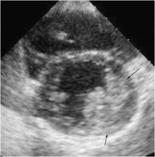
A healthy and asymptomatic 12-year-old girl presented for further evaluation of a murmur of increasing intensity. A murmur was originally auscultated 3 years ago, and the evaluation at that time included electrocardiography and echocardiogram. The electrocardiography was normal. The echocardiogram demonstrated mild mitral regurgitation but was otherwise thought to be benign. Physical examination revealed a grade 1–2/6 systolic murmur along the left sternal border without radiation and a 12-lead electrocardiogram was normal for her age. A repeat transthoracic echocardiogram revealed an ∼4.5 cm mass associated with the left ventricular free wall, with unobstructed left ventricular inflow and outflow and otherwise normal anatomy (Figure 1). In retrospect, the mass was visible but not clearly demonstrated on the prior echocardiogram. It appeared to be unchanged in size.
Parasternal short-axis projection demonstrating the mass involving the left ventricular free wall.
Diagnostic evaluation
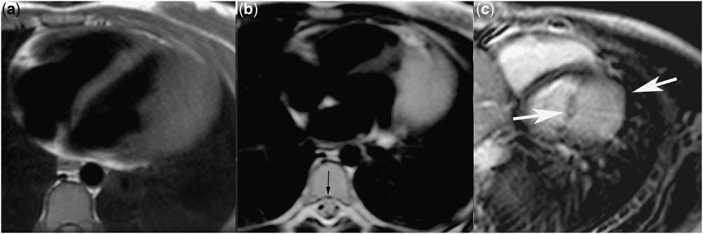
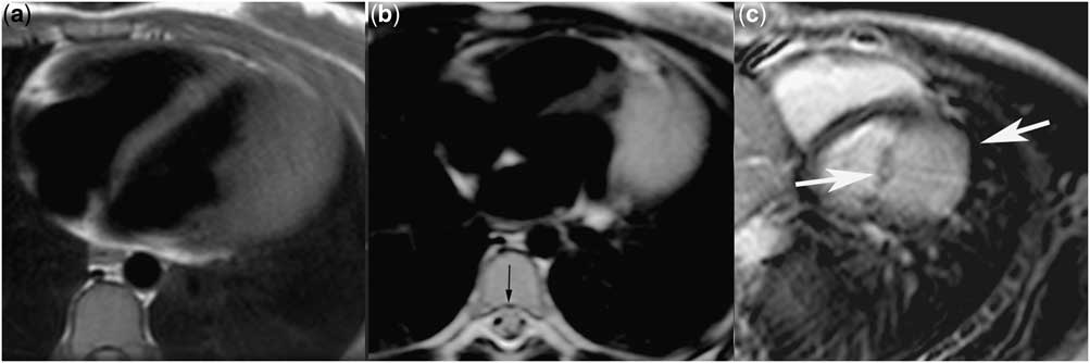
Magnetic resonance imaging revealed a 4.5 × 4.5 cm unobstructive mass within the posterior and lateral aspect of the left ventricular free wall. The mass was found to be hyperintense on T2-weighted images in comparison with the normal myocardium of the interventricular septum and was similar in intensity when referenced to the typically high-signal spinal fluid visualised in the spinal column (Figure 2B). The mass was of similar intensity to the normal myocardium on T1 turbo spin-echo acquisitions (Figure 2A). There was no signal loss on using fat-suppressed turbo spin-echo pulse sequences. First-pass perfusion imaging revealed marked contrast enhancement, which is suggestive of increased vascularity. Transmural gadolinium enhancement was observed on delayed imaging (Figure 2C, white arrows). The signal intensity of the enhanced tumour was not notably high but was similar to that of the normal blood pool, favouring enhancement of the blood pool within the tumour rather than fibrosis or necrosis. These magnetic resonance imaging characteristics have been documented in both benign and malignant vascular masses of the heart, such as angiosarcomas.Reference Sparrow, Kurian and Jones 1 , Reference Beroukhim, Prakash and Valsangiacomo Buechel 2 Given the concern for potential malignancy, both a chest and abdominal computed tomography were obtained and were found to be negative for evidence of metastatic disease.
( a ) Electrocardiography-gated T1-weighted image in axial plane (TR, 666.7 ms; TE, 10 ms; field of view, 20 cm; matrix, 256 × 256; 6 averages) showing that the tumour is isointense to the normal myocardium. ( b ) Electrocardiography-gated T2-weighted image in axial plane (TR, 1333 ms; TE, 100 ms; slice thickness, 7 mm; field of view, 30 cm; matrix, 448 × 448; 6 averages) showing a hyperintense mass involving the left ventricular free wall. Note that the signal intensity of the mass is similar to that of the cerebrospinal fluid (black arrow). ( c ) A late gadolinium enhancement image in the short-axis plane showing homogeneous enhancement of the tumour (white arrows) with the intensity similar to that of the blood pool.
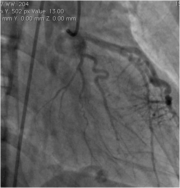
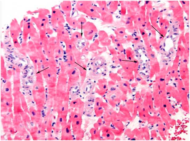
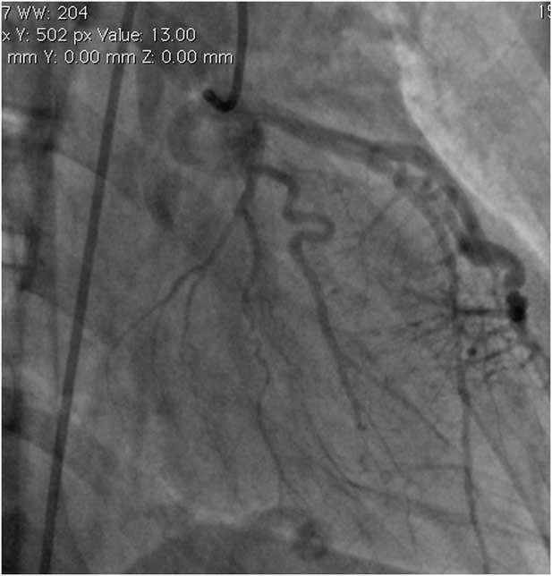
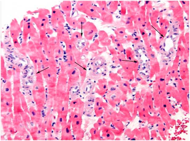
Angiography during left heart catheterisation revealed a convex distortion of the posterior and lateral wall of the left ventricular chamber bordered by an enlarged ramus of the left coronary system. Selective left coronary angiography revealed normal calibre left main, left circumflex, and left anterior descending coronary arteries. The ramus medialis was observed to be significantly enlarged, giving off numerous tortuous branches to a hypervascular and hypertrophied portion of the posterior and lateral left ventricle. In addition, there was evidence of a delayed washout of the contrast in the distribution of the mass (Figure 3). The biopsy specimens revealed myocardial tissues with increased small vascular channels, without evidence of nuclear atypia or mitotic activity (Figure 4). The findings were consistent with an intramuscular vascular malformation of the small vessel arteriovenous subtype – formerly referred to as intramuscular haemangiomas. Immunohistochemical analysis revealed the vascular cells to be negative for Glut-1, excluding an infantile haemangioma.
A left coronary artery angiogram demonstrating a markedly dilated medial ramus branch giving rise to numerous tortuous branches to the hypervascular left ventricular mass.
Haematoxylin and eosin staining of biopsy of the left ventricular mass demonstrating normal myocardium infiltrated with numerous small vessels (arrows).
Management
On the basis of our understanding of the biology of this mass, we identified three primary risks including the following: (1) ventricular arrhythmia, (2) enlargement via recruitment of vessels, potentially compromising myocardial function and causing obstruction or creating a clinically evident left-to-right shunt, and (3) bleeding by erosion into the pericardium. We concluded that both surgical resection and transcatheter embolisation techniques would present undue risk for myocardial and/or coronary artery injury in a currently asymptomatic patient. Medical therapies such as beta-blockers, corticosteroids, or novel antiangiogenic therapies such as vascular endothelial growth factor inhibitors were also considered. Currently, there is much interest in using beta-blockers to treat infantile haemangiomas, particularly in PHACE syndrome.Reference Léauté-Labrèze, Dumas de la Roque and Hubiche 3 – Reference Denoyelle, Leboulanger and Enjolras 5 Importantly, infantile haemangiomas are characteristically Glut-1 positive, and the effectiveness of beta-blocker therapy has not been demonstrated in Glut-1-negative lesions. In our opinion, the potential for myocardial thinning or infarction did not justify initiation of treatment in the absence of symptoms or a change in size of the mass over a 3-year period.
Subsequent to obtaining a tissue diagnosis, continued close surveillance of this patient over the past 30 months has included serial echocardiograms that have shown no change in the size or appearance of the mass or evolution of important haemodynamic compromise. In addition, yearly exercise testing and ambulatory electrocardiography monitoring have not demonstrated significant ectopy or important deviation from sinus rhythm. As such, we have not limited this patient from participation in high school-level competitive athletics. Accordingly, she has participated in competitive soccer and tennis. We have not pursued further medical or surgical interventions.
Discussion
Cardiac tumours are rare in children, with primary vascular tumours of the heart comprising <10% of these cases.Reference Mackie, Kozakewich and Geva 6 The differential diagnosis of this mass upon presentation spanned diagnoses seen in both children and adults and included cardiac rhabdomyoma, cardiac myxoma, cardiac fibroma, and vascular tumours including infantile or congenital haemangiomas and vascular malformations.Reference Mackie, Kozakewich and Geva 6 , Reference Beghetti, Gow and Haney 7 Malignant tumours such as angiosarcomas or rhabdomyosarcomas are less common than benign tumours and were also considered.Reference Sparrow, Kurian and Jones 1
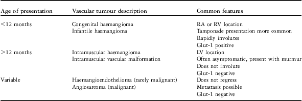
Vascular tumours of the heart are variable in their clinical and histopathological features, and imprecise nomenclature has contributed to the challenge of understanding their natural history and optimal management. A summary of the most common classifications found in the literature and their associated clinical features appears in Table 1. In an excellent review of the literature, Mackie et al summarised 36 reported cases of primary vascular tumours of the heart in children, adding six additional cases from their own institutions. This report emphasises the clinically important association between the age at presentation and both the anatomical and pathological features of the mass and response to therapy. Similar to our patient, Mackie et alReference Mackie, Kozakewich and Geva 6 reported that cardiac vascular tumours presenting after 12 months of age (n = 19) were more frequently found in asymptomatic patients presenting with a cardiac murmur (7/19), with the mass more commonly involving left heart structures (11/19).
Classifications of cardiac vascular tumours in children according to the age at presentation as cited in the literature.

RA = right atrium; RV = right ventricle; LV = left ventricle
Highlighting the rarity of these lesions, our own review of the literature has revealed eight subsequent case reports on primary vascular tumours of the heart in children.Reference Laga, Gewelling and Van Schoubroeck 8 – Reference Sharma, Hsu and Weinstein 13 Laga et al, Watanabe et al, and Sharma et alReference Laga, Gewelling and Van Schoubroeck 8 – Reference Sharma, Hirata and Mosca 10 have described five cases of neonatal haemangioma, two of which were associated with an evolving tamponade requiring emergent intervention. Moniotte et alReference Moniotte, Geva and Perez-Atayde 11 have described the case of a 12-year-old boy presenting with a heart murmur diagnosed by echocardiography and magnetic resonance imaging with a pedunculated haemangioma associated with the right ventricular outflow tract, which was successfully resected. Zerbo et al presented a dramatic case report of sudden cardiac death in a 15-year-old boy who was found on autopsy to have an intramural mixed small vessel and cavernous vascular malformation located within the cardiac apex, with evidence of intramuscular haemorrhage. The mechanism of death in this patient was not clearly proven but was hypothesised to be related to ventricular arrhythmia.Reference Zerbo, Argo and Maresi 12 Most recently, Sharma et alReference Sharma, Hsu and Weinstein 13 reported the successful resection of an obstructive haemangioma in the right ventricular outflow tract, which presented in adolescence.
Our patient represents one of a select few cases reported in the paediatric literature of an intramuscular vascular malformation of the small vessel arteriovenous subtype of the left ventricle. Traditionally, these lesions have been referred to as intramuscular haemangiomas. The small vessel arteriovenous subtype typically contains arterial and venous components with moderate-to-high intralesional flow, and the histopathological characteristics of this tissue are distinct from infantile or congenital haemangiomas. Vascular malformations are thought to represent a developmental anomaly, rather than an intrinsically proliferative tumour, and have a unique mode of progression involving enlargement via recruitment of additional collateral flow as opposed to deregulated cell division and growth, as demonstrated in haemangiomas. In addition, such arteriovenous malformations can result in clinically important arterial-to-venous shunting as seen with arteriovenous malformations located elsewhere in the body.Reference Buckmiller, Richter and Suen 14
The magnetic resonance imaging characteristics of the tumour were particularly useful in identifying the vascular nature of the mass and narrowing the differential diagnosis. Magnetic resonance imaging affords excellent soft-tissue characterisation to allow accurate assessment of the tissue type and extent of involvement of surrounding tissues. Enhancement properties with gadolinium aided in the exclusion of malignancy.Reference Sparrow, Kurian and Jones 1 , Reference Beroukhim, Prakash and Valsangiacomo Buechel 2 The ability to safely biopsy the lesion under fluoroscopic and echocardiographic guidance in the cardiac catheterisation lab ultimately facilitated the final diagnosis.
Conclusions
Primary vascular tumours of the heart are rare and varied in their presentation and classification. We present a case of a primary intramuscular vascular malformation of the small vessel arteriovenous subtype arising in the left ventricle in an asymptomatic 12-year-old girl. Magnetic resonance imaging played a crucial role in characterising the vascular nature of this mass, which was confirmed pathologically. We have chosen a “watchful waiting” approach to the ongoing management, with serial imaging and electrophysiological surveillance guiding our decision to avoid intervention or restricting athletic participation in this adolescent patient. During a period of more than 30 months of monitoring, there has been no change in the appearance of the mass or any concerning symptoms.
Acknowledgements
The authors would like to thank Dr Laurie Armsby for providing the catheterisation data and angiograms.