Excessive blood flow is a challenge in many congenital cardiac anomalies. A common instance is collateral pulmonary circulation in the setting of congenital pulmonary atresia or tetralogy of Fallot. These abnormal circulatory pathways should be occluded when total correction of the congenital cardiac anomaly is desired.Reference Boshoff and Gewillig 1 Another instance is excessive pulmonary circulation via a patent ductus arteriosus, which implies additional burden to the heart. Currently, these vessels are closed using dedicated devices or surgical methods. For congenital defects in which a single-stage correction is not possible, pulmonary artery banding is performed for lung protection and preparation for the next procedure. Banding entails a surgical approach.Reference Holzer and Cheatham 2 , Reference Yuan and Jing 3 In most situations, concomitant closure of the patent ductus arteriosus is performed to curb the abnormal aortopulmonary flow.
Radiofrequency energy is a type of electromagnetic energy commonly used for elimination of arrhythmic substrates in the heart. Delivery of radiofrequency causes irreversible cell death when the intracellular temperature rises beyond 50–55°C.Reference Haines 4 When radiofrequency is applied to vascular structures, it can cause narrowing of the vessel; pulmonary venous stenosis after pulmonary vein isolation in patients with atrial fibrillation is an example.Reference Holmes, Monahan and Packer 5 The other reported instance is coronary artery stenosis after radiofrequency application in the hearts of small children.Reference Castaño, Crawford, Yamazaki, Avula and Kalifa 6 The use of radiofrequency energy to voluntarily cause vascular closure or blood flow restriction has not been reported in the cardiology literature till date.
Method
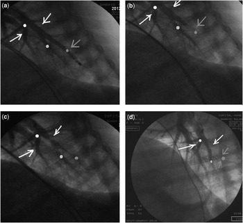
A 10-month-old sheep weighing 20 kg was chosen for the study. The sheep was sedated with intramuscular ketamine and laid on its right side on the fluoroscopy bed. Two accesses were obtained from the right femoral vein. A 6-Fr pigtail catheter (Boston Scientific Corporation, Boston, United States of America) was advanced into the heart and then into the left pulmonary artery. A 7-Fr ablation catheter (Osypka, Rheinfelden, Germany) was advanced into the terminal branch of the left pulmonary artery via the other venous access. An iodinated contrast injection into the main pulmonary artery (20 ml in 1 second) revealed the anatomy (Fig 1a). Radiofrequency energy of 80 W with a temperature limit of 70°C was delivered to the part of the branch having a diameter of about 3 mm for 3 minutes using a radiofrequency generator (Atakr® II, Medtronic, Minneapolis, United States of America). Re-injection of the contrast as above showed near cut-off at the distal vessel (Fig 1b). The ablation catheter was pulled more proximally to a point where the diameter of the vessel was about 7 mm, and radiofrequency energy was applied using the same settings as earlier. After 3 minutes, severe narrowing – about 80% stenosis – was observed at the site of radiofrequency application (Fig 1c). A total of five additional radiofrequency applications of 3 minutes each were administered at the same site to observe for additional effects or complications. After each application, angiography was repeated. No further narrowing or complete closure was observed, with additional burns at the second site. Vascular rupture or perforation did not occur. After the procedure, the sheep was transferred to the animal ward in good health.
Stepwise contrast injection before, during, early, and late after radiofrequency delivery. ( a ) Contrast injection in the main pulmonary artery revealed the exact size and shape of the terminal pulmonary artery branches; the ablation catheter is seen in a terminal branch. ( b ) After a 3-minute radiofrequency delivery, the terminal branch is nearly occluded (red arrow). ( c ) Radiofrequency application in the proximal larger part of the pulmonary artery branch resulted in severe narrowing (yellow arrow). ( d ) Angiography after 1 month revealed a totally occluded terminal branch (red arrow) and residual stenosis at the proximal part (yellow arrow). White arrow indicates a good contrast filling in the frames.

After 1 month, the sheep was re-evaluated. Its weight was 21 kg. A venous access was obtained, and a 6-Fr pigtail catheter was advanced into the heart and then into the pulmonary artery. An iodinated contrast injection in the main pulmonary artery (20 ml in 1 second) revealed that the site of the terminal branch cut-off remained occluded, but the stenosis had reduced to about 60% at the second site (Fig 1d).
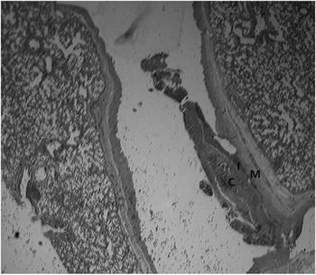
After 1 week of re-evaluation, the heart and lungs were excised and sent for histopathological analysis by a veterinary pathologist. Gross examination of the specimen revealed no significant pathological changes. Microscopically, organised thrombus formation was seen attached to the inner layer of the vessel at the site of energy delivery. The base of the thrombosis, which was attached to the intimal layer, showed migration and proliferation of fibroblasts and angioblasts, resulting in granulation tissue formation. The intimal layer of the vessel was destroyed, whereas the media, adventitia, and extra-vascular pulmonary parenchyma were intact (Fig 2).
Histopathology of the site of ablation: intimal cells (I) were damaged by radiofrequency application and a clot (C) was formed inside the vessel. The media layer (M) and extravascular tissue are intact.

Conclusion
Radiofrequency energy could be used as a potential tool for voluntary restriction of blood flow to the lung or other target parts of the body. Total closure of small vessels is possible. Similarly, partial flow restriction in larger vessels could perhaps be achieved. Further studies are needed on the regulation of blood flow when total occlusion is not desired. The maximal effect of the burn is achieved within the first few minutes. If this technique is confirmed to be safe on larger safety studies, it could be an effective alternative to pulmonary artery banding, closure of patent ductus arteriosus and collateral vessels, reduction of vascularity in highly vascular tumours, and restriction of blood flow in other desired parts of the body, thus serving as an effective alternative to embolisation therapies. The technique is relatively inexpensive, and at the end of procedure the patients would not have a foreign body within them. Evaluation of this technique in different vessels and scenarios would be undertaken.
Acknowledgements
Amir Darbandi Azar, veterinarian, is highly appreciated for preparing the animal and for its care during the procedure. Mohammad Javad gharagoozloo, professor of veterinary pathology, examined the lung for pathophysiological analysis of the procedure. The author thanks Dr Jayakeerthi Y Rao for his contribution toward preparation of the manuscript. Funding organisations: All costs were borne by the researcher. Rajaei Cardiovascular Medical and Research Center provided the animal laboratory and the required equipment.