Introduction
Deficits in multiple cognitive domains have been held to act as pathways to the core symptoms of attention deficit hyperactivity disorder (ADHD; Sonuga-Barke, Reference Sonuga-Barke2005; Durston et al., Reference Durston, van Belle and de Zeeuw2011; Castellanos and Proal, Reference Castellanos and Proal2012). Meta-analyses demonstrate associations between ADHD symptoms and deficits in working memory, general intelligence, and some attentional measures, along with the processing of rewards and emotionally charged stimuli (Lijffijt et al., Reference Lijffijt, Kenemans, Verbaten and van Engeland2005; Martinussen et al., Reference Martinussen, Hayden, Hogg-Johnson and Tannock2005; Willcutt et al., Reference Willcutt, Doyle, Nigg, Faraone and Pennington2005a; Huang-Pollock et al., Reference Huang-Pollock, Karalunas, Tam and Moore2012). While there is a large literature on cognitive deficits and their underlying brain function in ADHD (Cortese et al., Reference Cortese, Kelly, Chabernaud, Proal, Di Martino, Milham and Castellanos2012), there are fewer studies into the neuroanatomic substrates of these skills among those with the disorder. Thus, the first motivation for this study is the need to map the neuroanatomic substrates of cognitive skills pertinent to ADHD. Additionally, it is unclear if the neuroanatomic correlates of cognition overlap with the neuroanatomic correlates of the core symptoms of ADHD. This is an important question, as a high degree of overlap is not inevitable. The symptoms of ADHD are behaviorally complex, and the underlying neuroanatomic change may differ from the neural basis of more circumscribed cognitive abilities.
This study also aims to examine whether the cognitive profile associated with ADHD varies with age. One way to address this question is to use data from both multi-generational, extended and nuclear families that have members affected by ADHD. Families provide a relatively genetically and environmentally homogenous context for probing brain-cognition relationships. The inclusion of older individuals allows us to ask if the cognitive profile of ADHD is stable across multiple generations of the same family. In turn, this can partly address concerns that ADHD persisting into adulthood might be accompanied by accumulating cognitive deficits, as the disorder can entail a loss of educational and occupational opportunities (Doshi et al., Reference Doshi, Hodgkins, Kahle, Sikirica, Cangelosi, Setyawan, Erder and Neumann2012).
The third motivation for this study is the issue of whether lifetime use of psychostimulant medication as a treatment for ADHD might be associated with cognitive skills and their neural substrates. Again, the use of a multigenerational family design which includes both children and adults from relatively homogenous backgrounds provides a design suited to test for associations between the lifetime use of psychostimulant medication, cognition and neuroanatomy.
In summary, using family-based data, we first delineate the neuroanatomic correlates of cognitive abilities pertinent to ADHD and ask if they vary significantly with age. Second, we determine the degree of overlap between the neuroanatomy of these key cognitive abilities and the neuroanatomic correlates of the symptoms of the disorder. Finally, we test for associations between cognitive skills, the brain and the lifetime history of psychostimulant medication.
Methods
The study included 264 individuals, of whom 188 came from 25 extended multigenerational families and 76 from 35 nuclear families. Extended families were defined by the presence of a diagnosis of ADHD in at least 25% of family members, which represents a marked increase in background population prevalence rates of childhood and adult ADHD (Polanczyk et al., Reference Polanczyk, de Lima, Bernardo, Biederman and Rohde2007; Simon et al., Reference Simon, Czobor, Balint, Meszaros and Bitter2009). In the nuclear families, at least one member had ADHD. The study was approved by the institutional review boards of the National Institute of Mental Health and the National Human Genome Research Institute. Written informed consent was obtained from adult participants and parents; children gave written assent.
To make the diagnosis of ADHD in adults, the Conners’ Adult ADHD Diagnostic Interview for DSM-IV was used (Epstein and Johnson, Reference Epstein and Johnson2001). This clinician-administered structured interview ascertains symptoms of inattention and hyperactivity–impulsivity in adulthood and the childhood history of these symptoms. The Structured Clinical Interview for DSM-IV-TR Axis I Disorders, Research Version, Patient Edition was used to ascertain other psychiatric disorders (First et al., Reference First, Spetzer, Gibbon and Williams2002). The parental Diagnostic Interview for Children and Adolescents-IV (DICA) (Reich, Reference Reich2000) was used to obtain diagnoses in children. Interviews were conducted by two experienced clinicians (W.S. and P.S.). General exclusion criteria were an IQ < 80 (Wechsler, Reference Wechsler2001, Reference Wechsler2011), neurologic disorders affecting brain structure, current substance dependence, or psychotic disorders. Twenty-three of the 25 extended families and 24 of the 35 nuclear families were white, non-Hispanic (see online Supplementary Table S1). Details on medications and comorbidities are given in online Supplementary Table S2.
Neuropsychological testing was conducted by research fellows supervised by a clinical neuropsychologist. If subjects were taking psychostimulant medication, this was withdrawn at least 24 h before testing. Processing speed was assessed by two tests from the Woodcock–Johnson battery (Woodcock et al., Reference Woodcock, McGrew and Mather2001), visual matching, and decision speed. The visual matching task measures perceptual processing speed by asking subjects to make visual comparisons under time pressure. In this task, subjects must locate and circle two identical numbers in each row of six numbers; pairs become progressively more challenging to identify as single digits progress to double and triple digits. The decision speed task requires participants to analyze rows of six images and choose the two that are most closely related in each row under time pressure. Verbal working memory span was assessed through the number of correctly recalled digits in the original and reverse order. Number chains progressed from three digits up to nine in the original order, and two digits up to eight in the reverse order. Spatial working memory span was measured through the number of correctly repeated tapping patterns in the original and reverse order (Wechsler, Reference Wechsler2003). Intelligence was estimated from age-appropriate versions of the Wechsler intelligence scales (Wechsler, Reference Wechsler2001, Reference Wechsler2011). Attentional abilities were measured through the Conners’ continuous performance test (CPT) (Conners, Reference Conners2004). This task requires participants to press a key when presented with any stimulus letter, except the letter ‘X’. Based on prior factor analyses of the CPT, we calculated indices reflecting inattentive responding (reaction time variability and the standard error of hit reaction time, detectability, and omission errors), impulsive responding (commission errors, reaction time, response style, and perseverations), sustained attention (hit reaction time during block changes and its standard error) and vigilance (reaction time during inter-stimulus interval changes and its standard error) (Egeland and Kovalik-Gran, Reference Egeland and Kovalik-Gran2008).
Neuroimaging
A high-resolution (1.07 × 1.07 × 1.2 mm3) T1 weighted volumetric structural image was obtained using a magnetization prepared rapid gradient echo sequence (with ASSET preparation; 124 slices, 1.2 mm slice thickness, 224 × 224 acquisition metric, flip angle = 6°, field of view = 24 cm2) on a 3 T General Electric Signa scanner (USA) using an eight-channel head coil. Analyses were conducted on the National Institutes of Health High Performance Computer Cluster (Biowulf). Cerebral cortical reconstruction and cortical and subcortical volumetric segmentation were performed with the FreeSurfer image analysis suite version 5.3.0 (http://surfer.nmr.mgh.harvard.edu/). Technical procedures of this widely used method are described in the web link above. Analyses were conducted on the National Institutes of Health High Performance Computer Cluster (Biowulf). We chose volumes as our metric as these can be defined using FreeSurfer on both cortical and subcortical structures, unlike metrics such as thickness or gyrification that apply only to the cortex. All T1 images were visually inspected, and those with moderate or severe motion or other artifacts were excluded (~15%). Those judged by two raters to have no or minimal motion artifact proceeded to segmentation of the subcortical, cerebellar and cerebral cortical structures. Other artifacts that resulted in a scan not proceeding to segmentation included incomplete coverage of the brain; artifacts arising from dental procedures; high carotid pulsation artifact. These segmentations were inspected by two raters and scored as ‘1’ if no errors were detected; ‘2’ if minor errors were noted (e.g. poor segmentation of the inferior regions on one or two of the serial slices); ‘3’ if moderate errors were found (e.g. segmentation errors noted on three or more consecutive slices); ‘4’ if there were gross errors. If ratings differed by more than one point, the segmentations were re-inspected and a consensus rating reached. Only scans rated as 1 or 2 were retained, resulting in the further exclusion of around 15% of the segmentations. This study complements our earlier report on the heritability of the brain's structural connectivity defined through diffusion tensor imaging of white matter microstructure and functional connectivity defined through using functional magnetic resonance imaging (fMRI) resting state data (Sudre et al., Reference Sudre, Choudhuri, Szekely, Bonner, Goduni, Sharp and Shaw2017). A single scanner was used throughout the study, circumventing the problems inherent in trying to integrate data acquired on different platforms.
Analysis
To examine associations between ADHD, cognition and the brain we used linear mixed models, implemented in the nlme R package (Pinheiro and Bates, Reference Pinheiro and Bates2000; Pinheiro et al., Reference Pinheiro, Bates, DebRoy and Sarkar2014). All models included a random term which accounted for relatedness among individuals through a term for nuclear family identity which was nested within a term for extended family identity. In our neuroimaging analyses, we adjusted for multiple comparisons using the false discovery rate (FDR) procedure, declaring significance at an adjusted p value of 0.05 (meaning that 5% of the ‘discoveries’ were false) (Benjamini and Hochberg, Reference Benjamini and Hochberg1995).
We first consider associations between cognitive abilities and the brain. We asked if brain-cognition associations differed over the age range covered (i.e. testing for an interaction between diagnosis and the brain in the determination of cognition). No such interaction emerged that survived adjustment for multiple testing, and thus in further analyzes, we entered as fixed factors cognitive measures, age terms, and gender. We included quadratic and linear age terms as fixed effects, as our initial analyses showed these generally had a significant association with the brain measures. Thus, in the final model the jth brain region measure in the ith individual was modeled as:
 $$\eqalign{{\rm Brain}\,{\rm region}_{ij} & = {\rm intercept} + \beta _{\rm 1} \,{\rm cognitive}\,{\rm measure} + \beta _{\rm 2} \,{\rm age} \cr & \quad+ \beta _{\rm 3} \,{\rm age}^2 + \beta _{\rm 4} \,{\rm gender} + d_i + e_{ij}} $$
where d is a nested random effect modeling dependence within nuclear and extended family. The intercept and β terms are fixed effects and e ij represents the residual error. The association between regional brain volume and cognition is given by the β 1 term.
The same approach tested for associations between symptoms and the brain. Again, we first tested if age terms interacted with symptoms with respect to the brain regional volumes. No such interactions emerged as significant following adjustment for multiple comparisons and thus we used the same model as above, substituting symptoms for cognition. Finally, we calculated the lifetime history of psychostimulant medication by dividing years taking psychostimulant medication by years in the study and entered this term in the models above.
We used Dice's coefficient to quantify the degree of overlap between brain–cognition and brain–symptom relationships. First, brain–cognition and brain-symptom associations were binarized based on a p < 0.05. We determined the overlap between these two data sets (dividing twice the number of intersecting elements by the total number of elements in both sets). To determine the significance of this index we randomly shuffled (2000 permutations) the brain/cognition and brain/symptom labels to create a null distribution. The p value for an observed Dice coefficient is given by the number of Dice coefficients in the permuted sets that are larger than the observed coefficient, divided by the number of permutations.
Results
Cognition
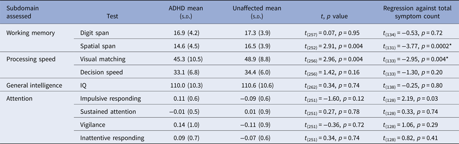
Several cognitive abilities showed associations with symptom severity: spatial span (t = −3.77, p = 0.0002, Bonferroni-adjusted p = 0.002), visual matching processing speed task (t = −2.95, p = 0.004, Bonferroni-adjusted p = 0.03) and impulsive responding from the CPT (t = 2.19, nominally significant only at p = 0.03) (see Table 1). The patterns of associations between inattentive and hyperactive–impulsive symptoms were similar for both working memory and information processing. In the CPT, the factor measuring impulsive responding associated with hyperactive–impulsive symptoms (t = 2.16, p = 0.03), whereas a vigilance factor associated with inattentive symptoms (t = 2.09, p = 0.04) (see online Supplementary Table S3). The associations between symptom severity and cognitive abilities did not vary significantly with age (i.e. there were no significant interactions between the age terms and symptoms with respect to cognition, all p > 0.05).
Performance on cognitive tasks in those with and without ADHD

* Bonferroni-adjusted p < 0.05.
Categorical contrasts are shown (ADHD v. non-ADHD), along with the results from a regression of total symptom counts against cognition (all analyses adjust for age terms and sex).
Brain-cognition associations
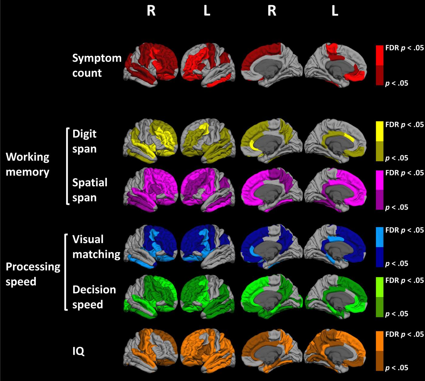
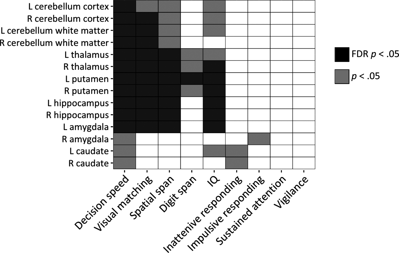
Multiple brain regions emerged as significantly associated with cognitive measures, and both nominal associations and those surviving an FDR adjustment are shown in Figures 1 and 2. Associations surviving FDR adjustment were found between spatial working memory and volumes of most of the prefrontal cortex, extending to the insula and lateral temporal regions. Verbal working memory showed more limited associations with the right orbitofrontal, middle frontal, cingulate and superior temporal gyri. The information processing speed measures both showed association with the precentral, anterior cingulate and orbitofrontal cortical volumes. General intelligence was associated with the volumes of the superior frontal, parietal (postcentral) and superior temporal gyrus. From the CPT, significant associations emerged between the factor reflecting impulsive responding and the right pericalcarine gyrus; and between vigilance and the right frontal pole – see online Supplementary Table S4 for FDR significant and nominal associations between CPT and subregional volumes.
Associations between symptoms of ADHD, cognitive abilities and cortical volumes (FDR adjusted and nominally significant associations are shown).

Associations between cognitive tasks and subcortical volumes (FDR-adjusted p < 0.05 and nominally significant associations at p < 0.05 are shown).

At a subcortical level, information processing and spatial, but not verbal, working memory were associated with the volumes of the thalami, hippocampus and right putamen (Fig. 2). Processing speed measures were additionally associated with cerebellar white matter volume. None of these associations between cognition and brain region volumes differed with age (i.e. there were no significant interactions between age and cognitive effects, following adjustment for multiple testing). The associations also did not differ by diagnostic group (see online Supplementary Results 1).
Impact of psychostimulant medication
Psychostimulant medication among those with ADHD (as represented by the proportion of life on the medication) was not associated with any cognitive measures (all FDR-adjusted p > 0.1). There were also no significant interactions between medication and the cognitive measures with respect to brain volumes: that is, the associations between cognitive abilities and the brain were not altered by psychostimulant medication history. Finally, we repeated analyses excluding those who were taking any psychotropic medication, and this did not alter the main pattern of results (online Supplementary Table S5). We also repeated analyses excluding those with comorbid disorders. The patterns of results held although some findings lost significance due to reduced sample size (online Supplementary Table S5).
ADHD symptoms and the brain
Symptom severity was associated (at FDR p < 0.05) with predominately prefrontal cortical volumes – specifically the middle, inferior, orbital and paracentral gyri, and the left inferior temporal gyri. Nominal associations were found in predominately lateral prefrontal and cingulate regions. Associations between regional volumes and hyperactive-impulsive symptoms were more extensive than those seen for inattentive symptoms, although the general pattern was very similar (see online Supplementary Figure S1).
We next determined the significance of the spatial overlap between the neuroanatomic substrates of symptoms and the cognitive measures that showed extensive brain associations (working memory, information processing, and IQ). The neuroanatomic substrates of working memory showed the most significant overlap with the neuroanatomic correlates of symptoms (Dice coefficient overlap with spatial working memory, p = 0.003; for verbal working memory p = 0.001). The overlap between the substrates for symptoms and information processing was found for the visual matching subtest only (p = 0.02). No other significant overlaps were found (for symptoms: IQ, p = 0.15; symptoms: decision speed, p = 0.48).
Discussion
There are three central findings in this study of families with ADHD. First, we find the cognitive profile tied to ADHD did not differ across generations. Second, lifetime history of treatment with psychostimulant medication in ADHD is associated with neither cognitive skills nor the relationships between cognition and the brain. Finally, extensive associations are found between brain structure and spatial working memory, information processing speed, impulsive responding and intelligence that centered on prefrontal and cortico–striato–thalamic regions. Further, there is a significant overlap between the neuroanatomic substrate of symptoms and the substrates for working memory and information processing.
Cognitive findings in ADHD
This work builds upon earlier studies of families affected by ADHD, which have mainly focused on young sibling pairs or parent–child dyads (Bidwell et al., Reference Bidwell, Willcutt, DeFries and Pennington2007; Casey et al., Reference Casey, Epstein, Buhle, Liston, Davidson, Tonev, Spicer, Niogi, Millner, Reiss, Garrett, Hinshaw, Greenhill, Shafritz, Vitolo, Kotler, Jarrett and Glover2007; Goos et al., Reference Goos, Crosbie, Payne and Schachar2009; Gau and Shang, Reference Gau and Shang2010). By including adults in middle age and beyond we find no differences in the ADHD cognitive profile across generations, suggesting there is no age-related cognitive decline in ADHD. However, as the study was cross-sectional it could only delineate how ADHD symptoms relate to cognition at one point in time, and we find that this cross-sectional association does not vary with the age of the participants. However, the study does not address the important but different question of whether a change in symptoms over time within a subject is tied to change in cognitive profiles. This requires a definitive developmental mapping of cognitive processes which awaits the collection of both cognitive and clinical longitudinal data. Secondly, we add to the growling literature associating ADHD with deficits in information processing, particularly perceptual processing speed as assessed by visual matching (Sergeant et al., Reference Sergeant, Oosterlaan and van der Meere1999; Willcutt et al., Reference Willcutt, Pennington, Olson, Chhabildas and Hulslander2005b; Rommelse et al., Reference Rommelse, Altink, De Sonneville, Buschgens, Buitelaar, Oosterlaan and Sergeant2007; Salum et al., Reference Salum, Sergeant, Sonuga-Barke, Vandekerckhove, Gadelha, Pan, Moriyama, Graeff-Martins, de Alvarenga and do Rosario2014a, Reference Salum, Sonuga-Barke, Sergeant, Vandekerckhove, Gadelha, Moriyama, Graeff-Martins, Manfro, Polanczyk and Rohde2014b). The finding echoes recent suggestions that processing deficits might be a core feature of the disorder (Salum et al., Reference Salum, Sergeant, Sonuga-Barke, Vandekerckhove, Gadelha, Pan, Moriyama, Graeff-Martins, de Alvarenga and do Rosario2014a, Reference Salum, Sonuga-Barke, Sergeant, Vandekerckhove, Gadelha, Moriyama, Graeff-Martins, Manfro, Polanczyk and Rohde2014b).
Psychostimulant medication
We examined chronic rather than acute effects of psychostimulant medication for ADHD as we tested during temporary medication cessation and found no significant associations between lifetime history of the medication and cognitive abilities. While psychostimulants have acute beneficial effects on core symptoms, the evidence of beneficial acute effects on cognition is more mixed (Advokat and Scheithauer, Reference Advokat and Scheithauer2013; Baroni and Castellanos, Reference Baroni and Castellanos2015). Looking to the brain, psychostimulant medication for ADHD has generally been associated with either no detectable change in brain structure or a slight shift toward more typical dimensions (Nakao et al., Reference Nakao, Radua, Rubia and Mataix-Cols2011; Frodl and Skokauskas, Reference Frodl and Skokauskas2012; Shaw et al., Reference Shaw, De Rossi, Watson, Wharton, Greenstein, Raznahan, Sharp, Lerch and Chakravarty2014a; Friedman and Rapoport, Reference Friedman and Rapoport2015). Here we further the field by demonstrating that psychostimulants do not uncouple the typical associations between cognition and brain structure, which were not significantly affected by the duration of psychostimulant treatment. These observational findings on psychostimulants should, however, be cautiously interpreted, as many factors that influence psychostimulant use could also impact on cognition and randomized control trials are required for causal inferences. Additionally, other neural measures, particularly of brain function, were not examined in this study and these may be more sensitive to the effects of psychostimulants.
The neuroanatomic substrates of cognition and symptoms
Working memory and information processing measures all showed associations with prefrontal cortical regions, with the exact regions varying by the task. Thus, while spatial working memory showed extensive links with most of the prefrontal cortex, the correlates of verbal working were more circumscribed. This is consistent with the literature, including both a meta-analysis and a recent large study of just over a thousand participants that found superior working memory to be associated with greater prefrontal cortical volumes, specifically the orbitofrontal, and lateral prefrontal cortex (pars orbitalis) (Yuan and Raz, Reference Yuan and Raz2014; Owens et al., Reference Owens, Duda, Sweet and MacKillop2018). Additionally, we report novel associations between information processing speed and multiple frontal cortical regions (the precentral, cingulate and orbitofrontal cortex). General intelligence was tied to prefrontal (superior, precentral), parietal (parietal lobule), and temporal (superior and inferior gyri) dimensions in line with previous reports (Colom et al., Reference Colom, Jung and Haier2006; Shaw, Reference Shaw2007; Deary et al., Reference Deary, Penke and Johnson2010). Notably, all the measures of information processing and working memory overlapped in their association with volumes of the right rostral anterior cingulate cortex, a region that has been consistently implicated in ADHD (Seidman et al., Reference Seidman, Valera, Makris, Monuteaux, Boriel, Kelkar, Kennedy, Caviness, Bush, Aleardi, Faraone and Biederman2006; Makris et al., Reference Makris, Seidman, Valera, Biederman, Monuteaux, Kennedy, Caviness, Bush, Crum and Brown2010).
In the subcortex, we find that information processing speed is also associated with thalamo–striato–cerebellar structures. We find stronger associations between cognitive skills and the putamen rather than the caudate. In this context, it is notable that a recent mega-analysis of neuroanatomic MRI studies finds the disorder is more strongly associated with a volume reduction in the putamen rather than the caudate (Hoogman et al., Reference Hoogman, Bralten, Hibar, Mennes, Zwiers, Schweren, van Hulzen, Medland, Shumskaya and Jahanshad2017). Additionally, the rostral putamen emerges as the most strongly correlated of all striatal subregions with fluid intelligence (Burgaleta et al., Reference Burgaleta, MacDonald, Martínez, Roman, Álvarez-Linera, Gonzalez, Karama and Colom2014). We find that the neuroanatomic correlates of attentional skills are more circumscribed although these have been extensively mapped using functional neuroimaging both in those with and without ADHD (Cortese et al., Reference Cortese, Kelly, Chabernaud, Proal, Di Martino, Milham and Castellanos2012; Petersen and Posner, Reference Petersen and Posner2012). We also find associations between the left amygdala, working memory and processing speed. While the amygdala is well recognized as a pivotal structure in the emotional brain, our finding is consistent with evidence for its role in higher cognitive processing (Schaefer and Gray, Reference Schaefer and Gray2007). For example, individual differences in amygdala activation predict processing speed during working memory tasks, and the amygdala acts to direct attention towards goal-relevant stimuli, that are often emotionally charged.
We also find associations between cerebellar volumes and processing speed. This is consistent with the role of the cerebellum in rapid information processing and the modification of behavior in response to feedback (Ivry, Reference Ivry1997). Faster information processing is associated with greater cerebellar grey matter volume (Genova et al., Reference Genova, Hillary, Wylie, Rypma and Deluca2009; Eckert et al., Reference Eckert, Keren, Roberts, Calhoun and Harris2010) and childhood change in cerebellar grey matter is tied to increases in processing speed (Moore et al., Reference Moore, D'Mello, McGrath and Stoodley2017).
Working memory showed the most significant overlap with the neural substrate of symptoms. This is in keeping with behavioral data which find prominent working memory deficits in children and adults with ADHD (Martinussen et al., Reference Martinussen, Hayden, Hogg-Johnson and Tannock2005; Kasper et al., Reference Kasper, Alderson and Hudec2012; Alderson et al., Reference Alderson, Hudec, Patros and Kasper2013) and experimental demonstrations that ADHD symptoms are exacerbated by increasing working memory demands, (Rapport et al., Reference Rapport, Bolden, Kofler, Sarver, Raiker and Alderson2009; Tillman et al., Reference Tillman, Eninger, Forssman and Bohlin2011; Hudec et al., Reference Hudec, Alderson, Kasper and Patros2014). It has been argued that boosting working memory might have some beneficial effect on symptom profile, though the evidence is mixed (Rapport et al., Reference Rapport, Orban, Kofler and Friedman2013). Combined these behavioral and imaging studies point to working memory as a core deficit in the disorder.
Limitations
There are several limitations. First, we did not assess some key cognitive domains pertinent to ADHD, such as processing of emotionally charged and rewarding stimuli (Plichta and Scheres, Reference Plichta and Scheres2014; Shaw et al., Reference Shaw, Stringaris, Nigg and Leibenluft2014b). We attempted to minimize the effects of comorbidity by excluding disorders known to impact brain structure, such as the psychoses, substance dependence and dementias. Other comorbidities, such as anxiety disorders, were too uncommon to evaluate separately but the general pattern of findings was unchanged when those with any current comorbidities were excluded. The inclusion criteria for the study centered on a high familial prevalence of ADHD and thus may limit the generalizability of the findings to the general population. Similarly, the main results held when those who were taking any psychotropics (including psychostimulants) were excluded. Finally, we consider only brain structure but note that the functional correlates of cognition in ADHD have already been extensively characterized (Cortese et al., Reference Cortese, Kelly, Chabernaud, Proal, Di Martino, Milham and Castellanos2012; Hart et al., Reference Hart, Radua, Nakao, Mataix-Cols and Rubia2013; Rubia et al., Reference Rubia, Alegria, Cubillo, Smith, Brammer and Radua2014).
Conclusion
We delineate anatomic alterations in fronto–striato–thalamo–cerebellar regions that are tied to the cognitive deficits found in the disorder; these associations are not significantly altered by psychostimulant medication history. The overlapping neural substrate of symptoms and cognition provide further evidence that working memory may be a core cognitive deficit in the disorder.
Supplementary material
The supplementary material for this article can be found at https://doi.org/10.1017/S0033291718001241
Financial support
The study was funded by the intramural programs of the National Human Genome Research Institute and the National Institute of Mental Health.
Conflict of interest
None.