Introduction
Leukaemia inhibitory factor (LIF) is a highly glycosylated 40–50 kDa glycoprotein with a wide range of biological functions (Haines et al., Reference Haines, Voyle and Rathjen2000). It is a member of interleukin (IL)-6 family, essential for successful implantation. LIF was first described as a factor that induced the differentiation of mouse myeloid leukemic M1 cells into macrophages (Gearing et al., Reference Gearing, Gough, King, Hilton, Nicola, Simpson, Nice, Kelso and Metcalf1987). The pleiotropic effects of LIF in many physiologic systems include proliferation, differentiation, and cell survival (Hilton, Reference Hilton1992). LIF is a multifunctional cytokine and has been considered as an essential factor for implantation and establishment of pregnancy (Lass et al., Reference Lass, Weiser, Munafo and Loumaye2001; Robb et al., Reference Robb, Dimitriadis, Li and Salamonsen2002). Implantation does not occur in the LIF gene knockout mouse and pregnancy is not possible in these mice (Stewart et al., Reference Stewart, Kaspar, Brunet, Bhatt, Gadi and Kontgen1992). Human LIF (hLIF) has been found to enhance development of IVF-derived bovine morula and blastocysts when cultured in synthetic oviduct fluid (SOF) supplemented with bovine serum albumin (BSA) or polyvinyl alcohol (PVA) (Fukui & Matsuyama, Reference Fukui and Matsuyama1994; Han et al., Reference Han, Lee, Mogoe, Lee and Fukui1995). In sheep, addition of hLIF to SOF-based embryo culture medium improved in vitro development of ovine embryos and pregnancy rates after transfer to recipient ewes (Fry et al., Reference Fry, Batt, Fairclough and Parr1992). Furthermore the presence of bovine LIF in the culture medium from days 5 to 10 improved hatching rate and cell numbers of trophectoderm of IVF-derived bovine embryos (Yamanaka et al., Reference Yamanaka, Amano and Kudo2001). In inbred female C57Bl mice, null for the LIF gene, embryos developed to the blastocyst stage but did not implant (Cheng et al., Reference Cheng, Rodriguez and Stewart2002). Addition of LIF to the undefined bovine embryo culture conditions has led to inconsistent results. It has been reported that the supplementation of LIF in embryo culture medium enhanced embryo development in human (Dunglison et al., Reference Dunglison, Barlow and Sargent1996), bovine (Yamanaka et al., Reference Yamanaka, Kudo, Kimura, Amano and Itagaki1999, Reference Yamanaka, Amano and Kudo2001) murine (Mitchell et al., Reference Mitchell, Swanson and Oehninger2002; Tsai et al., Reference Tsai, Hang, Hsieh, Lo, Hsu and Chang1999, Reference Tsai, Chang, Hsieh, Hsu, Chang and Lo2000), rabbit (Lei et al., Reference Lei, Yang, Xia, Gan, Chen, Yuan and Zhu2004) and ovine (Ptak et al., Reference Ptak, Lopes, Matsukawa, Tishner and Loi2006) models. Sirisathien et al. (Reference Sirisathien, Herandez,-Fonseca and Brackett2003) did not observe major beneficial effects on bovine embryos produced under chemically defined conditions by adding LIF. Rodriquez et al., (Reference Rodriquez, DeFrutos, Diez, Caamano, Facal, Duque, Garcia-Ochoa and Gomez2007) reported an increased hatching rates and trophectoderm (TE) cell counts in bovine blastocysts cultured with cloned bovine LIF but not with recombinant human LIF. LIF acts on cells by binding to the heterodimeric LIF receptor (LIFR), at the cell surface, LIFRβ binds with the glycoprotein gp-130 (the common signaling receptor for IL-6 family cytokines) to form a high affinity receptor through which LIF signaling is triggered (Heinrich et al., Reference Heinrich, Behrmann, Haan, Hermanns, Muller-Newen and Schaper2003).
In vitro culture conditions alter gene expression in the embryos (Lee et al., Reference Lee, Chow, Xu, Chan, Ip and Yeung2001; Wrenzycki et al., Reference Wrenzycki, Hermann, Carnewath and Neimann1999, Reference Wrenzycki, Herrmann, Keskintepe, Martins and Sirisathien2001), analysis of such differences in mRNA expression may explain the differences between in vivo and in vitro-produced embryos and allow the opportunity to modify gene expression through modification of culture systems (Rizos et al., Reference Rizos, Ward, Duffy, Boland and Lonergan2002). Defining expression pattern of species-specific genes critically involved in preimplantation development will also aid in selecting markers for determining embryo quality (Niemann et al., Reference Niemann, Wrenzycki, Lucas-Hahn, Brambrink, Kues and Carnwath2002). Human and murine embryos produced in vitro express LIF and LIFRβ mRNA transcripts throughout the preimplantation period (Chen et al., Reference Chen, Shew, Ho, Hsu and Yang1999). Expression of the LIF gene has been demonstrated in bovine preimplantation embryos. Perturbation of mRNA expression pattern of the specific LIF–LIFR system in embryos generated in vitro could lead to abnormal differentiation of the cell compartments forming the blastocyst (Eckert & Niemann, Reference Eckert and Niemann1998). Therefore, it is important to determine the expression of LIF-LIFR system during early embryogenesis in farm animals. The elucidation of the function of this cytokine and its receptor in in vitro-produced buffalo embryos is important in order to reveal the larger mechanisms of implantation and thus to improve the reproductive performance. There has been no investigation into the expression pattern of LIF and LIFRβ in preimplantation buffalo embryos. The objective of this study was to determine the effect of supplementation of rhLIF on development of blastocysts, total cell numbers, hatching rate and expression pattern of LIF and LIFRβ in preimplantation buffalo embryos using semi-quantitative RT-PCR.
Materials and methods
All reagents are the products of Sigma (St. Louis, MO, USA) unless otherwise stated.
In vitro maturation
In vitro maturation (IVM) was carried out as per the standard protocol followed routinely in our laboratory. Briefly, slaughterhouse ovaries were collected in 0.9% normal saline solution (NSS) at 35–37°C fortified with antibiotics and reached the laboratory within 4 h after slaughter. Cumulus–oocyte complexes (COCs) were aspirated from 3–8-mm follicles in oocyte collection medium and selected with regard to homogenous cytoplasm and multiple compact cumulus cell layers for IVM. The basic maturation medium was TCM199 with HEPES further supplemented with sodium pyruvate (0.2 mM), 10% FBS (fetal bovine serum, Gibco, Invitrogen Co.), 20% follicular fluid, BSA (3 mg/ml), epidermal growth factor (EGF; 20 ng/ml), penicillin G (100 IU/ml), streptomycin (100 μg/ml). COCs of good quality were matured in 50 μl of IVM medium in groups of 10–15 for 24 h, at 38.5°C in humidified air atmosphere containing 5% CO2. The IVM droplets were equilibrated for minimum of 2 h prior to the initiation of the oocyte maturation.
In vitro fertilization and embryo development
Every IVM experiment was followed by in vitro fertilization of oocytes and 8- 9 days of embryo culture. Briefly, after maturation, oocytes were inseminated with in vitro-capacitated buffalo bull semen. The frozen–thawed buffalo bull semen of the same batch was used throughout the study. Semen was washed twice in Fert-TALP media containing sodium pyruvate (2.2 mg/ml), 6 mg/ml fatty acid-free BSA (faf BSA) and heparin (10 μg/ml). The final pellet was reconstituted with Fert-TALP medium to get a final sperm concentration of 2 × 10⁶/ml. Progressively motile sperms were placed in 100 μl droplets of Fert-TALP medium under mineral oil, in vitro-matured oocytes washed in Fert-TALP media were co-incubated at 38.5°C with 5% CO2 in air with high humidity for 18h. After co-incubation with sperms, the cumulus cells were removed through repeated pipetting and the presumptive zygotes were cultured in vitro in the modified synthetic oviductal fluid (mSOF) media supplemented without (control) or with LIF (100 ng/ml) at 38.5°C and 5% CO2 in air with high humidity for development up to blastocyst stage. The cleavage rate was recorded on day 2 of culture and the stage of embryonic development was evaluated every 24 h until day 9 or blastocyst formation.
Total cell count
To evaluate the total cell number (TCN), early and expanding blastocysts collected from both the groups were subjected to Hoechst 33342 staining (DNA specific fluorochrome) as described by Pursel et al. (Reference Pursel, Wall, Rexroad, Hammer and Brinster1985) with slight modifications. The blastocysts washed in phosphate-buffered saline (PBS) for 3–5 times and transferred in 50 μl droplet of Hoechst solution, placed on siliconized glass slide, it was incubated at 37°C for 30 min, excess of stain was removed with fine pipette, blastocysts were immediately covered with 15–20μl of 100% glycerol which acts as a mountant, a coverslip was placed over the glycerol drop. The blastocysts were examined under epifluorescence microscope (Olympus, Japan) with an ultraviolet (UV) light excitation filter of 365 nm with barrier filter of 400 nm. Bisbenzimide-stained nuclei appeared blue enabling total cell count. TCN was done directly and was further confirmed by counting the cell number in pictures.
Isolation of total RNA
Pools of immature, in vitro-matured oocytes, 2–4 cell, 8–16 cell, morula and blastocyst stage embryos were collected from both the groups, control and LIF treated at different developmental stages. Total RNA was extracted from the pooled samples using RNeasy Micro Kit (Qiagen, USA), as per manufacturer's instructions. Next, 1 μl of total RNA was used for the quantification using NanoDrop ND1000 spectrophotometer and absorbance values at 260 nm wavelength (A260) was recorded against nuclease free ultrapure water as a blank. To check the purity of total RNA extracted, the absorbance value at 280 nm wavelength (A280) was also recorded. Only RNA having a ratio higher than 1.65 (OD260nm/280nm) were used for RT-PCR.
Reverse transcription and polymerase chain reaction on oocytes and preimplantation embryos
A reverse transcription-polymerase chain reaction (RT-PCR) procedure was carried out as per standard protocol (Sambrook, Reference Sambrook1989), for buffalo oocytes and embryos, the reverse transcription (RT) for first strand synthesis was carried out using a mix of random hexamer primer and Moloney murine leukaemia virus reverse transcriptase (MMLV-RT) enzyme to prime the RT reaction and to produce cDNA. Tubes were heated to 70°C for 5 min to denature the primer and template mix, and then the RT reaction was completed with the addition of 50 units of Superscript RT enzyme it was incubated at room temperature for 10 min, at 42°C for 60 min to allow the reverse transcription of RNA, followed by 99°C for 5 min to denature the RT enzyme.
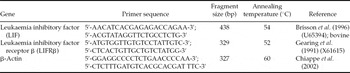
For PCR reaction, the primer pairs were designed from bovine LIF sequence (Accession Number U65394) and Human LIFRβ sequence (Accession Number X61615) using Beacon designer, the designed primers were obtained from Integrated DNA Technologies (IDT, USA). The sequences of primers used and the size of expected PCR fragments are shown in Table 1. Buffalo fetal tissue, uterine endometrium and ovarian tissue RNAs were used for the standardization of primer's annealing temperature for PCR. RT- negative and PCR-negative controls were also set for all PCR reactions to negate the DNA contamination in cDNA templates and for ascertaining the authenticity of the PCR. The resulted PCR products were visualized on 2% agarose gel in 1× TAE (Tris 10 mM, EDTA 1 mM) buffer that contained 0.5 μg/ml of ethidium bromide. β-Actin was included as an endogenous control to evaluate the quality of isolated total RNA, the extent of DNA contamination and the PCR procedure. To confirm the identity of the PCR products, the DNA was then used for cloning and sequencing.
Oligonucleotide primers used for gene-specific PCR reactions

Cloning and sequencing strategy
Amplification was carried out in a 50-μl reaction volume which contained 50 pmol of each forward and reverse primers, 3-μl template cDNA, 200 μM of dNTP mix, 1.0 mM MgCl2, and 3U proofreading DNA polymerase (MBI Fermentas, USA) in 1× Taq buffer. Amplification reaction was performed with following reaction condition: initial denaturation at 95°C for 3 min followed by 35 cycles of 45 s at 95°C, annealing at 51°C and 53°C (for primer 1 and 2 respectively), extension for 30 s at 72°C, and a final extension of 10 min at 72°C. The amplified products were resolved by agarose gel (1.5%) electrophoresis and visualized over a gel documentation system (Alpha Imager-2200, Alpha Innotech Corporation) by ethidium bromide (EtBr) staining under UV light. The amplicons were purified using Gel Cleanup kit (Advanced Microdevices (mdi), Ambala Cant, India) and cloned into pGEMT Easy vector (Promega, USA), following the manufacturer's instructions. Blue and white selection method was used to identify positive recombinant clones. The presence of insert was further confirmed by colony and plasmid PCR. Stab culture prepared from three positive clones was sent directly to Imperial Life Sciences (ILS BioServices, USA) for sequencing. The sequences were subjected to BLAST analysis (www.ncbi.nlm.nih.gov/BLAST). The nucleotides as well as deduced amino acid sequences were aligned with those of available species in the Genbank database using the ClustalW method of MegAlign Programme of Lasergene Software (DNASTAR, USA). Sequence was submitted to the NCBI GenBank and an accession number was obtained, which is now available in the public domain.
Immunocytochemical localization of LIF and LIFRβ in buffalo preimplantation embryos
In vitro-produced preimplantation embryos were fixed in 4% paraformaldehyde in PBS (pH7.4) for 20 min at room temperature and washed thrice with PBS. Embryos were then permeabilized in a 0.25% Triton-X 100 in PBS solution, then washed briefly in PBS. To minimize non-specific binding, samples were incubated for 1 h at 25°C in blocking solution (1% Tween 20, 0.5% BSA in PBS containing 5% serum, according to secondary antibody host). Following application of the primary antibody (LIF 1:200 dilution; and LIFRβ 1:200 dilution) overnight at 4°C in a humidified chamber, samples were washed in PBS and exposed for 1 h at room temperature to fluorescein isothiocyanate (FITC)-conjugated donkey-anti-goat IgG diluted 1:200 with blocking solution. Coverslips were mounted in the glycerol on cavity slides, and examined using an inverted phase contrast microscope. Control reactions were performed by: (1) omission of the primary antibody; (2) omission of the secondary antibody; and (3) omission of both the primary and secondary antibodies. A minimum of 10 samples was stained per treatment from each of six independent IVP replicates.
Statistical analysis
Statistical analysis of the means was carried out by one way analysis of variance (ANOVA), followed by Fisher's Least Squares Significant (F-test) analysis as described by Snedecor & Cochran (Reference Snedecor and Cochran1989).
Results
Effect of supplementation of rhLIF
The effect of rhLIF supplementation on blastocyst development, TCN and hatching rates are presented in Table 2. Different developmental stages of embryos produced up to hatched blastocyst are depicted in Fig. 1.
Comparison of effect of supplementation of LIF on development, total cell number and hatching of buffalo blastocysts

a,bValues (mean ± standard error (SE)) in the column bearing different superscripts differ significantly (P < 0.05).
Development and hatching of buffalo blastocysts produced in vitro. (a) Early blastocysts. (b) Expanded blastocyst. (c) Hatching blastocyst (formation of slit). (d) Hatching blastocyst (mass of inner cell comes out through slit. (e) Hatched blastocyst with the empty zona pellucida. (See online for a colour version of this figure.)

The cleavage rate in control and LIF-treated groups were 54.07% and 53.49%, respectively. The percentage of cleaved embryos and those which reached morula stage were not significantly different in both the groups. However, the development of blastocysts from morula was significantly higher (P < 0.05) in the LIF-treated group (23.13%) than in the control group (15.84%). Similarly higher numbers of expanding (16.57 versus 9.80%) and hatching (10.86 versus 5.38%) blastocysts were also noticed in LIF group when compared with the control. The mean TCN was also significantly higher (P < 0.05) in blastocysts obtained from the LIF-supplemented group (81.5) than from the control (70.5) group.
Expression of LIF and LIFRβ
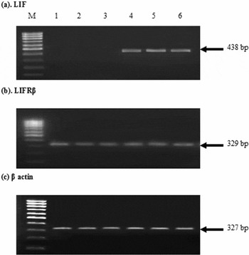
Expression pattern of LIF and LIFRβ mRNA transcripts was studied in buffalo oocytes and preimplantation embryos produced in vitro. Representative gel photos of mRNA expression in buffalo immature, in vitro-matured oocytes and different developmental stages of embryos for LIF and LIFRβ are shown in Fig. 2. A single 327 bp product for the housekeeping gene β-actin (Fig. 2c) was obtained in all the RNA samples used in the study indicating that there was no genomic DNA contamination during RNA extraction. PCR product of an expected 438 bp for LIF mRNA transcript (Fig. 2a) was recorded in 8–16-cell stage, morula and blastocyst stage of embryos, whereas no amplification was observed in the immature and in vitro-matured oocytes and 2–4-cell stage embryos. A 329-bp product of LIFRβ was detected in both immature and in vitro-matured oocytes and various developmental stages of embryos from the 2–4-cell stage to blastocyst stage (Fig. 2b).
Detection of mRNAs encoding leukaemia inhibitory factor (LIF) and leukaemia inhibitory factor receptor (LIFR)β in buffalo oocytes and preimplantation embryos by RT-PCR. (a) A single fragment of 438 bp encoding 438 bp encoding LIF. (b) A single fragment of 329 bp encoding LIFRβ. (c) A single fragment of 327 bp encoding β-actin. Lane M: 100 bp DNA ladder; lane 1: immature oocytes; lane 2: matured oocytes; lane 3: 2–4 cells; lane 4: 8–16 cells; lane 5: morula; lane 6: blastocyst.

Characterization of LIF Gene
The amplification reaction for partial sequence of LIF gene was performed from RNA (concentration 1.20 ng/μl and an optical density (OD) of 1.9) obtained from embryo stages. Total 3 μl per 25μl of final reaction volume was used for the PCR amplification of the LIF gene. The amplification reaction was carried out and agarose gel electrophoresis clearly revealed a 438-bp fragment of the LIF gene at 1.5% agarose gel (Fig. 2a). The partial coding sequence of the 438-bp sequence was submitted to GenBank, and the accession number EU926738.1 for the water buffalo LIF gene is now available in the public domain.
Sequence analysis
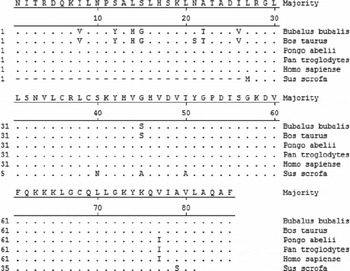
The nucleotide sequence and predicted amino acid sequence was aligned and compared with LIF cDNA sequences of different domestic species namely, Bos taurus (D50337.1), Sus scrofa (AY850174.1), Pongo abelii (XM_002830999.1), Pan troglodytes (XM_001140005.1) and Homo sapiens (M63420.1), using the ClustalW method of the MegAlign module in DNASTAR Version 4.0, Inc. USA, which revealed the nucleotide substitutions (Fig. X). The buffalo LIF gene shows 98.4% homology with bovine (Bos taurus), 85.8% with Sus scrofa, 98.1% with Pongo abelii, 98.4% Pan troglodytes and 88% with Homo sapiens, which indicates a close evolutionary relationship.
Alignment of predicted partial amino acid sequence of buffalo leukaemia inhibitory factor (LIF) with different domestic species and human. Identical sequence is indicated by a dot and differences by the corresponding one-letter symbol of the amino acid.

Phylogenetic analysis
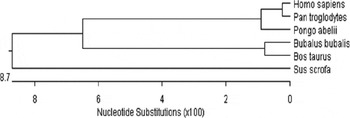
Phylogenetic analysis revealed that LIF is a highly conserved gene having a complete open reading frame (ORF) of 609 bp encoding 203 amino acids and of which we have aligned with buffalo partial sequence, which shows 85–99% homology amongst the mammalian species having a partial ORF of 258 bp encoding 86 amino acids. Based on the nucleic acid sequences of LIF partial ORF, a phylogenetic tree was drawn by DNASTAR Version 4.0, Inc. USA (Figure Y). It was found that bovine and humans are derived from different ancestors according to their closer evolutionary relationship. Among these, cattle, and buffalo, might have evolved from a common ancestor as expected; pig was positioned lowest and diverged early from the bovid ancestors. The buffalo LIF gene has an individual place closer to bovine but in a different lineage. However, all these species had similarity but had different lineage, a finding that suggested different ancestry.
Phylogenetic relationship of the leukaemia inhibitory factor (LIF) nucleotide sequences from different species using DNASTAR Version 4.0, Inc., USA following the alignment of the partial ORF sequences using ClustalW method (nucleotide p distance).

Immunocytochemical localization of LIF and LIFRβ protein
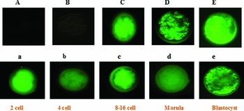
When fluorescently labelled antibodies were used, a uniform cytoplasmic localization of the protein was evident. The immunocytochemical localizations of LIF and LIFRβ protein in buffalo preimplantation embryos are presented in Fig. 3. The LIFRβ expression was abundant in the cytoplasm from the 2-cell embryo to the blastocyst (Fig. 3a–e). Strong signals of LIFRβ protein expression (shown in green) were found in preimplantation embryonic cytoplasm at the 2-cell, 4-cell, 8–16 cell, morula, and blastocyst stages. The 4-cell-stage embryos showed a relatively weak signal. Localization of LIF expression was not found in 2–4-cell-stage embryos, however, the protein could be localized from the 8- to 16-cell embryo, morula and blastocyst (Fig. 3A–E). Control experiments in the absence of specific primary antibodies did not show any signal.
Immunochemical localization of leukaemia inhibitory factor (LIF) and leukaemia inhibitory factor receptor (LIFR)β in buffalo preimplantation embryos. (A–E) LIF protein localization. (a–e) LIFRβ protein localization. (See online for a colour version of this figure.)

Discussion
Embryo co-culture is frequently employed to improve the rate of blastocyst production using IVM and IVF in several other species (Shamsuddin et al., Reference Shamsuddin, Larsson, Gustafsson and Rodriguez-Martinez1993; Yadav et al., Reference Yadav, Anil Saini, Kumar and Jain1998). In the present experiment, culture of presumptive zygotes in LIF significantly increased the buffalo blastocyst development and hatching rate compared with that of control. Madan et al. (Reference Madan, Chauhan, Singla and Manik1994) reported a significant increase in buffalo blastocyst developed in co-culture and stated that the significant increase in vitro may be due to the synthesis and secretions of some of the oviductal polypeptides during co-culture. Vero cells have also been shown to improve the blastocyst development rate and increased the pregnancy rate after embryo transfer in humans (Menezo et al., Reference Menezo, Hazout, Dumont, Herbaut and Nicollet1992). In the present experiment, there was no significant increase in cleavage and morula production rate when the presumptive zygotes were cultured in LIF media, but a significantly improved blastocyst production and hatching rate was observed when compared with the control group. Similar results were reported by Carnegie et al. (Reference Carnegie, Morgan, McDiarmid and Durnford1999) while using high LIF secreting co-culture system for bovine embryo production, which resulted in improvement of blastocyst formation and also in a better pregnancy rate after transfer of cryopreserved embryos into recipient cows. Kauma & Matt (Reference Kauma and Matt1995) reported that cells that expressed LIF when co-cultured with developing embryos enhanced blastocyst development in vitro by decreasing embryo fragmentation and degeneration in mice. Menck et al. (Reference Menck, Guyader-Joly, Peynot, Le Bourhis, Lobo, Renard and Heyman1997) found that Vero cells support bovine in vitro embryo development equal to or better than that obtained with bovine oviductal epithelial cells (BOECs). Co-culture improvement in bovine in vitro embryo development was reported by Dusewska et al. (Reference Dusewska, Reklewski, Piekowski, Karasiewicz and Modlinski2000) when cultured in Vero/BRL monolayer. Carnegie et al. (Reference Carnegie, Durnford, Algire and Morgan1997) found that proliferation-inactivated Vero cells supported the development of about 40% of IVM-/IVF-derived bovine embryos to hatched blastocysts whereas Pegoraro et al. (Reference Pegoraro, Thuard, Delalleau, Guerin, Deschamps, Marquant-Le Guienne and Humblot1998) reported a slight increase in the developmental potential of bovine embryos cultured with Vero cells. It was revealed that somatic cells might secrete embryotrophic factors, such as growth factors (positive conditioning), remove potentially harmful compounds and modify the concentration of the medium components to the levels more appropriate for embryo development (negative conditioning). Furthermore, somatic cells may lower the oxygen tension in proximity of the embryo, reducing oxidative damages caused by the oxygen species.
The advantage of LIF over that of somatic cell co-culture is its commercial availability, a safer and cell-free approach to promote embryo development in vitro. Most of the studies assessing the effect of LIF in bovine embryo cultures have used recombinant human LIF (rhLIF), as there is no commercially available bovine LIF. The rationale is that the human cytokine shares greater sequence homology with bovine LIF than mouse LIF. In the present study, we examined the effect of addition of 100 ng/ml of rhLIF in culture medium and observed a significant increase in the blastocyst development, hatching rate and TCNs. The increase in embryonic development by the addition of rhLIF in embryo development media is in agreement with Yamanaka et al. (Reference Yamanaka, Amano and Kudo2001) in bovines. Furthermore Cheung et al, (Reference Cheung, Leung and Bongso2003) reported beneficial effect of LIF and EGF on mouse blastocyst development in vitro and outcome of offspring. Jeon et al. (Reference Jeon, Oh, Park, Kim, Roh and Yoon2001) reported increased inner cell mass with reduced cell mortality and increased Oct-4 gene expression in mouse embryos, after addition of IGF-I, LIF and transforming growth factor (TGF)α in the culture media. LIF conditioned medium significantly increased the blastocyst formation rates of human cryopreserved embryos (Hsieh et al., Reference Hsieh, Tsai, Chang, Hsu, Chang and Lo2000). Reinhart et al. (Reference Reinhart, Dubey, Mummery, van Rooijen, Keller and Marinella1998) reported that oviductal cells synthesize LIF to promote and condition the embryos for implantation. Additionally, Cai et al. (Reference Cai, Cao and Duan2000) reported that LIF enhanced the murine blastocyst formation and outgrowth in culture. In rabbits, the transfer of embryos to LIF-treated recipients significantly increased pregnancy and implantation rate when compared with controls (Liu et al., Reference Liu, Yuan, Wang and Lu1999), similar effects were also found by Vogiagis et al. (Reference Vogiagis, Fry, Sandeman and Salamonsen1997) in ewes. Recently, Ptak et al. (Reference Ptak, Lopes, Matsukawa, Tishner and Loi2006) reported that LIF supplementation in sheep IVM and in in vitro culture (IVC) media exerted a beneficial effect on oocytes and embryos in vitro at stages concomitant with the steroid hormone surge. Based on these observations and our results, it is suggested that the observed improvement in blastocyst production and hatching is probably due to positive effect of LIF.
Studies employing RT-PCR methods have shown revealed differences between the relative abundance of some developmentally important gene transcripts between in vivo- and in vitro-produced embryos (Niemann & Wrenzycki, Reference Niemann and Wrenzycki2000). In addition, it is known that the conditions of in vitro culture can also alter gene expression in the embryo (Lee et al., Reference Lee, Chow, Xu, Chan, Ip and Yeung2001; Wrenzycki et al., Reference Wrenzycki, Hermann, Carnewath and Neimann1999). The analysis of such differences in mRNA expression may explain the differences between in vivo- and in vitro-produced embryos and allow the opportunity to modify gene expression through modification of culture systems (Rizos et al., Reference Rizos, Ward, Duffy, Boland and Lonergan2002). The partial nucleotide sequence and predicted amino acid sequence were aligned and compared with all LIF cDNA sequences of different domestic species namely Bos taurus, Sus scrofa, Pongo abelii, Pan troglodytes and Homo sapiens. The buffalo LIF gene shows an 85–98% homology with bovine and other mammalian species and shows close evolutionary relationship with them.
The results indicate that buffalo LIF nucleotide and deduced amino acid sequence are highly conserved across the species. Buffalo LIF also shows an 85–98% identity at nucleotide level and 90–100% identity at amino acid level with other domestic animals.
The data presented here document detection of LIF and LIFRβ mRNA expression for the first time in buffalo preimplantation embryos. The transcript for the LIF was detected from 8–16-cell stage, morula and blastocyst in buffaloes. Whereas LIFRβ transcripts were detected in immature, matured oocytes and from 2–4-cell stage up to blastocyst stage. These observations confirm those made by van Ejik et al. (Reference Van Ejik, Mandelbaum, Salat-Baroux, Belaisch-Allart, Plahcot, Junca and Mummery1996) and Fu et al. (Reference Fu, Jin-Yuh, Hong-Nerng, Wei-Li and Yu-Shih1999) in human and Eckert & Niemann (Reference Eckert and Niemann1998) in bovine. Nichols et al. (Reference Nichols, Davidson, Taga, Yoshida, Chambers and Smith1996) demonstrated that LIF is exclusively expressed in the trophectoderm, whereas the mRNA of LIFRβ is primarily localized in the inner cell mass in mice. In cattle, Eckert and Niemann (Reference Eckert and Niemann1998) reported differences in the expression of LIF and LIFRβ between in vitro- and in vivo-derived embryos. Some perturbations of the mRNA expression patterns of the specific LIF/LIF receptor system occur during the in vitro development of embryos in bovine. This situation may lead to abnormal development of the inner cell mass and trophectoderm in the blastocyst. Rizos et al. (Reference Rizos, Gutierrez-Adan, Perez-Garnelo, De La Fuente, Boland and Lonergan2003) detected LIF and LIFRβ mRNAs at higher levels in in vitro-produced bovine blastocysts, irrespective of culture system, than in in vivo-derived blastocysts. Niemann & Wrenzycki (Reference Niemann and Wrenzycki2000) detected LIFRβ mRNA from immature oocytes up to blastocyst stage, whereas the LIF transcripts in morula only not in from matured oocytes upto 16-cell stage, as well as in blastocyst. Although LIF is mainly provided by the maternal uterus for preimplantation embryos (Vogiagis & Salamonsen, Reference Vogiagis and Salamonsen1999), the expression of this cytokine is only dependant on the embryo in IVF. Because blastocyst implantation depends on maternal expression of LIF (Jurisicova et al., Reference Jurisicova, Ben-Chetrit, Varmuza and Casper1995), LIF gene mutation may give rise to decreased availability or biological activity of LIF in the uterus and cause implantation failure.
In this study, the appearance of LIF mRNA at the 8–16-cell stage onwards but not at the oocyte level could be due to polyadenylation of the LIF mRNA before onset of the embryonic genome activation as was reported by Shim et al., Reference Shim, Lee and Song1997. The start of embryonic transcription of LIF at the morula and at the onset of blastocyst stage, suggested that this secreted cytokine is probably involved in the critical developmental stages of compaction and cavitation through an autocrine/paracrine manner during early embryogenesis of buffalo. The expression of the LIFRβ transcripts in buffalo preimplantation embryos indicated that these embryos may be responsive to LIF originating either from the surrounding environment or from the embryos themselves and exerting its function in a paracrine or autocrine manner.
In this study, the LIFRβ was localized in all developmental stages of in vitro-produced buffalo embryos and that of LIF protein from 8–16-cell stage onwards up to blastocyst stage.
In conclusion, our data indicate that conditions of post fertilization culture, the period of development in vitro influence the mRNA expression in the resulting embryos. The alterations in mRNA expression are directly linked to the quality of blastocysts. The challenge for the future is to modify the conditions of in vitro culture during this critical window of development in order to try and mimic the pattern of mRNA expression as it occurs in vivo, and in that way, produce embryos of higher quality.
Acknowledgements
The authors express their personal appreciation of the valuable assistance and funding given them in their research by the ICAR and NFBSRA.