INTRODUCTION
Bladder preservation with radical radiotherapy during the treatment of muscle-invasive bladder cancer (MIBC) is an increasingly attractive treatment option. It offers comparable outcomes to radical cystectomy with the added benefit of the patient retaining their own bladder.Reference Kotwal, Choudhury, Johnston, Paul, Whelan and Kiltie1,Reference Cowan, McBain, Ryder, Wylie, Logue, Turner, Van der Voet, Collins, Khoo and Read2 The current gold standard for bladder preservation requires a tri-modal approach, combining a maximal transurethral resection of bladder tumour (TURBT), neoadjuvant chemotherapy and radiotherapy. There is increasing evidence to support the concurrent use of chemotherapy or carbogen and nicotinamide as radiosensitisers.3–Reference James, Hussain and Hall6 Patient selection for radical radiotherapy is influenced by stage of disease, baseline bladder function and patient preference. The scope of xthis review is to focus on the technological advances in radiotherapy treatment delivery.
The bladder is a mobile, dynamic organ varying in shape and size day by day, subsequently impairing the precision of conventional bladder radiotherapy and posing a challenge for the radiation oncologist when planning and delivering radiotherapy.Reference Pos and Remeijer7 Radiotherapy is delivered to a precisely defined volume and is, therefore, reliant on accurate imaging to confidently identify the tumour and bladder. Accurate organ delineation and subsequent response to its varying physical characteristics is crucial to enable dose escalation to the bladder. This entails delivering a higher dose to the target, thereby maximising the likelihood of tumour response and improving patient outcomes. It is only feasible if the target is confidently defined and tracked and adjacent normal tissues are spared.Reference Sterzing, Engenhart-Cabillic, Flentje and Debus8
Radiotherapy planning and delivery
The radiotherapy treatment pathway begins with imaging from which to define the treatment volume. CT simulation is currently the gold standard for radiotherapy planning.Reference Logue and McBain9 It is usually performed post-voiding with the patient lying supine in the treatment position. The entire bladder outline is delineated as the clinical target volume (CTV) which accounts for macro- and microscopic disease. Conventionally a population-based isotropic planned target volume (PTV) margin of 1.5–2cm is applied to the CTV to account for random and systematic patient positioning and set-up errors.Reference Button and Staffurth10
Outlining a patient’s radiotherapy treatment volume from a single CT scan performed prior to the initiation of treatment has flaws. This snapshot of the bladder’s volume, shape and anatomical location fails to recognise the organ’s dynamism, the likely, but unpredictable, movement and volume variation as the treatment progresses and the subsequent impact on dose heterogeneity. The accuracy of the generic PTV growth algorithm is limited by the unpredictable nature of inter- and intra-patient bladder movement. Bladder motion is most variable in the cranial direction compared with other planes and anisotropic PTV margins can be considered to ensure therapeutic coverage of the bladder dome.Reference Logue and McBain9,Reference Burridge, Amer, Marchant, Sykes, Stratford, Henry, McBain, Price and Moore11–Reference Muren, Redpath, Lord and McLaren13
Once the patient is placed in the treatment position they are imaged according to the treatment centre’s protocol, for example, days 1–5 and thereafter weekly during treatment. This is to monitor for systematic set-up errors rather than internal organ movement. Radiotherapy regimens typically deliver 60–66 Gy in 30–33 fractions or 52.5–55 Gy in 20 fractions to the whole bladder (PTV) using 3–4 conformal fields.Reference Logue and McBain9,Reference Murthy, Master, Adurkar, Mallick, Mahantshetty, Bakshi, Tongaonkar and Shrivastava14 Increasing the dose conformality of a radiotherapy treatment plan enables optimal target volume definition, steep dose gradients and sparing of adjacent healthy tissue (organs at risk (OARs)). The approach optimises dose delivery to the target and reduces treatment toxicity. This is increasingly important in an era of concurrent chemotherapy or radiosensitiser administration because of the heightened toxicity associated with combined treatment. Intensity Modulated Radiotherapy (IMRT) demonstrates a major technological advance in the delivery of 3-dimensional conformal radiotherapy with rapid dose fall-off beyond the target volume. It is therefore vital that the target volume is accurately delineated and relatively fixed in anatomical location. The use of IMRT in the treatment of bladder cancer may be limited by organ movement and the unacceptable risk of geographical miss due to bladder movement beyond the PTV.Reference Logue and McBain9,Reference Murthy, Master, Adurkar, Mallick, Mahantshetty, Bakshi, Tongaonkar and Shrivastava14
Bladder volume variability during treatment
The PTV must cautiously balance adequate dose delivery to the mobile, distensible bladder with the need to minimise the dose delivered to adjacent OARs, most notably the rectum and small bowel. The inconsistent bladder volume, due to the varying emptying and constant filling, associated with its unpredictable mobility produces geometric uncertainty.Reference Muren, Redpath, Lord and McLaren13,Reference Viswanathan, Yorke, Marks, Eifel and Shipley15 Bladder voiding immediately prior to treatment is one approach when attempting reproducibility. This is often an easier instruction for patients to follow instead of maintaining a ‘full’ bladder which is subjective, may be difficult to maintain due to the patient’s bladder function and symptoms and difficult in the practical setting because of the timing implications imposed on treatment radiographers. Pre-treatment voiding may help reduce inter-fractional variation, but does not affect the intra-fractional bladder volume changes due to filling. The use of drinking and voiding protocols in addition to the adoption of a low fibre diet are aimed at limiting the bladder’s geometric variability, but are inadequate to stabilise and replicate the target volume over the treatment duration.
Studies have repeatedly demonstrated the inter- and intra-fractional variability of patient bladder volumes during radiotherapy.Reference Burridge, Amer, Marchant, Sykes, Stratford, Henry, McBain, Price and Moore11,Reference Muren, Redpath, Lord and McLaren13,Reference McBain, Khoo, Buckley, Sykes, Green, Cowan, Hutchinson, Moore and Price16,Reference Turner, Swindell, Bowl, Marrs, Brookes, Read and Cowan17 Turner et al. demonstrated considerable inter-fractional variability of CT measured bladder dimensions among 20 patients (mean bladder size = 36.9 cm2, range 16.2–80.9 cm2) with a median area change of 11.1cm2 over the course of treatment. A maximum area difference ranging from 3.3cm2 to 29.1cm2 reflecting an area variation of 7–55% was reported.Reference Turner, Swindell, Bowl, Marrs, Brookes, Read and Cowan17 Muren assessed variations in patients’ bladder volumes (cm3) by performing regular CT scans during their treatment course and comparing their bladder volumes to their baseline planning scan, to calculate a relative bladder volume (RBV = repeat CT scan bladder volume divided by the planning CT scan bladder volume). He reported an RBV range of 0.27–1.78 in an observational setting of 20 patients and RBV of 0.36–1.65 in a subsequent investigational study involving 8 patients. Fluid intake restriction prior to treatment resulted in a reduction of bladder volume variability.Reference Muren, Redpath, Lord and McLaren13
Patient imaging
Regular imaging during the treatment schedule can be undertaken to address concerns relating to the target positioning of the dynamic bladder. Technological advances have enabled the integration of imaging modalities directly into the linear accelerator.Reference McBain, Henry, Sykes, Amer, Marchant, Moore, Davies, Stratford, McCarthy, Porritt, Williams, Khoo and Price18 A megavoltage Electronic Portal Imaging Device (EPID) enables 2-dimensional visualisation of the bony anatomy. Bony landmarks can act as a surrogate for the target to help confirm correct patient set-up, but EPID cannot reliably locate soft tissue structures like the bladder and subsequently is not appropriate for monitoring internal organ motion. An alternative imaging method, such as cone beam computed tomography (CBCT) is required to visualise the bladder organ although the tumour itself is unlikely to be visualized.Reference Turner, Swindell, Bowl, Marrs, Brookes, Read and Cowan17,Reference McBain, Henry, Sykes, Amer, Marchant, Moore, Davies, Stratford, McCarthy, Porritt, Williams, Khoo and Price18 CBCT involves the reconstruction of a 3-dimensional data set from multiple cone-beam X-ray transmission projections through the patient and provides adequate resolution to allow visualisation of the bladder and rectum.Reference Button and Staffurth10,Reference Muren, Redpath, Lord and McLaren13 Soft-tissue definition is dependent on the patient size, organ distension, amount of intra-luminal air and organ boundaries.Reference McBain, Henry, Sykes, Amer, Marchant, Moore, Davies, Stratford, McCarthy, Porritt, Williams, Khoo and Price18 The enhanced soft tissue definition offered by CBCT in comparison with EPID improves patient alignment and has been associated with reduced treatment related genitourinary toxicity in a pelvic malignancy setting.Reference Ost, De Gersem, De Potter, Fonteyne, De Neve and De Meerleer19
The aim of treatment is to maximise the ‘therapeutic ratio’, the ratio between the radiotherapy effects on the tumour against the effects on normal tissue. Without target monitoring during the treatment course the patient may be subjected to ineffective therapy in addition to inappropriate radiation exposure to OARs. Image Guided Radiotherapy (IGRT) involves the direct integration of imaging into the linear accelerator to verify the target and subsequently aid treatment plan adaptation to ensure optimum dose coverage. Accurate delineation of the varying bladder target volume during the treatment course enables dose escalation to the tumour (exploiting the dose response relationship of the bladder) whilst reducing exposure to neighbouring radiosensitive tissues.Reference Sterzing, Engenhart-Cabillic, Flentje and Debus8,Reference Faroudi, Wong and Kron20
Adaptive radiotherapy
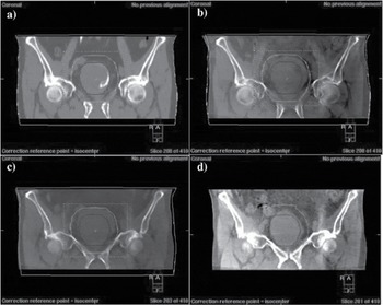
Image acquisition during the course of treatment enables an adaptive radiotherapy (ART) approach to treatment delivery by modifying, personalising and re-optimising the target volume coverage according to variation in size, shape and geographical location of the CTV as the treatment progresses. ART can be performed online, an approach which necessitates imaging to be performed and reviewed with appropriate treatment plan adjustment in real time. Alternatively, an offline approach can be adopted, whereby patient images from a previous timepoint are assessed and the plan is amended prior to further treatment (see Figure 1).Reference Varadhan, Hui and Way21 The optimum frequency of imaging is an on-going debate.Reference Button and Staffurth10 From a planning perspective, the optimum ART approach would involve daily, online imaging with daily plan modification. Whilst this would maximise precision and dose conformality it is limited by the time, financial and technical requirements it necessitates. An offline approach may offer a compromise between optimal conformality and practicability, but to date no study has compared online versus offline adaptive approaches. Compared with conventional radiotherapy, offline ART offers improved target volume conformality.Reference Foroudi, Wong, Haworth, Baille, McAlpine, Rolfo, Kron, Roxby, Paneghel, Williams, Duchesne and Tai22 In the practical setting, it is an easier approach to adopt compared with online plan adaptation, because of the minimal impact on treatment times and the lesser time constraints imposed on technical staff. However, precision is limited by the restrictive ability to correct systematic errors only, compared with an online approach which can also address random daily movement and volume variations. Consequently, online ART is at the forefront of technological advances in delivering a personalised, conformal radiotherapy plan, albeit, with considerable resource demands.
Coronal images of the bladder CTV and PTV. Figure (a) CT planning scan with the bladder outline representing the CTV with a 15mm expansion to create the PTV. (b) Day 1 CBCT. The bladder volume had increased and subsequently the PTV coverage of the bladder was compromised. (c) Day 2 CBCT confirmed a systematic increase in bladder volume and the PTV required enlargement. (d) Offline plan adaptation was performed and an anisotropic PTV margin applied. Whilst the lateral and caudal margins remained unchanged, the cranial margin was increased by 5mm to 20mm, subsequently ensuring adequate target volume coverage.

The concept of ‘plan of the day’ was initially presented by Burridge et al. providing a practical approach to online ART. During the treatment planning stages 3 PTVs were grown from the CTV with anisotropic margins—15 mm uniform margin in all directions except cranially, where either a 5 mm, 10 mm or 15 mm margin was created. Pre-treatment CBCT was performed on the first 5 days of treatment and weekly thereafter. The appropriate PTV, or ‘plan of the day’, providing bladder coverage with 2-mm clearance was selected. Burridge concluded that a choice of three plans of the day was found to be sufficient for 75% of patients, while the remaining 25% of patients had systematically smaller or larger bladders, suggesting they needed to be re-planned.Reference Burridge, Amer, Marchant, Sykes, Stratford, Henry, McBain, Price and Moore11 The number of PTVs available for online adaptation must balance accurate delineation with time, economical and practical constraints. 3–6 PTV options are generally accepted.Reference Burridge, Amer, Marchant, Sykes, Stratford, Henry, McBain, Price and Moore11,Reference Murthy, Master, Adurkar, Mallick, Mahantshetty, Bakshi, Tongaonkar and Shrivastava14
Several studies have demonstrated the benefits of ART in minimising radiation exposure to OARs. In Burridge’s cohort of 20 patients the volume of small bowel spared with a ‘plan of the day’ adaptive technique was calculated by subtracting the selected PTV volume from the standard PTV volume. On average, 31 ± 23 cm3 (± 1 S.D) of small bowel was spared, assuming all tissue in the larger PTV was small bowel.Reference Burridge, Amer, Marchant, Sykes, Stratford, Henry, McBain, Price and Moore11 Foroudi et. al. recruited 27 patients with muscle invasive bladder cancer and delivered the first seven treatments conventionally with a CTV to PTV margin of 1.5 cm. Adaptive plans were created from the first five daily CBCT scans, and small, medium and large adaptive plans were subsequently created from these images. For the remaining treatment, fractions 8–32, daily CBCTs were performed and the most appropriate adaptive PTV plan was selected to ensure CTV coverage by the 95% isodose line with the smallest possible margin. The small, medium, large and conventional PTV plans were used in 9.8%, 49.2%, 39.5% and 1.5% of treatment fractions respectively. Over the duration of treatment, the mean bladder volume was noted to have reduced by 27%, which they attributed to the increased frequency of voiding secondary to treatment induced bladder irritation. Importantly, they also demonstrated the mean volume of normal tissue receiving greater than 45 Gy was 29% less (95% CI, 24–35%) with ART compared with conventional radiotherapy.Reference Faroudi, Wong and Kron20 Whilst these studies both demonstrate the potential to spare normal tissue with ART, there have been no published clinical results comparing ART with non-adaptive treatment delivery, either in terms of tumour control or treatment toxicity. These questions are currently being investigated by several groups and their results are awaited.
Whilst IGRT can offer improved delineation and personalised treatment delivery, its benefits are limited by the practical and financial implications. Daily imaging results in a prolonged treatment time and increased requirement for specialist support. It is also inevitably associated with additional radiation exposure.Reference Burridge, Amer, Marchant, Sykes, Stratford, Henry, McBain, Price and Moore11 IGRT may negatively impact on intra-fraction variability due to prolongation of the interval between voiding and treatment delivery, thereby contributing to the risk of a geographical miss.Reference Murthy, Master, Adurkar, Mallick, Mahantshetty, Bakshi, Tongaonkar and Shrivastava14 It is also accepted that PTV selection is subjective. However, Burridge’s planning study did not demonstrate a clinically significant inter- or intra-observer variability; 70% of PTV selections were consistent and no plans differed by more than 5mm.Reference Burridge, Amer, Marchant, Sykes, Stratford, Henry, McBain, Price and Moore11
The choice of imaging modality must also be considered. Compared with kilovoltage (kV) CT, kV CBCT is associated with increased scatter contribution resulting in reduced image contrast, increased image noise and the subsequent possibility of reconstruction error.Reference Varadhan, Hui and Way21 However these online images can be quickly reconstructed with relative ease, providing adequate images to confidently define the CTV.Reference McBain, Henry, Sykes, Amer, Marchant, Moore, Davies, Stratford, McCarthy, Porritt, Williams, Khoo and Price18 Helical tomotherapy combines megavoltage (MV) CT imaging with IMRT. The online imaging offers an image guided IMRT approach and may provide a practical solution to the target movement issues which have previously limited the use of IMRT in the bladder setting. However, kVCT and CBCT offer better contrast resolution compared with MVCT.Reference Varadhan, Hui and Way21 Magnetic Resonance Imaging (MRI) offers optimal soft tissue imaging and a hybrid MRI/linear accelerator is currently being developed.Reference Lagendijk, Raaymakers, Raaijmakers, Overweg, Brown, Kerkhof, Van der Put, Hårdemark, Vulpen and Van der Heide23
Partial bladder radiotherapy
The majority of tumour recurrences occur at the original site of disease.Reference Cowan, McBain, Ryder, Wylie, Logue, Turner, Van der Voet, Collins, Khoo and Read2 The discussion so far has assumed that the CTV encompasses the entire bladder volume. The bladder demonstrates a dose-response relationship, correlating increasing dose intensity with increasing toxicity. This relationship is also true of small bowel toxicity.Reference Pos and Remeijer7 Therefore, it seems logical that reducing the total bladder volume irradiated would be associated with a lower risk of toxicity. By reducing the target volume to the tumour site alone rather than incorporating the entire bladder volume, the dose delivered to the tumour could be escalated. This offers potential improvements in outcome providing the target volume is adequately covered, whilst global bladder injury and OAR exposure could be minimized.Reference Pos and Remeijer7,Reference Viswanathan, Yorke, Marks, Eifel and Shipley15
A partial bladder approach either involves treating the whole bladder to a lower dose with subsequent boost to the tumour volume or treating the partial tumour volume with limited margins alone. Partial bladder radiotherapy is therefore an attractive option given the dose-response relationship exhibited by the bladder, but does not account for associated bladder field changes nor overcome the issues relating to organ motion or volume variability and the risk of geographical miss.Reference Pos and Remeijer7,Reference Coen, Zietman, Kaufman and Shipley12 There is no consensus regarding the margins required for microscopic spread and TURBT prior to radiotherapy may add to the difficulty localising the tumour on CT imaging.Reference Pos and Remeijer7,Reference Button and Staffurth10 In view of these difficulties, the optimal role of partial bladder radiotherapy may be in the delivery of a radiotherapy boost to the tumour following whole bladder radiation. This will enable dose escalation to a limited field, with the potential benefit of maximising tumour control.
The partial approach is reliant on accurate tumour demarcation and further strengthens the need for IGRT. Fiducial markers may help to identify the tumour location. The markers, for example, titanium clips or gold seeds, are implanted into the bladder wall at sites of residual disease or suspected areas of microscopic disease. By improving target delineation these markers aid IGRT. However the bladder wall is thin and subject to distension, causing fiducial marker displacement and possible marker loss during the course of treatment. This therefore limits their practical application in positioning verification and ART.Reference Hulshof, van Andel, Bel, Gangel and van De Kamer24,Reference Mangar, Thompson, Miles, Huddart, Horwich and Khoo25 Recently a radio-opaque marker has been developed. Using flexible cystoscopy, Lipiodol is injected into the sub-urothelial region around the tumour and overcomes the issue of marker displacement. Lipiodol is visible on CT and CBCT and should be used in conjunction with imaging to plan the target volume, rather than being solely used to delineate the tumour.Reference Pos, Bex, Dees-Ribbers, Betgen, van Herk and Remeijer26
A prospective randomised trial performed at The Christie, Manchester, hypothesised that delivering an increased radiotherapy dose to the tumour-bearing region of the bladder alone may improve local control and overall survival without increased toxicity. Patients were randomised to receive whole bladder radiotherapy (52.5 Gy in 20 fractions) versus dose escalated partial bladder radiotherapy using two different dose schedules: a 4-week regime (57.5 Gy in 20 fractions) or an accelerated regime (55 Gy in 16 fractions). Partial bladder radiotherapy resulted in a 61% reduction in the mean PTV, which enabled an increased dose to be delivered without increased treatment related toxicity being observed. There was no difference in local control or overall survival, suggesting that a smaller PTV did not compromise tumour control. Interestingly, all recurrences among patients receiving whole bladder radiotherapy occurred at the primary site, compared with the partial bladder group, which demonstrated 33% of relapses occurring in the un-irradiated bladder volume.Reference Cowan, McBain, Ryder, Wylie, Logue, Turner, Van der Voet, Collins, Khoo and Read2
Advances in biological markers to predict radiotherapy response
The ultimate goal of any clinician is to predict response or toxicity of treatment for their patients. Currently there are no validated predictive markers for response to radiotherapy. The success of radiotherapy is dependent upon inducing fatal DNA damage, especially double-stranded DNA breaks, and therefore the expression levels of DNA repair proteins may correlate with radiosensitivity. A number of different DNA repair genes and proteins have been studied within different cancers, although there are no validated predictors of radiation response in bladder cancer.Reference Parliament and Murray27,Reference West, McKay, Hölscher, Baumann, Stratford, Bristow, Iwakawa, Imai, Zingde, Anscher, Bourhis, Begg, Haustermans, Bentzen and Hendry28. The MRN complex (MRE11/RAD50/NBS1) is responsible for the repair of ionising radiation induced DNA damage. Choudhury et al. demonstrated high MRE11 expression was associated with improved outcomes following radiotherapy, reporting a 3-year cancer specific survival of 68.7% versus 43.1% among patients with high compared with low tumour MRE11 expression. This finding was validated in a second cohort of patients undergoing radical radiotherapy. There was no correlation between MRE11 expression levels and cancer specific survival observed among patients undergoing cystectomy, suggesting its role in predicting radioresponse only. Patients with high MRE11 expressing tumours undergoing radiotherapy demonstrated a 16% absolute improvement in cancer specific survival compared with their cystectomy counterparts.Reference Choudhury, Nelson, Teo, Chilka, Bhattarai, Johnston, Elliott, Lowery, Taylor, Churchman, Bentley, Knowles, Harnden, Bristow, Bishop and Kiltie29 These results suggest the emergence of MRE11 as a predictive marker for patients being considered for radical radiotherapy.
CONCLUSION
Radical radiotherapy to the bladder is an attractive treatment option, offering comparable outcomes to surgery whilst allowing organ preservation. Ongoing developments, including improved patient selection with the emergence of potential predictive biomarkers and the use of concurrent chemotherapy and radiosensitisers may maximise patient outcomes and further strengthen the role of radiotherapy in the radical setting. Ensuring optimal dose conformality is maintained over the treatment duration, enables dose escalation to the target volume potentiating tumour control and minimising normal tissue toxicity. However, this theoretical advantage is limited by the practical challenges relating to bladder mobility and volume variation over the treatment course, which subsequently limit the conformality that can be imposed on a treatment plan. Adopting an adaptive radiotherapy technique to identify and respond to the organ’s varying geometry offers a dynamic approach to treatment delivery and ensures a personalised, adjustable treatment plan is delivered to the tracked target volume. The technical, economic and time implications associated with ART remain a major hurdle restricting its implementation in daily practice. Continuing advances in online patient imaging, accurate delineation of the appropriate target volume and developments in adaptive radiotherapy techniques will further maximise precision.