Introduction
Although extrapyramidal signs (EPSs), including resting tremor, rigidity, postural abnormalities, and akinesia/bradykinesia, are characteristic of Parkinson's disease (PD), they also frequently accompany clinically diagnosed Alzheimer's disease (AD) (Merello et al., Reference Merello1994). Clinically, AD patients with EPSs display significantly more severe deficits in executive functions, faster cognitive decline, and higher mortality than those without (Mayeux et al., Reference Mayeux, Stern and Spanton1985; Merello et al., Reference Merello1994).
The anatomical and pathological basis for EPSs in AD has been poorly understood. Studies have shown neuropathological similarities in the substantia nigra between PD and AD with EPSs, including α-synuclein aggregation, hyperphosphorylated tau accumulation, and neuron loss, which was a defining pathologic feature of PD (Burns et al., Reference Burns, Galvin, Roe, Morris and McKeel2005). However, the fact that not all AD patients with EPSs have shown substantia nigral pathology or response to levodopa treatment suggests that extranigral factors may involve in manifestation of EPSs in AD.
Clinically, the vascular risk factors such as high blood pressure, dyslipidemia, diabetes mellitus, smoking, and the presence of atherosclerotic disease have been associated with an increased risk of AD (de la Torre, Reference de la Torre2004). The link between AD and vascular risk factors was suggested by significant cerebral microvascular pathology and atherosclerosis, and deficient clearance of amyloid across the blood–brain barrier in AD, all of which indicate that vascular risk factors are important features of chronic neurodegeneration in AD (Honig et al., Reference Honig, Kukull and Mayeux2005). However, it was reported that patients with PD have a lower risk of atherosclerosis. This has been accompanied by various hypotheses as to why a low risk of atherosclerosis would accompany PD. The autonomic dysfunction in PD or the hypotensive effect of L-dopa, the primary medication for PD treatment, may act to provide protection from atherosclerosis (Lee et al., Reference Lee2007). The dopamine deficiency in PD may provide additional protection from atherosclerosis (Jacobsson and Fowler, Reference Jacobsson and Fowler1999). It was also found that cigarette smoking is inversely associated with PD risk, given that nicotine induces neurotrophic factors and has an antiparkinsonian effect (Maggio et al., Reference Maggio1998).
Little is currently known, however, about how the pattern of vascular risk factors differs across EPSs in AD. If AD patients with EPSs were to differ on vascular risk factors, as well as white matter lesions (WMLs), such findings would contribute to improving our clinical characterization of AD pathophysiology.
Methods
Subjects
Subjects were recruited using a registration system established by the Clinical Research for Dementia of South Korea (CREDOS). To set up a registry for dementias, a league of neurologists and psychiatrists who are specialized in dementia clinics established the CREDOS with the support from the Korean government in 2005. This online dementia registration system mandates a standardized protocol for subject registration of a nationwide multicenter-based cohort in South Korea. This study was retrospectively analyzed with data obtained from the CREDOS database on 13 July 2009. Data were based on 1,187 patients diagnosed as having incident AD (clinically probable AD; McKhann et al., Reference McKhann, Drachman, Folstein, Katzman, Price and Stadlan1984) and 333 controls without cognitive impairment or EPSs. The controls were either healthy individuals or subjects who had no cognitive impairment on neuropsychological tests despite memory complaint. Patients were excluded if they had other clinical diagnoses of mild cognitive impairment by Petersen's criteria (Petersen et al., Reference Petersen2001), PD according to the U.K. Parkinson's Disease Society brain bank criteria (Gibb and Lees, Reference Gibb and Lees1988), PD with dementia (Emre et al., Reference Emre2007) and dementia with Lewy bodies (McKeith et al., Reference McKeith1996).
Clinical evaluation
Each subject was evaluated using the same protocol including a clinical history, a physical and neurological examination, a standardized neuropsychological battery including Clinical Dementia Rating (CDR) (Morris, Reference Morris1993), a laboratory workup including genotyping of the APOE polymorphism, and a brain MRI evaluation. Classical vascular risk factors such as hypertension, diabetes mellitus, dyslipidemia, body mass index (BMI), and smoking status were also assessed. We classified the APOE ε4 (ApoE4) polymorphism as present (homozygous or heterozygous) or absent.
The extent of WMLs was evaluated using a semi-quantitative scale, as previously described (Seo et al., Reference Seo, Lee, Park, Park and Lee2008). In brief, WMLs were considered present if there were high signal intensities on both T2-weighted and FLAIR images. WMLs were divided into those in deep WML (dWML) and those in periventricular WML (pvWML). The dWML was defined as WMLs that were found in the second cut or above from the upper margin of the ventricle on FLAIR images, or as those that were beyond 10 mm from the margin of the lateral ventricle. The dWML was rated as D1 (small and large foci, <10 mm), D2 (beginnings of a confluent lesion, 10–24 mm), or D3 (confluent lesion, >25 mm). When a periventricular cap or band was found, those lesions were defined as pvWML. The size of the cap was measured along the longest length from the anterior or posterior horn on a perpendicular axis. The size of the band was measured along the longest length from the lateral wall of the ventricle on the horizontal axis. The pvWML was classified as P1 (cap and band <5mm), P2 (5mm ≤cap or band<10mm), and P3 (10mm ≤cap or band). The dWML and pvWML were combined to give out the final white matter rating score as minimal (D1P1, D1P2), moderate (D1P3, D2, D3P1, D3P2), or severe (D3P3). In this study, we defined the present of WMLs when there was moderate to severe in the state of WMLs, the present of dWML when there was more than D2 in the state of dWML, and the present of pvWML when there was more than P2 in the state of pvWML.
EPSs evaluation
EPSs were assessed by a physician during the neurological examination. The subjects with AD and associated EPSs were classified as AD/EPS+; the subjects having AD without EPSs were labeled as AD/EPS-. The EPS subtypes were classified as postural instability gait difficulty (PIGD), tremor-dominant (TD), or indeterminate subtype (IND), following modified criteria based on a previous method (Jankovic et al., Reference Jankovic1990; Williams-Gray et al., Reference Williams-Gray, Foltynie, Brayne, Robbins and Barker2007) as follows. The tremor domain was assessed as the presence of the following items: tremor of the face, lips, chin, or all four limbs while at rest, and postural tremor in both arms as determined by the neurological examination. The complex of postural instability gait difficulty (PIGD) was assessed during examination as the presence of the following items: rigidity at extremities or axial muscle, bradykinesia, parkinsonian gait such as decreased arm swing, stooped posture, short step gait, festination, shuffling or multi-step turning. The IND group was defined as subjects with both a tremor domain and a complex of PIGD.
Statistical analysis
To analyze the similarity of clinical and demographic characteristics among subjects, these were compared using an unpaired Student t test or ANOVA for continuous variables and by χ 2 test for categorical variables. In order to identify the factors associated with EPSs in AD as well as vascular risk factors, we performed a multiple binary logistic regression, using the AD/EPS+ as the dependent variable. Potential factors such as demographics (age, gender, level of education), cognitive status (CDR), and vascular risk factors (hypertension, diabetes mellitus, dyslipidemia, BMI and smoking status) were included as independent variables in the final regression models with enter methods, if the literature suggested that the covariate was an important predictor of vascular risk factors, and that the variable had a significantly different distribution between AD and control, they were included in the model. Because of the limited evaluation of ApoE4 (only 631 subjects; 41.5% of a total of 1,520 subjects), an additional regression analysis was performed on the subjects who had been evaluated for ApoE4. For the analysis of the relationship of EPS subtypes in AD to WMLs, we performed a multiple binary logistic regression, using the WMLs as the dependent variables and the EPS subtypes as additional independent variables. The level of significance was set at p < 0.05. The analysis was performed using SPSS for Windows, version 10.0 (SPSS Inc., Chicago, IL).
Results
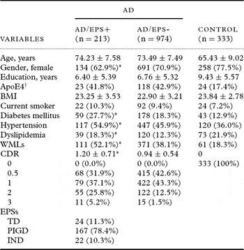
Demographic characteristics and clinical data are presented in Table 1. For AD patients, age (73.62 ± 7.51 years), female gender (69.5%), level of education (6.70 ± 5.34 years), BMI (22.96 ± 3.28 kg/m2), ApoE4 (42.8%), diabetes mellitus (20.0%), hypertension (47.5%), dyslipidemia (13.4%), WMLs (40.6%), and CDR (0.99 ± 0.58) displayed different distributions to controls (p < 0.05). The smoking status, however, did not different significantly between AD patients and controls (9.6% vs. 7.2%, p > 0.05). The evaluation of ApoE was performed on only 41.5% of all subjects (41.5% of a total of 1,187 AD patients and 41.4% of a total of 333 controls). Subjects with known ApoE status had more PIGD subtypes of EPSs (14.5% vs. 6.0%, χ 2 = 29.746, df = 3, p < 0.001) and lower educational level (6.94 years vs. 7.55 years, F = 0.266, p = 0.03) than those without known ApoE status, but were similar with respect to age (p = 0.92), gender (p = 0.21), BMI (p = 0.79), current smoker (p = 0.52), diabetes mellitus (p = 0.52), hypertension (p = 0.14), dyslipidemia (p = 0.06), CDR (p = 0.14), and WMLs (p = 0.376).
Demographic and clinical characteristics

Values represent means ± SD or number (%).
*p < 0.05 between AD/EPS+ and AD/EPS-
†ApoE4 data were analyzed for 631 subjects.
AD = Alzheimer's disease; EPSs = extrapyramidal signs; ApoE4 = ApoE ε4 allele; BMI = body mass index; WMLs = white matter lesions; CDR = Clinical Dementia Rating; TD = tremor dominant; PIGD = postural instability gait difficulty; IND = indeterminate.
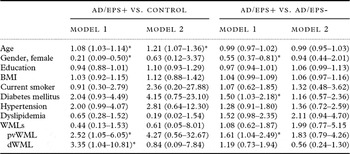
Of 1,187 AD patients, EPSs were present in 213 (AD/EPS+, 17.9%). When AD/EPS+ and AD/EPS- groups were compared, vascular risk factors such as male gender, hypertension, diabetes mellitus, and dyslipidemia, and WMLs were more frequently found in AD/EPS+ (p < 0.05); however, other variables such as age, level of education, ApoE4, BMI, and smoking status did not differ between the two groups(p > 0.05). Accordingly, we constructed a multivariate regression analyses adjusting for potential covariates (Table 2). Compared with controls, AD/EPS+ was significantly associated with age, male gender, pvWML, and dWML. Compared with AD/EPS-, AD/EPS+ was significant associated with male gender, diabetes mellitus, and pvWML.
Relationship between vascular risk factors and EPSs in AD

Values represent odd ratios (95% confidence intervals); *p < 0.05.
In model 1, the binary logistic regression analyses, used age, gender, education, BMI, smoking status, diabetes mellitus, hypertension, dyslipidemia, WMLs, pvWML, dWML, and CDR as independent variables.
In model 2, the binary logistic regression analyses, used ApoE4 as an additional independent variable.
BMI = body mass index; WMLs = white matter lesions; pvWML = periventricular white matter lesion; dWML = deep white matter lesion.
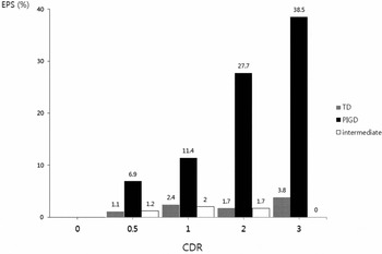
For the EPS subtypes, the majority of patients with AD/EPS+ (78.4%) were classified to be of the PIGD subtype (Table 1). According to CDR scores, PIGD was more prevalent in higher CDR scores, but tremor dominant (TD) and intermediate were not associated with CDR (Figure 1). Among the EPS subtypes in AD/EPS+, the distributions of ApoE4 were not significantly different (36.4% in TD subtype, 42.1% in PIGD subtype, and 50.0% in IND subtype) (χ 2 = 0.301, p > 0.05). Although the distributions of WMLs (37.5% in TD, 55.7% in PIGD, and 40.9% in IND; χ 2 = 4.016, p > 0.05) and pvWML (54.2% in TD, 70.7% in PIGD, and 54.5% in IND; χ 2 = 4.350, p > 0.05) were not different among the EPS subtypes, the dWML were significantly different (16.7% in TD, 41.3% in PIGD, and 27.3% in IND; χ 2 = 6.478, p = 0.039). In the regression analyses, adjusting for potential covariate, the PIGD subtype was significantly associated with pvWML, dWML, and WMLs (Table 3).
The prevalence of EPSs according to CDR scores.

The relationship between EPS subtypes in AD to WMLs

*p < 0.05.
The binary logistic regression analyses used age, gender, education, BMI, smoking status, diabetes mellitus, hypertension, dyslipidemia, and CDR as independent variables.
AD = Alzheimer's disease; EPS = extrapyramidal signs; WMLs = white matter lesions; pvWML = periventricular white matter lesion; dWML = deep white matter lesion; TD = tremor dominant; PIGD = postural instability gait difficulty; IND = indeterminate.
Discussion
The main outcome of this study was that EPSs were present in 17.9% of AD patients and that there was an association between EPSs in AD and some vascular risk factors. Moreover, the PIGD subtype was the major subtype of EPS found in AD, and it was also associated with WMLs.
Although EPSs are a main clinical feature of PD patients who have a lower risk of atherosclerosis, EPSs in AD were found to be associated with vascular risk factors. This suggests that with respect to the biological basis or aspects of risk, the pathophysiology of EPSs in AD may be different from that of EPSs in PD. Neuropathologically, the presence of EPSs may be a surrogate indicator of AD neuropathologic severity (Burns et al., Reference Burns, Galvin, Roe, Morris and McKeel2005). Interestingly, neuron loss best correlates with the presence of EPSs, suggesting that neuron loss may be the most important neuropathologic substrate for the generation of EPSs in AD (Burns et al., Reference Burns, Galvin, Roe, Morris and McKeel2005). Because WMLs were clinicopathologically indicated as cell loss or dysfunction of neurons, we hypothesized that WMLs could contribute to EPSs in AD. In this study, patients with AD and EPSs were associated with WMLs. Thus, it was hypothesized that extranigral factors, such as WMLs, were one of the vascular risk factors associated with the clinical symptoms of EPSs in AD.
This study demonstrated a relationship between EPSs and pvWML but not between EPSs and dWML in AD. There was dissimilarity in the clinicopathological mechanism between pvWML and dWML. It was suggested that pvWML might be more hemodynamically determined, whereas dWML might be more attributable to small vessel disease (Kim et al., Reference Kim, MacFall and Payne2008). Clinically, the risk of dementia and severity of cognitive impairment in demented patients were preferentially associated with pvWML rather than dWML, whereas the risk and outcomes of mood disorders and the severity of cognitive impairment in depressed patients were preferentially associated with dWML rather than pvWML. Given the results of this study with additional consideration of the association between pvWML and AD (Kim et al., Reference Kim, MacFall and Payne2008) and the possible contribution of WMLs to EPSs, we hypothesized that pvWML could be more closely associated with EPSs in AD.
This study showed that the PIGD subtype was the most frequently observed in AD subjects. This finding builds on a previous study demonstrating that rigidity and akinesia but not tremor were the most prevalent EPSs in subjects with AD (Merello et al., Reference Merello1994). For the association between the EPS and cognitive status, it was reported that the development of PIGD was a strong predictor of incident dementia (Verghese et al., Reference Verghese, Lipton, Hall, Kuslansky, Katz and Buschke2002) and this study showed that greater prevalence of PIGD was associated with higher CDR scores. In previous studies for PD, it was reported that the non-tremor dominant subgroup had significant cognitive impairment and a temporal relationship between the development of PIGD symptoms and occurrence of dementia (Emre et al., Reference Emre2007; Williams-Gray et al., Reference Williams-Gray, Foltynie, Brayne, Robbins and Barker2007). Pathophysiologically, it is possible that PIGD and dementia have overlapping etiologies, with distinct loci of dysfunction different from those underlying tremor-dominant PD. In particular, the PIGD tends to be refractory to dopaminergic therapy, and may relate to dysfunction within the cholinergic system (Burn et al., Reference Burn2003).
This study demonstrated that the PIGD in AD was significantly associated with WMLs. The mechanism of how WMLs affect the symptoms of PIGD in AD remains unclear. To link both bradykinesia and gait disturbance to the frontal lobe function (Watts and Mandir, Reference Watts and Mandir1992) and the relationship between WMLs and gait disturbance by disruption of the frontal-subcortical circuit or cortical–subcortical connections that serve such cognitive systems (Hennerici et al., Reference Hennerici, Oster, Cohen, Schwartz, Motsch and Daffertshofer1994; Baloh et al., Reference Baloh, Ying and Jacobson2003; Snijders et al., Reference Snijders, van de Warrenburg, Giladi and Bloem2007), as well as the association between relation parkinsonism and WMLs (Piccini et al., Reference Piccini1995), we considered that PIGD could be associated with WMLs. It remains to be clarified whether the effect of WMLs on PIGD is influenced by their topographical location or whether progression of WMLs leads to EPSs.
It was suggested that EPSs were observed more commonly in those without ApoE4 (Stern et al., Reference Stern1997; Scarmeas et al., Reference Scarmeas2004). It has been suggested that the presence of ApoE4 may predispose to AD-predominant pathology, thereby reducing the likelihood of concomitant pathologies such as Lewy bodies that may more strongly influence the presence of EPSs (Scarmeas et al., Reference Scarmeas2004). However, the current study reported no association between ApoE4 and EPSs. We considered that EPSs in AD were not entirely influenced by specific neuropathologies such as PD pathology or nigral factors; however, given our results, it is suggested that extranigral factors such as WMLs may exert more influence on EPSs.
Limitations of this study must be acknowledged. First, we did not evaluate subjects with PD, which was the major neurodegenerative disorder, having EPSs. However, studies of PD had reported a similar association of the non-tremor dominant subgroup of PD with significant cognitive impairment (Emre et al., Reference Emre2007; Williams-Gray et al., Reference Williams-Gray, Foltynie, Brayne, Robbins and Barker2007). Second, in terms of methodology, it was very difficult to distinguish between EPSs of AD and those of PD. There was a possibility that the subjects with AD contaminated the subjects with both AD and PD. However, the subjects of this study were recruited from the comprehensive dementia database using structured instruments. Third, conventional neurological examinations were utilized to assess EPSs rather than more rigorous scales without assessment of reliability such as the Unified Parkinson's Disease Rating Scale (Fahn and Elton, Reference Fahn, Elton, Fahn, Marsden, Goldstein and Calne1987). These methodological limitations are likely to reduce the power to discern existing associations and may increase the risk of spurious observations. Fourth, we did not precisely exclude those subjects taking neuroleptics. This methodological defect might be confounding our results given that iatrogenic EPSs can develop through the treatment of patients with neuroleptic medications. However, it was difficult to separate the relative contribution of neuroleptic use to EPSs in AD, and it has been reported that idiopathic EPSs occur in AD in the absence of psychotropic medications, particularly neuroleptics (Portet et al., Reference Portet, Scarmeas, Cosentino, Helzner and Stern2009). Fifth, we could not evaluate the ApoE4 status in all subjects. This might make a precise assessment of the relationship between ApoE4 and EPSs somewhat challenging. Sixth, the number of control subjects was relatively small and so might be heterogeneous for inclusion of subjects with no cognitive dysfunction without regard to subjective memory impairment. Seventh, the retrospective cross-sectional design is certainly the main limitation of these results. A prospective study design will allow us to report the temporal and functional dynamics of EPSs and the vascular risk factors in AD.
Despite these limitations, this study is one of the largest studies using a complete analysis of the clinical, neuropsychological, and brain MRI data. It allows a detailed analysis of the correlation between EPSs and clinical features. In conclusion, this study showed that EPSs in AD were associated with vascular risk factors as well as WMLs.
Conflict of interest
None.
Description of authors' roles
M.H. Park undertook the literature searches and the data analyses, edited all author contributions and had main responsibility for producing the paper. J.Y. Min, D.Y. Kwon and S.H. Lee undertook the data collection, participated in the data analysis and contributed to final drafts of the paper. H.R. Na and S.T. Cho participated in the data analysis. D.L. Na contributed to revisions and final drafts of the paper.
Acknowledgments
This study was supported by a grant from the Korea Healthcare Technology R&D Project, Ministry for Health, Welfare & Family Affairs, Republic of Korea (A050079).