Monitoring the presence of antibiotic residues above the maximum residue limits (MRLs) in foodstuffs of animal origin is mandatory in the European Union (Regulation (EC) No 853/2004). Microbial inhibitor tests are widely used for screening antibiotics in raw milk as they are relatively inexpensive, user-friendly and have a high sample throughput. However, they are non-specific and several factors related to milk composition (natural inhibitors or somatic cell counts, among others), have been associated with the occurrence of false positive results (Andrew, Reference Andrew2001; Althaus et al. Reference Althaus, Torres, Peris, Beltrán, Fernández and Molina2003). Also, the presence of contaminants in milk related to regular farming practices (detergents, disinfectants or antiparasitic substances) may interfere with the microbial test responses (Romero et al. Reference Romero, Beltrán, Althaus and Molina2014, Reference Romero, Beltrán, Reybroeck and Molina2015). However, available information about this aspect is rather limited.
Antiparastic treatments are often applied to prevent and control various parasitic diseases. Sarcoptic mange, caused by the ectoparasite Sarcoptes scabiei var. caprae, is a common parasitic disease in caprine livestock causing considerable economic losses as milk production is decreased (Walton & Currie, Reference Walton and Currie2007). Additionally, it can have repercussions on public health because of its potential transmission; therefore the antiparasitic treatment of goats infested with sarcoptic mange becomes necessary.
Ivermectin (IVM) is one of the most effective drugs against scabies and other parasitic diseases (McKellar & Benchaoui, Reference McKellar and Benchaoui1996). However, its use is not authorised in animals from which milk is produced for human consumption (Commission Regulation (EU) No 37/2010) due to the high persistence of IVM residues in milk (Alvineire et al. Reference Alvineire, Sutra and Galtier1993; Fernanda et al. Reference Fernanda, Imperiale, Margarita and Lanusse2004). Accordingly, the MRL for this substance in milk has not been established and, therefore, the health impact of the illegal use of this drug in lactating dairy goats in unknown. The European Medicine Agency (EMA, 2014) indicates that the Codex Alimentarius establishes an MRL for IVM residues in milk at a concentration of 10 µg/kg, although more studies about its potential effects are necessary to set the MRL in the European legislation.
The aim of this study was to evaluate the response of microbial inhibitor tests in milk from dairy goats treated with IVM during lactation.
Material and methods
Animals, antiparasitic treatment and milk sampling
Twenty-eight Murciano-Granadina goats in the fifth month of lactation (body weight 55–65 kg) from the Institute of Animal Science and Technology of Universitat Politècnica de València (UPV, Valencia, Spain) were used. Animal procedures and management were approved by the Ethics Committee of UPV.
Animals naturally infested with sarcoptic mange were treated with Ivomec® (Merial Laboratorios S.A., Madrid, Spain; IVM 1·0%) by subcutaneous injection following the manufacturer's recommendations for ovine species. The dose administered was 0·2 ml/10 kg b.w. (200 µg of IVM per kg b.w.). To prevent re-infestation, a second dose was applied 7 d after the first treatment.
The animals were milked in a milking parlour (08:00 a.m.) and the sampling was carried out for 15 d after the first administration. Milk samples were analysed by microbial screening tests and aliquots were frozen (−80 °C) for ivermectin quantification by HPLC.
Microbial inhibitor tests
Goat's milk samples were analysed in triplicate by microbial inhibitor tests: BRT MRL (Analytik in MilchProduktions-und Vertriebs-GmbH, Munich, Germany), Delvotest SP-NT MCS (DSM Food Specialties, Delft, Netherlands) and Eclipse 100 (ZEULAB S.L., Zaragoza, Spain). The tests were used according to each manufacturer's instructions. A negative control (antimicrobial-free goat milk) and a positive control (antimicrobial-free goat milk spiked with 4 µg/kg of benzylpenicillin) were included on each plate. Visual interpretation of the test results was carried out by three technicians assessing the colour change in the medium; classifying milk samples as ‘positive’ when the colour remained purple/blue and ‘negative’ when the colour changed to yellow.
Ivermectin quantification
Extraction and derivatisation procedures to quantify IVM were carried out following the technique described by Imperiale et al. (Reference Imperiale, Lifschitz, Sallovitz, Virkel and Lanusse2004). IVM was analysed using a Waters HPLC system (Milford, MA, USA), which included two pumps (Mod. 515, Waters), an autosampler (Mod. 717 plus, Waters), and a fluorescence detector (Mod. 474, Waters) set at an excitation and emission wavelength of 365 and 475 nm, respectively. The separation in isocratic form was achieved using a Nova-Pack C18 column (Waters, 4 µm, 3·9 × 150 mm) coupled with a guard column Nova-Pack C18 (3·9 × 2 mm) at 35 °C. The mobile phase consisted of a methanol/water mix (97 : 3, v/v) using a flow rate of 1·8 ml/min. IVM was identified by comparison with retention time of a reference standard and IVM concentration was calculated using an internal standard (abamectin, ABM). The standards of IVM (I8898) and ABM (31732) were provided by Sigma-Aldrich (Sigma-Aldrich Química, S.A., Madrid, Spain). The detection limit (LOD) and the quantification limit (LOQ) calculated for IVM were 0·13 and 0·43 ng/ml, respectively. Average ivermectin recovery was 92·7% and the coefficient of variation (CV) values were ranging 4·8–8·9%.
Statistical analysis
The non-parametric Kruskal–Wallis test (K–W) was applied to analyse statistical differences (P < 0·05) of IVM concentrations with respect to days post-treatment. Subsequently, the Bonferroni test was performed to establish the differences along the days post-treatment. Statgraphics Centurion XVI software (16.1.03) (StatPoint Technologies, Inc., Warrenton, VA, USA) was used for statistical analyses.
Results and discussion
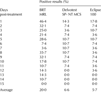
As shown in Table 1, a large number of positive outcomes were obtained when antibiotic-free milk samples from goats treated with IVM were analysed. Although the occurrence of positive results was observed throughout most of the experimental period (up to 14 d), the highest percentage was recorded, in all cases, on the first day post-treatment (BRT MRL = 46·4%; Delvotest SP-NT MCS = 14·3%; and Eclipse 100 = 17·8%), the BRT MRL being the most affected test throughout the experimental period. These results could be related to the different composition of the culture medium of the tests (BRT MRL: Mueller–Hinton agar and black brilliant as redox indicator; Delvotest SP-NT MCS and Eclipse 100: Plate count agar and bromocresol purple as pH indicator).
Positive results in microbial inhibitor tests in milk from goats (n = 28) treated with ivermectin

Microbial screening tests present a lower false-positive rate (0·6–4·3%) in milk from individual goats (Beltrán et al. Reference Beltrán, Berruga, Molina, Althaus and Molina2015) and, therefore, results obtained herein suggest that the administration of IVM may be related to these interferences.
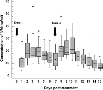
A significant increase (P < 0·05) of the concentration of IVM residues in goat's milk along the 3 d following the application of the drug (Fig. 1) was observed. However, the concentration of IVM ranged from 8·9 to 24·3 ng/ml, values rather lower than the inhibitory concentration producing 5% of positive results in microbial tests (IC5) calculated by Romero et al. (Reference Romero, Beltrán, Reybroeck and Molina2015) in an in vitro study (BRT MRL = 41; Delvotest SP-NT MCS = 83 and Eclipse 100 = 109 mg/l). In addition, the highest occurrence of positive results was not related to an elevated concentration of IVM residues in milk, suggesting that factors associated to drug administration other than residues in milk could be the cause of the interferences found. Thus, for example, IVM has been evaluated as immunomodulatory showing a favourable potential use as positive modulator (Stankiewicz et al. Reference Stankiewicz, Cabaj, Jonas, Moore, Millar and Chie1995; Sajid et al. Reference Sajid, Iqbal, Muhammad, Sandhu and Khan2007). Moreover, several studies indicate that sarcoptes mange does not only produce visible dermatological symptoms in mite-infested goats, but is also associated with physiological changes such as highest concentration of biochemical indicators of oxidative stress in the blood (Ujiwal & Dey, Reference Ujjwal and Dey2010), high total protein levels, γ-globulin and IgG in the serum plasma (Lastras et al. Reference Lastras, Pastor, Marco, Ruiz, Viñas and Lavin2000), and increased pro-inflammatory cytokine levels (Mullins et al. Reference Mullins, Arlian and Morgan2009) as well as high levels of acute phase proteins in the blood (Rahman et al. Reference Rahman, Lecchi, Fraquelli, Sartorelli and Ceciliani2010). The potential changes in the immunological system of the animals as a result of the IVM activity or the immune response of goats to combat the parasitic disease, could result in the presence of inhibitory substances in milk capable of interfering with the response of microbial inhibitor tests for screening antibiotics.
Box and whisker plots illustrating the concentration of ivermectin (IVM) in goat milk on the post-treatment days. The plots show the 25th and 75th percentiles (box), median (—), mean (+), 10th and 90th percentiles (whisker bars) and outliers (□).

It can be concluded that the illegal application of IVM in lactating dairy goats infested with sarcoptes mange produces persistent drug residues in milk and could also cause false-positive results in microbial inhibitor tests for screening antibiotics.
This work is part of the AGL-2009-11524 funded by the Ministry of Science and Innovation (Madrid, Spain) and the Generalitat Valenciana (ACOMOP/2012/164, Valencia, Spain).