Introduction
Most of the current brachytherapy dose calculation engines rely on a number of precalculated parameters obtained from single dose distributions in an infinite water medium to estimate dose distributions in a patient’s body. The current brachytherapy dose calculations are based on the superposition of single source dose distributions. These dose distributions are precalculated for each source centered in a homogeneous water phantom of certain geometry and, most commonly, used in the form of the TG-43 dosimetric formalism.Reference Rivard, Coursey and DeWerd1–Reference Rivard, Butler and DeWerd3 Although the use of TG-43 formalism in brachytherapy dose calculations has the advantage of a fast dose calculation mechanism, the accuracy of calculated dose values by such an algorithm is usually not at the desired level. This is because there are some effects ignored by the TG-43 method such as human tissue densities, material compositions, body interfaces and dose perturbations; due to the use of applicators, these factors have a significant impact on dose distributions in the patient’s body. All these effects can be significant in the brachytherapy photon energy rangeReference Landry, Reniers and Murrer4, Reference Hsu, Wu and Lee5 and can be included in modern treatment planning systems (TPS) for brachytherapy by using model-based dose calculation algorithms (MBDCAs). The American Association of Physicists in Medicine TG-186 issued guidelines toward implementing TPS, which can take the abovementioned complexities into account.Reference Beaulieu, Carlsson Tedgren and Carrier6
Some of the MBDCAs use Monte Carlo (MC) particle transport codes, which offer a high accuracy for dose calculations. Yegin et al. have developed BrachyDose, an MC code for rapid brachytherapy dose calculations.Reference Yegin and Rogers7 The code is based on EGSnrc,Reference Mainegra-Hing, Rogers and Tessier8 an MC particle transport code for the simulation of photons and electrons, and uses the Yegin’s Multi-Geometry packageReference Yegin9 to model the geometry of a brachytherapy source and the surrounding environment. BrachyDose has the capability of performing dose calculations in a voxelized phantom, and such a phantom can be even constructed from a series of CT images of a particular patient.Reference Taylor, Yegin and Rogers10
In addition to MC-based MBDCA algorithms, there are also some other deterministic MBDCA’s for brachytherapy dosimetry such as deterministic solvers of the differential linear Boltzmann transport equation (LBTE). This approach involves calculation of the angular and energy-dependent photon fluence at selected points in any computational domain through solving the LBTE by discretizing its six variables: Energy; using the multigroup approximation, angle; using discrete ordinates methods (DOMs), and space (3D); using finite difference or finite element methods (FEMs). A commercial TPS (Varian, Eclipse Acuros®BV version 8 software; Transpire Inc., Gig Harbor, WA) with an LBTE is now available for use with a number of 192Ir source models.Reference Daskalov, Baker and Rogers11–Reference Gifford, Price, Horton, Wareing and Mourtada14
There are studies showing that the difference between dose distributions of calculation algorithms increases when the computing medium is heterogeneous. In a study using 11 different heterogeneous configurations of Virtual Water™—Med-Cal, Inc. (VM-low density polyester foam), Verona, WI BR50/50 ™—CIRS, Inc., Norfolk, VA (equivalent to breast tissue), fungus and aluminium materials to ensure heterogeneous conditions, TG-43 and Acuros®BV were compared.Reference Moura, Micka and Hammer15 When heterogeneous materials (particularly aluminium and cork) were placed in the experimental phantom, relative differences were as high as 11·5% compared with that of the homogeneous phantom. The inconsistency dose distribution in TPS between TG-43 and Acuros®BV in BrachyVision has exceeded 12% in phantom geometries when the material after source is selected aluminium.
Acuros®BV and MC results were found to be 2% for 98% of the voxels of the scoring volume compatible with the AMIGOBrachy interface tool used for brachytherapy dose calculations, and Acuros®BV and MC had 20% difference with the TG-43U1 in the phantom.Reference Fonseca, Antunes, Yoriyaz, Reniers and Verhaegen16 In another study where the same module was used, there was no difference in tissue components between LTBE and MC. In clinical cases, up to 25% difference has been found in MC results compared with TG-43.Reference Fonseca, Reniers and Landry17
BrachyGuide—MATLAB (MathWorks, Natick, MA), a similar software for brachytherapy, has mentioned that treatment plans can be obtained in DICOM-RT (Digital Imaging and Communications in Medicine) format and can compare calculation results.Reference Pantelis, Peppa, Lahanas, Pappas and Papagiannis18 Compared with the results of Acuros®BV and MC, the differences between the near source and the longitudinal axis of the source are within ±2%. Differences between Acuros®BV and MC are ±3% in the TG-43 water sphere and inhomogeneous patient plans.
In a study of 26 cervical carcinoma patients who received 192Ir intracavitary brachytherapy, the heterogeneous dose calculation effect of the Acuros®BV and TG-43 algorithms was evaluated. The tandem applicators used are CT/ MRI compatible and not shielded. D2cc was left 5% in bladder for TG-43 and Acuros®BV. Rectum D2cc, sigmoid D2cc and ICRU19 Rectum doses differ by >5%. When the rectal and bladder balloon were contrasted, the dosimetric parameters remained within the difference of 5%.Reference Mikell, Klopp and Gonzalez20
In this work, we compared dose distributions of an HDR 192Ir source in different scenarios using Eclipse TG-43 formalism, Acuros®BV, which are relevant in routine TPSs and BrachyDose code, which have not previously been used for clinical dose calculations and can be used in the near future.
Methods and Materials
GammaMed HDR 192Ir model 12i source
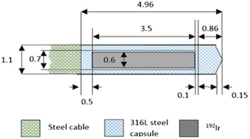
The model GammaMed HDR 12i source that Eclipse TPS-Transpire Inc., Gig Harbor, WA, uses consists of an iridium core of 3·5 mm in length and 0·7 mm in diameter, encapsulated in a stainless steel wire as shown in Figure 1. The distance from the physical tip of the source to the distal face of the active source core is 0·86 mm.Reference Perez-Calatayud, Ballester and Das21
Materials and dimensions (mm) of the Varian Medical Systems GammaMed HDR 12i source.19

For BrachyDose MC calculations, the same source is modelled by using Yegin’s Multi-geometry package, and the physical dimensions of the source are the same as the model used in Eclipse TPS. The source has a cylindrical pure Ir core, 3·5 mm long and 0·70 mm diameter. This core is enclosed in a 1·10 mm diameter AISI 316L stainless steel capsule (ρ = 7·8 g/cm3). There is a conical section at the distal end of the capsule that has 0·143 mm height and 75° half angle. Attached to the cone, there is a 0·717 mm long cylindrical section that is followed by a hollow section of 3·60 mm long and 0·70 mm diameter. After the hollow section, there is a 0·50 mm long solid cylindrical section. In this geometry, a total of 6·00 cm of AISI 304 stainless steel cable (effective density of ρ = 5·6 g/cm3) is included. The active length of this source is 3·50 mm long.Reference Taylor and Rogers22
Dose calculation scenarios
To evaluate various dosimetric capabilities of BrachyDose MC code for HDR brachytherapy (such as tissue densities, material compositions, medium volumes and medium interfaces), we prepared three different dose calculation scenarios. These are called ‘Scenario-1: Homogeneous water phantom scenario’, ‘Scenario-2: Extreme case heterogeneous media scenario’ and ‘Scenario-3: A patient with a cervical cancer scenario’. The aim of the Scenario-1 is to perform ideal geometry reference dose calculations, in which the dose perturbation effects caused by media heterogeneity is completely ignored. In these calculations, water was chosen as the environment medium. In Scenario-2, an unusual heterogeneous geometry consisting of three different materials (i.e., water, aluminium and air), which have different densities and dose absorption properties, was created and a number of dose calculations were performed in this geometry. The purpose of this scenario is to determine the algorithmic limitations by measuring possible dose perturbations that may occur during the propagation of photons in the interface of two materials with different absorption and scattering properties. In Scenario-3, a treatment plan using a series of CT images of a real patient was established, and dose calculations were performed according to this plan.
Three different dose calculation algorithms were used in dose comparisons namely, BrachyDose MC code, Eclipse Acuros®BV and TG-43 dose calculation tool. Although it is known that TG-43-based brachytherapy dose calculation tools cannot adequately calculate dose values in heterogeneous environments, the Eclipse TG-43 code is included in this study as a comparison tool considering that it will help us to see more clearly the accuracy of dose distributions calculated by the other MBDCAs. For each scenario, we modelled a special geometry design either in the Linux-based EGSnrc code system or in the Windows-based Eclipse TPS.
In the MC calculations in this study, electrons were not transported, and the photon cutoff energy was set to 1 keV. Rayleigh scattering, Compton scattering, photoelectric absorption and fluorescence emissions from X-rays were taken into account. In all calculations, the photon cross-sections were taken from the Photon Cross Sections on A Personal Computer (XCOM) databaseReference Mainegra-Hing, Rogers and Tessier8 and the mass energy absorption coefficients (μ en/ρ) calculated by using the EGSnrc user code ‘g’. In Acuros®BV dose calculations, cross-sections were prepared using Coupled Electron-Photon Cross Section (CEPXS)Reference Lorence, Morel and Valdez23 that includes all photon interactions except for Rayleigh scattering. All dose values reported in this study are calculated as dose to water. That is because the available version of Eclipse TPS produce for dose as dose to water (i.e., D m,w) only. Dose to water means that particles were transported within the given media but dose is scored in a small water medium at the centre of each voxel. BrachyDose calculates as dose to medium, which is estimated dose by scoring the collision kerma in voxels via a tracklength estimator. Due to this reason, in every particle step, kerma values were multiplied by (μ en/ρ)water (as described Landry et al in [Ref. Reference Landry, Reniers, Pignol, Beaulieu and Verhaegen24]) to obtain dose to water value.
Some dose comparisons in this study require that a point by point dose ratio is calculated at various points within the geometry. On the other hand, it is not always possible to produce the dose distributions on the same grid from the outputs of any two programs utilized in this study. For example, BrachyDose requires that dose to be calculated in a very small voxel. Since, over a certain limit of voxel dimensions, the accuracy of the dose estimated by the program at the centre of any voxel is effected by the large size of it. The maximum limit of the voxel size required by the BrachyDose code may not always be achieved in other programs. In such a case, the dose values are calculated in different grids. For these situations, the voxel dimensions were reduced to 0·1 cm in both dose data sets by applying a tri-cubic spline interpolation on the dose grid, and then, the dose comparisons were performed. We chose the tri-cubic spline interpolation because it always produces exact values at the points of the value grid as long as the derivatives of the interpolation curve higher than the second are negligible. In our dose comparisons, the sensitivity of the dose values to the smoothness of the derivatives was also investigated in a number of selected dose plans, and no significant differences were found.
Scenario-1: homogeneous water phantom scenario
In the first scenario, a homogeneous virtual water phantom with dimensions of (10 cm)3 was modelled in Eclipse TPS. The phantom consists of 39 × 39 × 51 cm slices and 0·25 × 0·25 × 0·20 cm voxel thickness. The mass density (ρ) of water in the phantom is chosen as 1·0 g/cm3. Its relative electron density is equal to 1, and the Hounsfield number value (HU) is chosen as 0.
For BrachyDose dose calculations, a cubic water phantom of (10 cm)3 consisting of 101 × 101 × 101 slabs with 0·1 × 0·1 × 0·1 cm voxels were used as the calculation medium. The number of histories for the simulations was 5 × 10⁹.
Two different dose calculation plans were prepared for the same geometry. In the first plan, a single HDR brachytherapy source is placed at the centre of the phantom and dose is scored in the environment water medium. In the second plan, there are five HDR brachytherapy sources in the phantom. These sources were placed in the phantom as follows: one source is at the centre and the other sources were placed symmetrically with 1 cm spacing on the same axis and on both sides of the source at the centre of the phantom.
For each plan, three separate dose calculations were performed by using either BrachyDose, Acuros®BV or TG-43. In each plan, the relative dose was normalized to 1·0 Gy at a point that is located at 1 cm away from the source centre and on the transverse axis of the source.
Scenario-2: extreme case heterogeneous media scenario (water- aluminium-air-water)
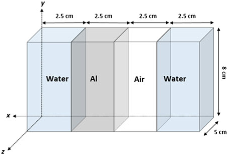
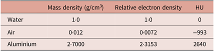
In the second scenario, a virtual rectangular phantom of 10 × 5 × 8 cm3 is designed in Eclipse TPS, which consists of 39 × 19 × 41 voxels with 0·25 × 0·25 × 0·2 cm thicknesses. The whole phantom was subdivided into four equal volumes, each has 2·5 × 5 × 8 cm3, to have heterogeneity in the phantom (see Figure 2 and Table 1). For dose calculations with the BrachyDose MC code, the same phantom, designed for Eclipse TPS, was used with the exception that the voxel dimensions were 0·1 × 0·1 × 0·1 cm. The number of histories for the simulations was 5 × 10⁹.
Extreme Case Heterogeneous Media Scenario. The virtual rectangular phantoms of size 10 × 5 × 8 cm3 and consisting of 41 slices with 0·2 cm thickness of CT created in Eclipse TPS. The phantom was divided into equal volumes of 2·5 × 5 × 8 cm3, and different densities were assigned to these volumes (Scenario-2).

Density and HU of materials used in extreme case heterogeneous media scenario for Eclipse TPS calculations (Scenario-2)

Two different dose calculation plans were prepared for the same geometry. In the first plan, a single HDR brachytherapy source is placed at the centre of the phantom and dose is scored in the environment water medium. In the second plan, there are five HDR brachytherapy sources in the phantom. These sources were placed in the phantom as follows: one source is at the centre and the other sources were placed symmetrically with 1 cm spacing on the same axis and on both sides of the source at the centre of the phantom.
For each plan, three separate dose calculations were performed by using either BrachyDose, Acuros®BV or TG-43. In each plan, the relative dose was normalized to 1·0 Gy at a point which is located at 1 cm away from the source centre and on the transverse axis of the source.
Scenario-3: a patient with a cervical cancer scenario
In the last scenario, a series of CT images of a patient with cervical cancer was studied. In these images, clinical tumour volume (CTV) and structures of the critical organs such as rectum, sigmoid, bladder and femur heads were drawn in Eclipse TPS. A treatment plan was formed by defining 26 dwell positions with 0·5-cm steps to provide optimal dose distribution in CTV. For dose calculations, the whole phantom is divided into 178 × 106 × 137 voxels, which has a voxel grid of 0·25 × 0·25 × 0·375 cm.
For BrachyDose plan, the same CT geometry and source positions defined above for Eclipse TPS were used. Also, the volume of each organ is extracted from the proper DICOM file, and material assignments were done accordingly. The mass density of any voxel is estimated from the HU value assigned to the same voxel in the DICOM file. For the conversion from HU to ρ, we used a CT calibration curve defined in an EGSnrc document.Reference Walters, Kawrakow and Rogers25 The phantom is divided into 150 × 106 × 151 voxels, which is of the size 0·2 × 0·2 × 0·2 cm. The number of histories is 5 × 10⁹ in MC simulations. In every dose calculation, the relative dose was normalized to 1·0 Gy at a point within the tumour region, which is same for all repeated dose calculations.
Results
For the phantom scenarios, isodose curves and dose difference were examined for dose distributions calculated with any of the abovementioned algorithms in each plan. For the latter scenario, CTV and critical organ doses were evaluated by comparing dose–volume histograms.
Scenario-1
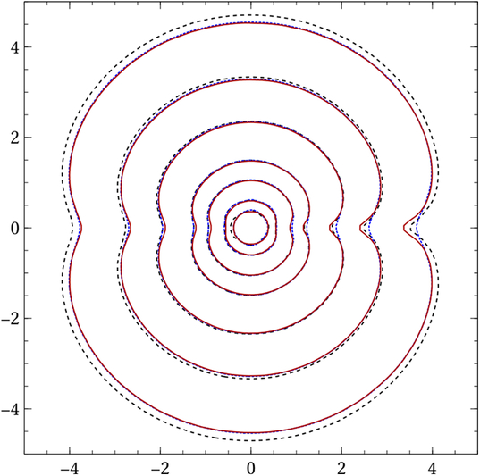
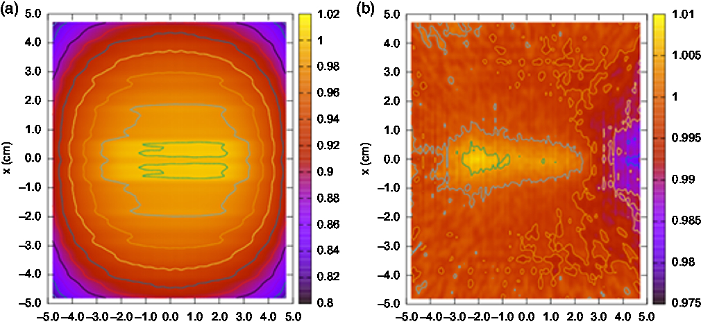
In the homogeneous water phantom scenario, three dose distributions, calculated with different algorithms, give consistent results in all points as expected. BrachyDose and Acuros® BV are in excellent agreement in the single source plan (see Figure 3). Relative dose differences for the five source plans in the homogeneous water phantom are given in Figure 4. It has been observed that BrachyDose and Acuros® BV are in agreement with 1% dose difference in almost all points. This can be taken as compatible with within statistical uncertainties.
Comparison of isodose curves for the single source plan in the Scenario-1. Red line is BrachyDose, blue line is Acuros®BV and dashed line is Eclipse TG-43 isodose curves.

Relative dose difference for the five sources plans a) Acuros®BV and TG-43. b) Acuros®BV and BrachyDose algorithms in homogen water phantom (Scenario-1).

Scenario-2
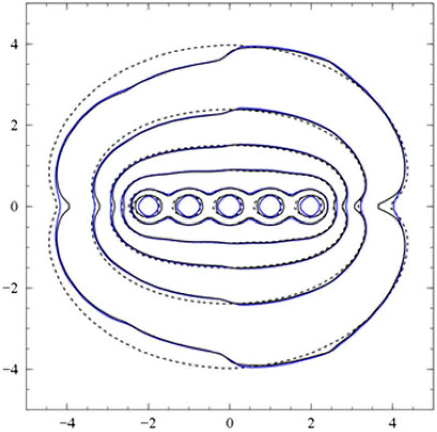
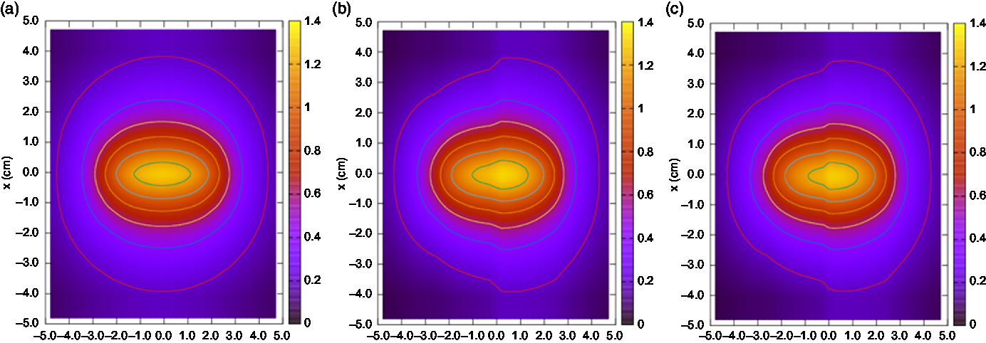
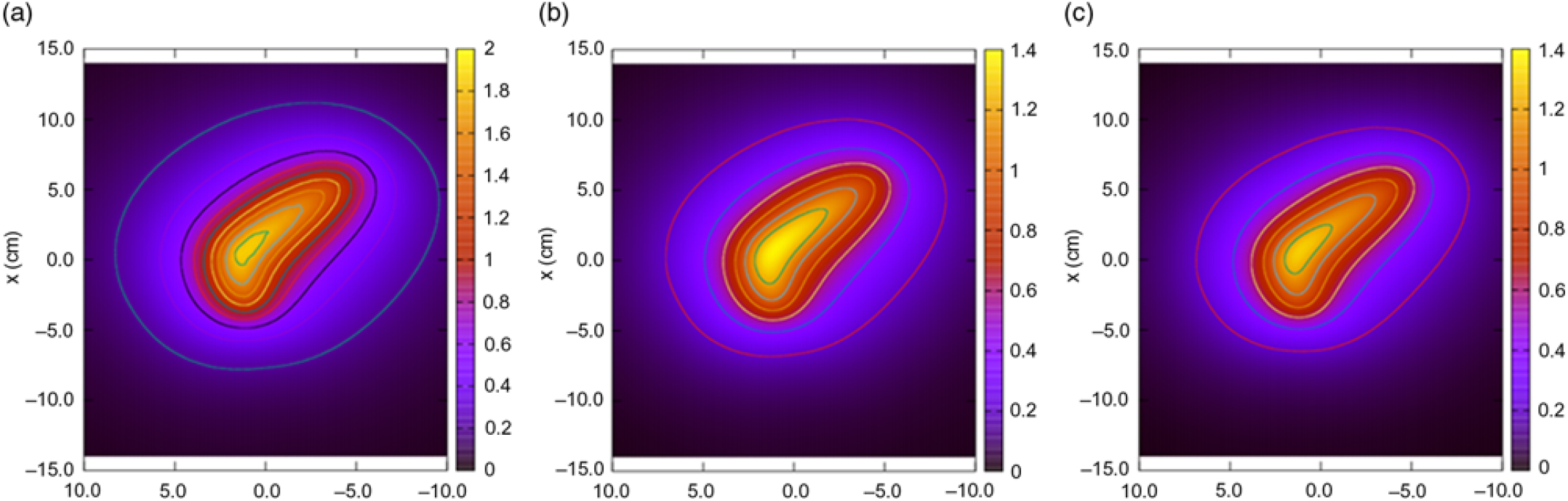
In this scenario, the dose distributions obtained from either BrachyDose, Acuros®BV or Eclipse TG-43 in heterogeneous media, which is formed by four regions (i.e., water-air-aluminium-water), were investigated. When the isodose curves were examined, it was found that the change of material composition did not make any difference for TG-43-based dose plan at any interfaces of two medium, as expected. On the other hand, the shapes of isodose curves are considerably distorted at the aluminium–air interface for both BrachyDose and Acuros®BV plans. The degree of the distortion was found to be more pronounced in the BrachyDose plan than Acuros®BV (Figures 5 and 6).
Comparison of isodose curves for the five source plans in Scenario-2. Black line is BrachyDose, blue line is Acuros®BV and dashed line is Eclipse TG-43 isodose curves.

Isodose curves of the five source plans in Scenario-2: (a) Eclipse TG-43 plan, (b) Acuros®BV plan and (c) BrachyDose plan. In isodose curves, the material did not make a difference for TG-43 at the interfaces, but the isodose curve breaking at the aluminium–air interface was found to be more pronounced in BrachyDose than Acuros® BV.

Scenario-3
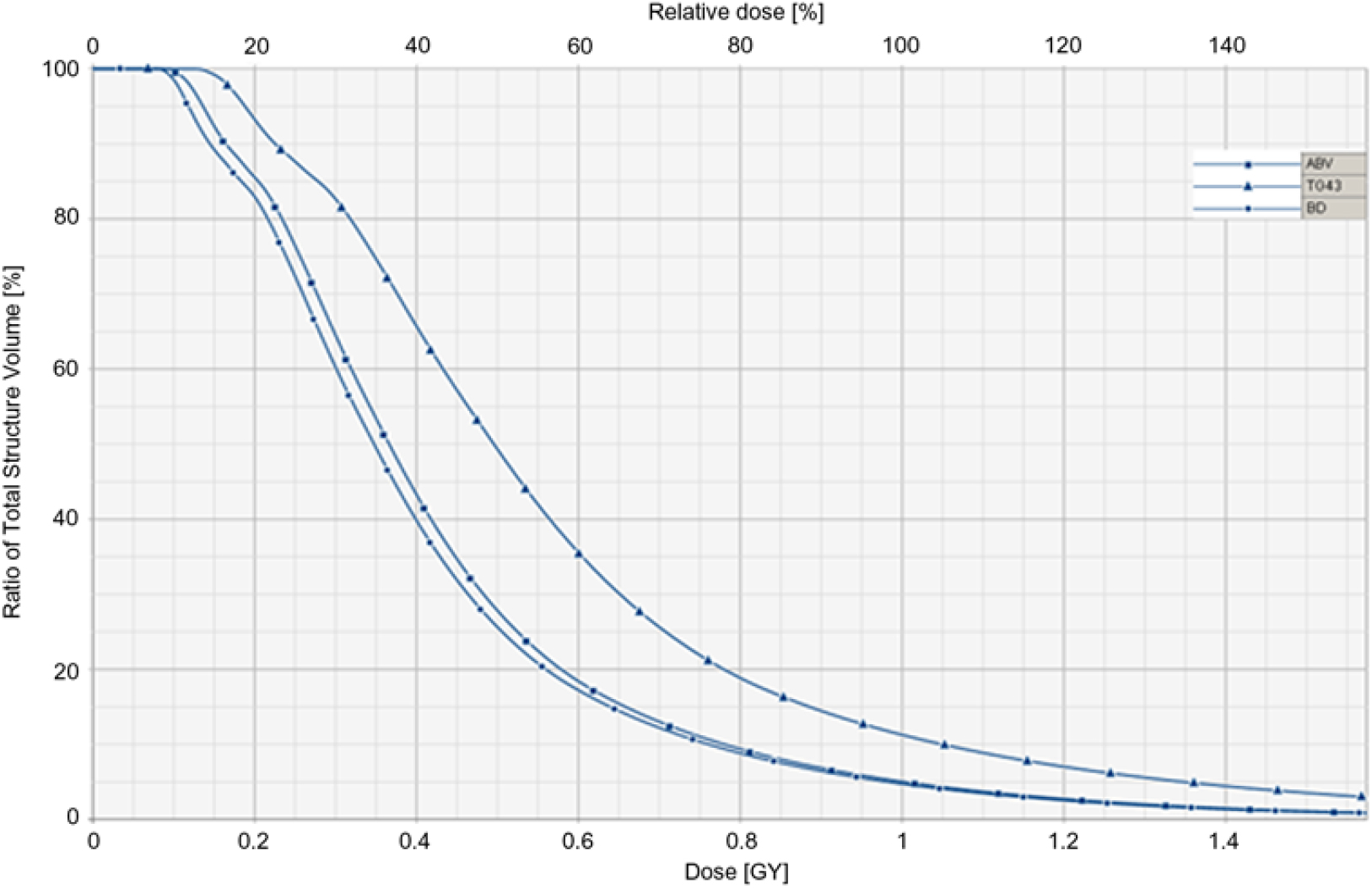
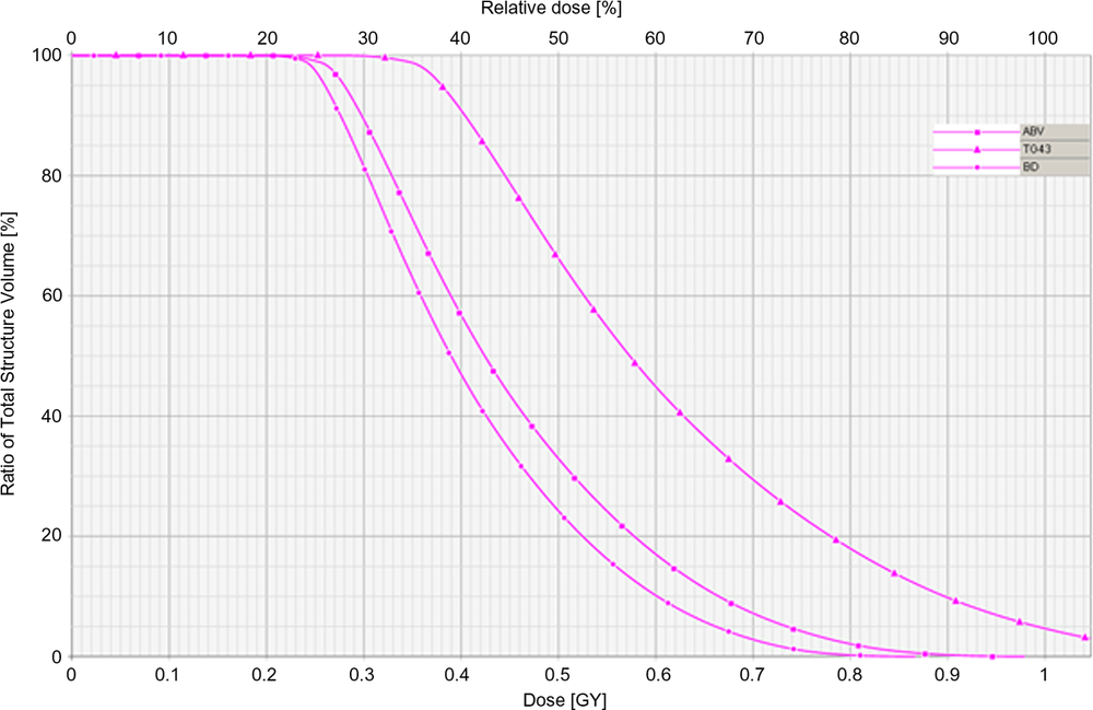
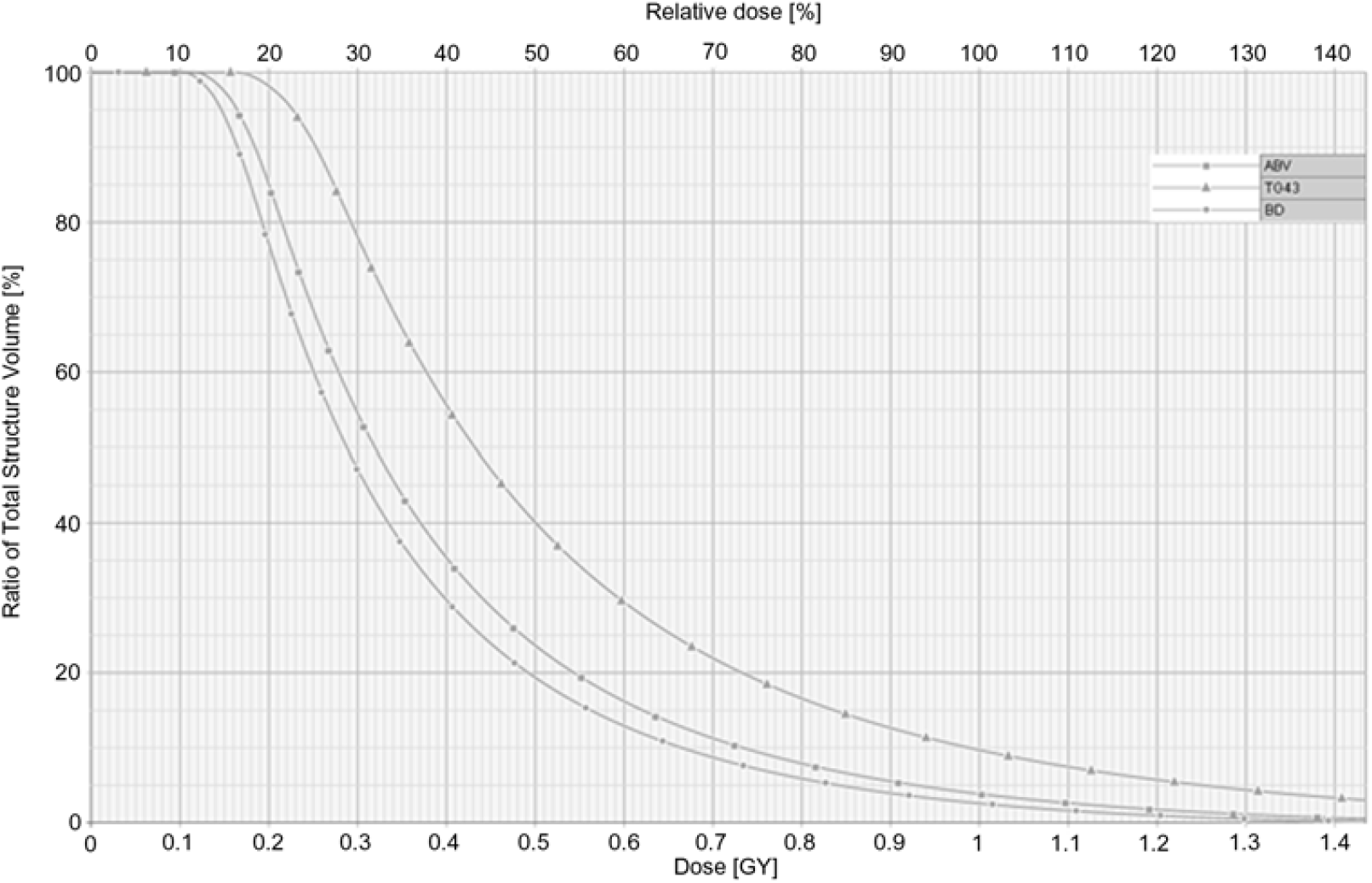
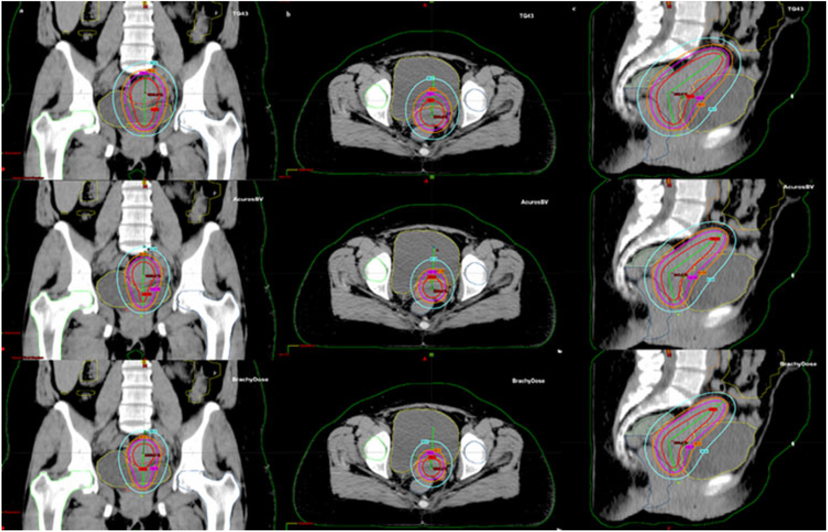
In the last case of our study, CT images obtained from a patient with cervical cancer were studied. Dose distributions obtained from either BrachyDose, Acuros®BV or Eclipse TG-43 were evaluated in the following organs: sigmoid, rectum, bladder and femoral heads. In the evaluation process, the minimum dose in the most irradiated 2cc volume (D2cc) was calculated for selected organs. For rectum, the value of D2cc from BrachyDose plan matched that from Acuros®BV plan within 2%, but the difference between Eclipse TG-43 and BrachyDose was around 37% (Figure 7). The sigmoid dose difference of D2cc is 35·7% between TG-43 and Acuros®BV and 50·8% between TG-43 and BrachyDose, and the difference between Acuros® BV and BrachyDose is around 11% (see Figure 8). For bladder, the greatest difference in the D2cc dose is observed between TG-43 and BrachyDose (see Figure 9). While the mean doses of the right femur head do not differ between Acuros® BV and BrachyDose, the differences between the model-based algorithms and the TG-43 were 75%. When the isodose curves were examined on these patient images, MBDCAs and TG-43 plans have shown significant differences (Figures 10 and 11).
Dose–volume histogram for rectum in Scenario-3. Dotted line is BrachyDose, square line is Acuros®BV and triangle line is Eclipse TG-43 formalism.

Dose–volume histogram for sigmoid in Scenario-3. Dotted line is BrachyDose, square line is Acuros®BV and triangle line is Eclipse TG-43 formalism.

Dose–volume histogram for bladder in Scenario-3. Dotted line is BrachyDose, square line is Acuros®BV and triangle line is Eclipse TG-43 formalism.

The isodose curves in Eclipse TPS for a patient with cervical cancer (a) frontal section and (b) the axial section. (c) The sagittal section of the pelvic region in Scenario-3. Isodose curves from the innermost to the outermost is 50% (blue), 80% (orange), 100% (magenta) and 150% (red), respectively.

The isodose curves for a patient with cervical cancer (a) Eclipse TG-43 plan, (b) Acuros®BV plan and (c) BrachyDose plan. Model-based algorithms and TG-43 plans have shown significant differences.

Discussion
In the homogenous water phantom scenario, two MBDCAs and the TG-43 based code gave similar results as previously found in the literature for the 192Ir source.Reference Fonseca, Reniers and Landry17, Reference Pantelis, Peppa, Lahanas, Pappas and Papagiannis18
In our study, for the extreme case heterogeneous media scenario, the difference between BrachyDose and Acuros®BV plans is usually around ~2% in all media, and the maximum differences were observed in the aluminium medium and in five source plans.
In the scenario, ‘a patient with a cervical cancer’ dose differences of the bone structure between TG-43 and the other two model-based algorithm outputs in the patient plan reached a value up to 75%. The maximum dose difference between BrachyDose and Acuros® BV is within 2%, 11%, 11% and 1% in rectum, bladder, sigmoid and bone, respectively.
Conclusions
In this study, the results of dose distributions produced by BrachyDose MC code were evaluated in various geometry conditions including a homogeneous water phantom, a heterogeneous phantom and a phantom constructed from CT data of a patient for the GammaMed HDR 12i model 192Ir source. For dose comparisons, Eclipse Acuros®BV and TG-43 were used. When the whole calculation medium is homogeneous water, dose values calculated by any algorithm given above were in excellent agreement in most of the regions in the phantom for the single source plan. For the five source plan, however, dose differences between TG-43 and the other model-based algorithms are slightly different, and the differences between BrachyDose and Acuros®BV are negligible.
In brachytherapy dose calculations, calculation medium characteristics, patient size, applicator characteristics and the number of sources used in the plan affect dose distribution significantly. It has been observed that the lack of TG-43 formalism in dose calculation and the use of model-based algorithms can lead to more reliable results. After the accumulation of sufficient data by using MBCDAs, the dose determined for the target tumour tissue can be given more accurately, while taking less dose to critical tissues, side effects that may occur in the patient can be reduced. In the ‘a patient with a cervical cancer’ plan, we observed significant dose distributions between BrachyDose and Acuros®BV.
We think that these differences are mostly due to the fact that CT-calibration curves used in the transformation of HU values to mass density in the CT data are different. In a future study, we are going to investigate the effects of CT data conversions on MC brachytherapy dose distributions.
Author ORCIDs
Ayse Dagli 0000-0003-1219-8870; Gultekin Yegin 0000-0002-0727-7569