It has long been known that the patterns of origin of the coronary arteries from the aortic root can vary markedly when the heart is congenitally malformed. Many have attempted to provide classifications for the variations, most notably in the setting of hearts with transposed arterial trunks. Some of these classifications have been sufficiently complex to deter all but the most adventurous code-breakers.Reference Shaher and Puddu 1 Others have been unduly simple, failing to cater for all known variations.Reference Yacoub and Radley-Smith 2 Still others have attempted to avoid the complication produced by the known variation in the location of the aortic and pulmonary roots.Reference Gittenberger-de Groot, Sauer, Oppenheimer-Dekker and Quaegebeur 3 None have achieved universal recognition, perhaps because insignificant attention has been paid to the importance of the location of the aortic root within the cardiac base.Reference Chiu, Chu and Wang 4 – Reference Huang, Chiu and Lee 6 Recent advances in understanding the development of the heart have now provided added impetus to incorporating knowledge of aortic position into description of coronary arterial origin. This is because, rather than budding out from the aorta, as had originally been the concept accepted for coronary arterial development,Reference Hackensellner 7 the major coronary arteries grow into the developing aortic valvar sinuses,Reference Bogers, Gittenberger-de Groot, Poelmann, Peault and Huysmans 8 with the major arteries themselves developing within the atrioventricular and interventricular grooves.Reference Hutchins, Kessler-Hanna and Moore 9 The known random disposition of the origin of the coronary arteries in the setting of common arterial trunkReference Crupi, Macartney and Anderson 10 – Reference Bogers, Bartelings and Bokenkamp 12 provides additional evidence of the added importance of the process of septation in guiding the coronary arteries to their optimal positions within the arterial roots. In this review, we show how attention to both aortic sinusal origin and the position of the aortic root is essential if we are to fully understand and predict the variable patterns of coronary arterial morphology in congenitally malformed hearts.
The normal anatomy of the coronary arteries
The major coronary arteries in the normal heart occupy the atrioventricular and interventricular grooves. Hence, there are four major coronary arteries. In most normal individuals, only two coronary arteries take their origin from the aortic valvar sinuses, although it is frequent to find accessory arteries arising within the sinuses.Reference Angelini 13 In the typical situation, it is the right and left arteries that arise from the aortic sinuses. In nine-tenths of the population, the right coronary artery, which runs within the right atrioventricular groove (Fig 1, left-hand panel), then gives rise to the artery that nourishes the inferior component of the muscular ventricular septum. It is regrettable that, currently, this inferior interventricular artery is described as being posterior and descending. It fulfills these alleged qualities only when the heart is removed from the thorax and examined in the manner of the Saint Valentine heart. When described attitudinally, a feature now seen with increasing frequency subsequent to the development of computed tomographic angiography, there can be no question but that the artery is inferior and interventricular (Fig 1, middle panel). It has long been recognised and taught that pathological disease of this artery produces inferior myocardial infarction. In the other one-tenth of the population, with the exception of a few circumstances where there are duplicated inferior interventricular arteries, the artery nourishing the inferior part of the muscular ventricular septum arises from the circumflex coronary artery. The circumflex artery itself is a branch of the main stem of the left coronary artery. The main stem is typically very short. It branches in the space between the subpulmonary infundibulum and the left atrial appendage (Fig 1, right-hand panel), giving rise to the circumflex and anterior interventricular arteries.
The computed tomographic angiograms show the essential features of the major coronary arteries. The left-hand panel shows the course of the right coronary artery, which in nine-tenths of the population gives rise to the inferior interventricular artery (middle panel). The box provided to show the orientation of the heart in space (lower right-hand corner of the panel) shows that this artery supplies the inferiorly positioned diaphragmatic surface of the heart. The main stem of the left coronary artery (LCA – right-hand panel) branches to become the anterior interventricular and circumflex arteries.

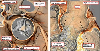
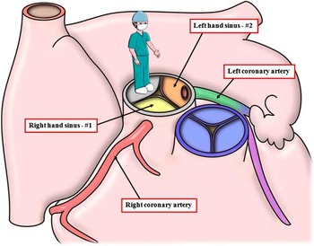
Although there are four major coronary arteries, only two arteries typically arise from the aortic root. It is the variation in these aortic sinusal origins that are of most clinical significance.Reference Kurosawa, Imai and Kawada 14 Here again, the advent of computed tomographic angiographic has served to clarify the normal and abnormal variants of sinusal origin.Reference Hlavacek, Loukas, Spicer and Anderson 15 In most instances, the coronary arteries arise within the aortic valvar sinuses, proximal to the sinutubular junction (Fig 2, left-hand panel). Almost always, the arteries arise one each from the valvar sinuses that are adjacent to the pulmonary infundibulum (Fig 2, right-hand panel). When the arterial pedicles are themselves in their anticipated position, it is then an easy matter for the artery to arise from the right-sided and posterior aortic sinus to pass directly into the right atrioventricular groove, whereas the artery arising from the anterior and left-sided sinus passes into the space immediately beneath the left atrial appendage, where it divides to supply the circumflex and anterior interventricular branches (Fig 1, left-hand panel). In malformed hearts, however, particularly those with abnormal ventriculo-arterial connections, the aortic valvar sinuses do not always adopt these normal relationships relative to the pulmonary root. Irrespective of the relationships, nonetheless, it is the rule that the coronary arteries continue to arise from the valvar sinuses that are adjacent to the pulmonary root. If these two sinuses are then viewed, figuratively speaking, from the stance of the observer standing in the non-adjacent aortic sinus, and looking towards the pulmonary root, it is possible to distinguish between the two other aortic sinuses as being to the right-hand or the left-hand side. This perception holds good irrespective of the arterial relationships themselves. It has now become conventional to define the sinus to the right-hand side as being #1, and the left-handed sinus as being #2. In the normal heart, it is sinus #1 that gives rise to the right coronary artery, whereas sinus #2 supports the main stem of the left coronary artery (Fig 3).
The left-hand panel shows a computed tomographic angiogram illustrating the aortic root as seen from above and the right. It shows how the coronary arteries take their origin from the two aortic sinuses that are adjacent to the pulmonary root. As shown in the right-hand panel, usually the coronary arteries arise within the valvar sinuses, athough as shown here, one artery, in this instance the left coronary artery, can be directly adjacent to the sinutubular junction.

The cartoon shows how, figuratively speaking, and when the coronary arteries arise from the aortic valvar sinuses adjacent to the pulmonary root one of the sinuses will always be to the right hand of the observer standing in the non-adjacent sinus. This sinus is now defined as being #1. The other sinus, defined as #2, is to the observer's left hand. In the normal heart, it is sinus #1 that gives rise to the right coronary artery, and sinus #2 that supports the left coronary artery. When defined in this manner, it is always possible to distinguish the left-handed and right-handed sinuses, irrespective of the interrelationships of the arterial trunks.

Development of the coronary arteries
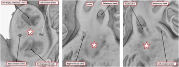
Much has been learnt within the last three decades regarding the embryological development of the coronary arteries. It is now well established that both the arteries and the coronary veins are derived from the epicardial tissues, these in turn being formed from the pro-epicardial organ. This structure is located within the inferior atrioventricular groove adjacent to the transverse septum of the developing embryo.Reference Virágh and Challice 16 Cells migrate from the organ and grow over the epicardial surface of the heart, extending as far as the distal outflow tract. Under the influence of Wt1 and Raldh1 genes,Reference Pérez-Pomares, Phelps and Sedmerova 17 the epicardial cells become transformed to mesenchyme, penetrating the developing myocardial walls to produce the fibrous matrix of the compact myocardium and the smooth muscular walls of the coronary arteries and veins. It was initially believed that these epicardial coronary arteries obtained their aortic connection by fusion with channels that had budded out from the developing aortic valvar sinuses.Reference Hackensellner 7 In reality, the proximal components of the developing epicardial coronary arteries grow into the aortic root (Fig 4),Reference Bogers, Gittenberger-de Groot, Poelmann, Peault and Huysmans 8 subsequent to the completion of its separation from the pulmonary root.Reference Angelini 18 The very fact that the epicardial coronary arteries, almost without exception, take their origin from the aortic sinuses adjacent to the pulmonary root suggests that the developmental processes separating the roots one from the other is also instrumental in guiding the epicardial coronary arteries to their appropriate aortic origin. It has now also been established that, during its development, the ventricular musculature supporting the pulmonary root is impervious to the passage and development of epicardial coronary arteries.Reference Theveniau-Ruissy, Dandonneau and Mesbah 19 Owing to the fact that the coronary arteries are formed within the epicardial tissue planes before achieving their connection to the developing aortic valvar sinuses, it follows that the location of the sinuses themselves will play a significant role in determining the definitive morphology of the coronary arterial patterns. Coupled with the knowledge that the subpulmonary myocardial domains are impervious to the passage of the developing epicardial coronary arteries, appreciation of these facts can bring order to the understanding of the potential random patterns seen in various congenital cardiac malformations, in particular those found in the setting of transposition and common arterial trunk.
The images are from an episcopic data set prepared from a mouse at embryonic day 15.5. The left-hand panel is a short-axis section across the developing arterial roots. The ends of the major outflow cushions are cavitating to form the sinuses and leaflets of the arterial valves. The fused central parts of the cushions are shown by the star. The coronary arterial primordiums have penetrated the muscular turret surrounding the developing roots, but as yet have not penetrated into the developing aortic sinuses. The middle panel, a frontal section, shows the proximal part of the right coronary artery, with the star showing the fused proximal parts of the cushions, while the right-hand panel shows the epicardial proximal component of the left coronary artery, the star again showing the fused proximal cushions. The images are produced by kind permission of Dr T.J. Mohun, MRC National Institute for Medical Research, London, United Kingdom.

The arrangement of the coronary arteries in the setting of transposition
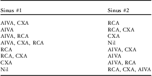
It is now generally accepted that the optimal surgical procedure for patients having the combination of concordant atrioventricular and discordant ventriculo-arterial connections, in other words the commonest variant of transposition, is the arterial switch procedure. It is also accepted that, although all known patterns of coronary arterial origin are amenable to operative switching, pre-operative knowledge of the specific patterns is of great advantage to the surgeon.Reference Kurosawa, Imai and Kawada 14 , Reference Lacour-Gayet and Anderson 20 When attempts are made to combine knowledge of sinusal origin with variations in the epicardial course of the coronary arteries, however, the resulting categorisation is remarkably complex.Reference Shaher and Puddu 1 On the other hand, attempts to simplify the classification produced a system that proved incapable of coping for all the variations encountered.Reference Yacoub and Radley-Smith 2 If attention is directed to those cases in which the three major coronary arteries, namely, the right, circumflex, and anterior interventricular arteries, arise from both, or exclusively one, of the adjacent aortic valvar sinuses, then it follows that, simply in terms of sinusal origin, there are only eight possible combinations (Table 1).
The table shows how there are eight possible combinations when it is presumed that the three major coronary arteries arise either from both adjacent aortic valvar sinuses, or exclusively from one of the sinuses.

AIVA = anterior interventricular coronary artery; CXA = circumflex artery; RCA = right coronary artery
All options have now been observed apart from the one highlighted in gray shade
This basic premise, emphasised by Quaegebeur in discussions with one of the current authors (R.H.A.), spawned the approach now known as the Leiden convention.Reference Gittenberger-de Groot, Sauer, Oppenheimer-Dekker and Quaegebeur 3 This convention was itself incorporated as one of the major features of the classification proposed by the combined study groups of the Society of Thoracic Surgeons and the European Association of Cardiothoracic Surgery.Reference Jaggers, Cameron, Herlong and Ungerleider 21 Another of the current authors has long championed the importance of aortic position in classifying the coronary arterial patterns.Reference Chiu, Chu and Wang 4 – Reference Huang, Chiu and Lee 6 Aortic position in itself, however, cannot serve as a surrogate for defining the various combinations seen. The location of the aorta, nonetheless, provides the key to understanding why certain patterns are more frequent in particular subsets of patients with transposed arterial trunks. Thus, the significance of aortic position becomes immediately evident when we consider the fact that when 100 hearts were selected at random from the archive of congenitally malformed hearts held at Children's Hospital of Pittsburgh it proved possible to identify seven of the eight possible patterns predicted by Quaegebeur (Table 1). When this occurrence was first observed, the question was not asked as to why one of the eight possibilities should be absent. On further investigation,Reference Chiu and Anderson 22 it transpires that this missing option has never been reported within the worldwide literature concerning coronary arterial patterns in patients with transposition. The missing option would be found if the right coronary artery, together with the circumflex artery, arose from sinus #1, while the anterior interventricular artery arose from sinus #2. Attention to the influence of aortic position shows why it is highly unlikely that this pattern can occur in patients with transposition.
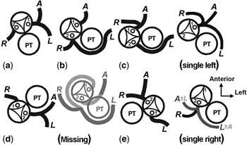
In most patients with transposition, the aortic valve is positioned anteriorly and rightward relative to the pulmonary valve. Indeed, it is because of this frequent finding that transposition itself is often described as “d-transposition”. In certain instances, the aortic valve can be positioned anterior and leftward, even when the atrial chambers are normally positioned and there is right-handed ventricular topology. This is the combination described as “transposition {S,D,L}”. More frequently, particularly when there is deficient ventricular septation, the aortic valve is positioned rightward and side by side relative to the pulmonary valve. Very rarely, the aortic valve can be positioned posteriorly and to the right of the aortic valve. In all of these circumstances, if the anterior interventricular artery was to take its origin from the left-handed sinus (#2), while the other two arteries were arising from the right-handed sinus (#1), then the proximal part of the anterior interventricular artery would be required to cross the territory already occupied by the proximal parts of one of the other coronary arteries (Fig 5, missing option). This is unlikely to happen. Indeed, the only aortic position that would favour such an arrangement is for the aortic root to be positioned posteriorly and leftward relative to the pulmonary trunk, such that sinus #1 is adjacent to the proximal parts of the right and circumflex arteries, while the anteriorly located sinus #2 is directly adjacent to the proximal part of the anterior interventricular artery. This situation has yet to be described in the setting of transposition. Taken together, the evidence suggests that there is a marriage of convenienceReference Chiu, Wu and Chen 5 , Reference Huang, Chiu and Lee 6 , Reference Wernovsky and Sanders 23 , Reference Landolt, Anderson, Zorn-Chelton, Guyton, Hatcher and Williams 24 between the proximal parts of the epicardial coronary arteries and the aortic valvar sinuses. This marriage emphasises the importance of the location of the aortic root relative to the pulmonary trunk in determining the patterns seen in the various subsets of patients with transposed arterial trunks.
The cartoon shows the potential origins of the coronary arteries relative to the relationship of the arterial valve. The orientation is as seen echocardiographically, examing the base of the heart as seen from the ventricular apex. The missing option is shown in grey shading. It is an impossible arrangement, as the anterior interventricular artery would need to cross the territory of another epicardial artery so as to reach sinus #2. This pattern could theoretically exist should the aortic valve be positioned posteriorly and leftward relative to the pulmonary trunk (PT), but to the best of our knowledge, this relationship has never been encountered in a patient with transposition. Note that pattern (d) shows so-called “posterior” transposition, in which the arterial valves are normally related even though the trunks are discordantly connected.

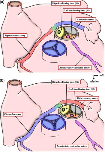
When considered in terms of this marriage of convenience, coupled with the fact that the subpulmonary myocardium is known to be impervious to the passage of epicardial coronary arteries, it proves no longer to be coincidental that the situation in which the circumflex coronary artery takes a retropulmonary course through the transverse sinus to arise from sinus #2 is seen most frequently when the arterial roots are aligned in side-by-side manner, with the aorta to the right (Fig 5b and c). It is similarly no coincidence that, when the aortic valve is leftward relative to the pulmonary root, the right coronary artery usually passes in front of the aortic infundibulum and arises, along with the anterior interventricular artery, from sinus #2 (Fig 5e). The marriage of convenience explains well why, in those rare circumstances when the aortic valve is transposed but posterior and rightward, the right coronary artery arises from sinus #1, while the circumflex and anterior interventricular arteries arise from sinus #2 (Fig 5d). The marriage of convenience also explains why the option that is missing in patients with regular transposition can be found in patients with concordant ventriculo-arterial connections but parallel arterial trunks in whom the aorta is positioned posteriorlyReference Huang, Chiu and Lee 6 (Fig 6a), and in those patients with congenitally corrected transposition having a leftward and posterior aortaReference Chiu, Wu and Chen 5 (Fig 6b).
The cartoons show the two situations in which the “missing option” for transposition has been encountered. The upper panel ( a ) shows the arrangement when the arterial trunks are concordantly connected, but have a parallel arrangement as they extend into the mediastinum – so-called “anatomically corrected malposition”. The lower panel ( b ) shows the situation with congenitally corrected transposition, which is the combination of discordant atrioventricular and ventriculo-arterial connections. In both reported instances,Reference Chiu, Wu and Chen 5 , Reference Huang, Chiu and Lee 6 the aortic root was posterior and left-sided, with marriage of convenience making the possible the missing option.

The arrangement of the coronary arteries in common arterial trunk
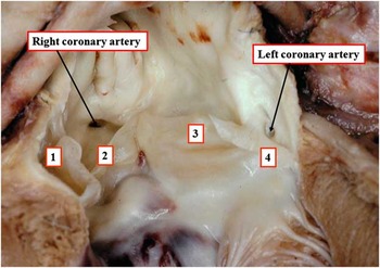
It is now generally accepted that a common arterial trunk is a solitary vessel leaving the ventricular mass that supplies directly the systemic, pulmonary, and coronary arterial circulations.Reference Collett and Edwards 25 The morphogenesis of the entity is more controversial. It is intuitive to argue that the entity is the consequence of failure of septation of the developing outflow tract. It has been argued, however, that it represents failure of growth of the subpulmonary conus.Reference Van Praagh and Van Praagh 26 The arrangement of the coronary arteries is helpful in arbitrating this debate. If there had been failure of growth of the subpulmonary infundibulum, the truncal valve would, in essence, be an aortic valve. The disposition of the coronary arteries would then be standardised as for the normal heart. In fact, there is no standard pattern for the sinusal origin of the coronary arteries in patients with common arterial trunk.Reference Crupi, Macartney and Anderson 10 – Reference Bogers, Bartelings and Bokenkamp 12 Nor is the arrangement of the valvar sinuses as would be expected in the valve was simply aortic in pattern, as a good proportion of patients have quadrifoliate truncal valves. In those with truncal valves having four sinuses and leaflets, the coronary arteries arise most frequently from the opposite right and left-sided valvar sinuses (Fig 7), as would be anticipated had the embryonic outflow tract failed to septate.
The left ventricular outflow tract in this heart from a patient with common arterial trunk has been opened from the front. It can be seen that the truncal valve has four sinuses, each supporting a valvar leaflet (1 through 4). The coronary arteries arise from the right- and left-sided opposite valvar sinuses. This is the usual arrangement when the truncal valve has four leaflets.

Even when the truncal valve has three sinuses, it is the exception rather than the rule to find the coronary arteries arise in the manner that would be expected had the valve been aortic.Reference Crupi, Macartney and Anderson 10 Furthermore, the left anterior circumference of the truncal root has been shown to be devoid of coronary arterial orifices.Reference Bogers, Gittenberger-de Groot, Poelmann, Peault and Huysmans 8 , Reference Chiu, Wu, Chen, Chen and Wang 27 This is not unexepected, as this anterior component is the potential subpulmonary infundibular musculature, which, as we have discussed, is known to be impervious to the passage of epicardial coronary arteries.Reference Theveniau-Ruissy, Dandonneau and Mesbah 19 The circumstantial evidence from the location of the coronary arteries, therefore, strongly supports the notion that common arterial trunk is the consequence of failure of septation of the developing outflow tract. Scientific evidence to support this notion had long since been provided by Van Mierop et al.Reference Van Mierop, Patterson and Schnarr 28 This has now been endorsed by studies of genetically modified mice.Reference Anderson, Chaudhry and Mohun 29 All the evidence, therefore, points strongly to an additional influence of septation of the outflow tract, over and above the position of the aortic root relative to the cardiac base, in guiding the ingrowing epicardial coronary arteries to their appropriate sinuses.
Conclusions
There are two primary features that should be considered by those seeking to provide all-encompassing classifications for the origin of the coronary arteries in congenitally malformed hearts. The first is the sinusal origin of the major coronary arteries. This, of course, is a feature that can readily be established by the surgeon in the operating room. To be forewarned, however, is to be forearmed. The second feature, which thus far has received less attention in its own right, is the location of the aortic root relative to the cardiac base. It is this feature that produces the marriage of convenience between the aortic sinuses adjacent to the pulmonary root and the proximal epicardial components of the major coronary arteries. The marriage of convenience provides rational explanations not only for the arrangement of the coronary arteries in transposition and common arterial trunk, but also lesions such as tetralogy of Fallot.Reference Chiu, Wu and Wang 30 The concept provides insights into additional features that we have been unable to discuss because of the constraints of space. These include the notions of dual as opposed to single sinus entry to the aortic root,Reference Chiu and Anderson 22 and intramural origin of the coronary arteries.Reference Chiu, Wu and Chang 31 It is our belief that, in time, the notion of marriage of convenience will underscore an all-embracing categorisation for the disposition of the coronary arteries in the congenitally malformed heart. To prove successful, however, such an advance will require uniform description of the location of the aortic root within the cardiac base.Reference Chiu, Wang and Wu 32
Acknowledgements
We are indebted to Dr Tim Mohun, from the MRC National Institute for Medical Research in London, for permitting us to use the images from the episcopic data sets he prepared from developing hearts of mice.