Introduction
Meningoceles are formed when meningeal membranes herniate through defects in the skull base. They may remain clinically silent until a dural breach permits spontaneous cerebrospinal fluid (CSF) leakage. If present in the anterior skull base, this results in CSF rhinorrhoea.Reference Gutiérrez-González, Boto, Pérez-Zamarrón and Rivero-Garvía1 In cases where CSF leakage is present without herniation of the dural membranes, CSF may accumulate, under constant pulsation and pressure, behind the sinonasal mucosa; this is known as a pseudomeningocele.Reference Vaezi, Snyderman, Saleh, Carrau, Zanation and Gardner2 Both lesions are encountered rarely within the skull base.
Here, we present the first case of an asymptomatic left sphenoid sinus lesion mimicking a mucocele, which confounded the true underlying diagnosis of pseudomeningocele.
Methods
We conducted a literature search using the Medline, EMBASE, CINAHL and AMED databases and the Cochrane Database of Systematic Reviews using the Ovid Collection, to identify scientific publications relevant to pseudomeningocele.
Case report
A 55-year-old woman was referred to our otolaryngology department following the discovery of an incidental lesion in the left sphenoid sinus, identified by computed tomography (CT) performed for dental evaluation. The patient's general health was good although her body mass index was 45 kg/m2. She reported no previous head injury or sinonasal surgery. The patient had previously been reviewed three years earlier in the ENT clinic with symptoms of intermittent, left-sided otalgia and dizziness. At that time, otological examination had been unremarkable, and audiography had revealed mild, symmetrical, high frequency, sensorineural hearing loss requiring no further treatment.
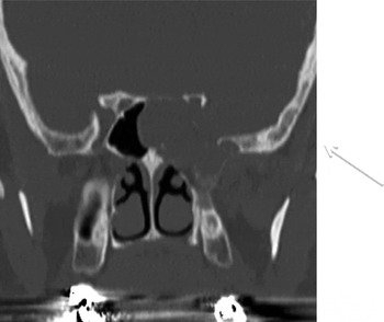
During the current presentation, otological and neurological examinations were normal. Fibre-optic nasoendoscopy revealed a stenosed left sphenoid ostium with expansion of the face of the sphenoid sinus. A CT scan demonstrated a 2.6 × 2.0 cm, smooth, soft tissue mass situated within the left sphenoid sinus, extending inferolaterally to the pterygopalatine fossa (Figure 1). There was a dehiscence of the lateral wall of the left sphenoid sinus and also of the left optic canal. No intracranial pathology was present and ventricles were within normal limits.
Coronal computed tomography image demonstrating a soft tissue mass within the left sphenoid sinus.

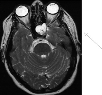
A contrast-enhanced magnetic resonance imaging scan was performed to delineate further the soft tissue lesion. This confirmed the presence of a lobulated mass in the left sphenoid sinus, expanding into the posterior ethmoid air cells, the medial wall of the left optic canal, the left cavernous sinus and the floor of the pituitary fossa (Figure 2). Features were consistent with a mucocele. The case was discussed at the regional skull base multidisciplinary team meeting, where the diagnosis of a left sphenoid sinus mucocele was supported.
Axial, T2-weighted magnetic resonance image confirming the presence of a lobulated mass in the left sphenoid sinus, diagnosed radiologically as a mucocele.

Due to the unilateral nature of the lesion, a left endoscopic sphenoidotomy was scheduled to drain the mucocele and to examine the sphenoid sinus cavity. During the procedure, macroscopic appearances were consistent with a mucocele, with expansion of the face of the sphenoid sinus confirmed. Anatomical identification of the stenosed sphenoid ostium in the sphenoethmoidal recess was mapped and a sphenoidotomy performed. This, however, produced an immediate discharge of CSF. The face of the sphenoid sinus was excised to facilitate identification of the site of the CSF leak. This was confirmed to be in the dehiscent lateral wall of the sphenoid sinus, which was noted to be denuded of mucosa.
The defect was immediately repaired using a free septal mucosal graft and Tisseel fibrin tissue adhesive (Baxter, Deerfield, Illinois, USA), plus Nasopore nasal packing (Polganics, Groningen, The Netherlands) to support the repair.
The patient received three post-operative doses of augmentin (GlaxoSmithKline, London, UK). Post-operative recovery was unremarkable, with no evidence of CSF leakage or meningitis. She was discharged the following day with advice to avoid bending, lifting and straining for three weeks, having been commenced on a stool softener.
Six months following surgery, the patient remained leak-free with a healed lateral wall of the sphenoid sinus.
Discussion
Pseudomeningoceles occur when arachnoid membranes are breached and the extravasated CSF is contained within the soft tissues or mucosa. They most commonly occur within the spine, in particular the thoracic and lumbar regions.Reference Gutiérrez-González, Boto, Pérez-Zamarrón and Rivero-Garvía1 Occurrence within the head and neck is rare, and exceptionally rare within the anterior skull base. Commonly, the aetiology is trauma or iatrogenic injury, and the usual presentation includes CSF rhinorrhoea or nasal obstruction.Reference Smith, El Sayed, Pafundi and Dolan3 Spontaneous, asymptomatic occurrence is very unusual. Pseudomeningoceles differ from meningoceles in their absence of meningeal herniation through the bony defect. In the case of a meningocele, CSF has a propensity to leak through the dura, resulting in CSF rhinorrhoea if occurring within the anterior skull base.
Only two similar cases have been reported in the literature.Reference Vaezi, Snyderman, Saleh, Carrau, Zanation and Gardner2 Both, however, were symptomatic and presented with symptoms of nasal obstruction.
The first case presented with acute nasal obstruction in the absence of rhinorrhoea. No further demographic information was provided. Clinical examination revealed a ‘polyp’ originating from the lateral recess of the sphenoid sinus and posterior maxilla and filling the posterior nasal cavity. Computed tomography revealed an intact skull base and appearances consistent with a mucocele. The patient underwent an endoscopic transnasal transpterygoid approach to the lateral recess of the sphenoid. Uncapping of the mucosa revealed a 2-mm dehiscence in the lateral recess of the sphenoid sinus, with no meningeal herniation. The defect was successfully repaired with a free fascia and fat graft. Initial post-operative recovery was uneventful, but no further follow-up details were provided.
The second case was a 55-year-old woman presenting with an acute exacerbation of chronic nasal obstruction without rhinorrhoea. She denied any previous head injury. Fibre-optic nasoendoscopy revealed an ipsilateral, right, sphenoethmoidal, polypoid mass and coexistent deviated nasal septum. Cranial nerve examination was unremarkable. Computed tomography revealed a right-sided nasal polyp fully occupying the sphenoid sinus, obstructing the sphenoethmoidal recess and extending to the postnasal space. Magnetic resonance imaging supported the diagnosis of an inflammatory polyp. The patient underwent septoplasty and endoscopic excision of the unilateral sphenoethmoidal polypoid tissue. Excision revealed clear fluid, post-operatively confirmed to be CSF, requiring repair with a second procedure. The initial repair included a two-layer mucoperichondrial flap adhered with fibrin glue, which proved unsuccessful. Revision balloon sphenoidotomy and repair of the 3 × 3 mm, inferolateral, bony defect with fascia lata, septal cartilage and mucoperichondrium sealed with fibrin glue and Gelfoam (Baxter) proved successful. The patient recovered well, with no further CSF leakage at four-month follow up.
In all three cases described, there was a dehiscent lateral wall of the sphenoid sinus. This is not an unusual site for spontaneous CSF leakage: in fact, it is one of the commonest sites for such pathology.Reference Gutiérrez-González, Boto, Pérez-Zamarrón and Rivero-Garvía1 However, it is highly unusual to encounter a pseudomeningocele in the sphenoid sinus, with the mucosa of the sinus lifted off the underlying bone by the pressure and pulsation of the CSF collection. Bony dehiscence is a consistent finding in all cases of pseudomeningocele and meningocele, permitting an aberrant communication between the intracranial and sinonasal cavities. When bony dehiscence is observed on pre-operative imaging, it is important to exclude intracranial pathology. The precise aetiology of both pathologies, however, remains unknown.
Uncertainty exists over whether the bony erosion is a consequence of the CSF leak or vice versa.Reference Tóth, Selivanova, Schaefer and Mann4 An association has been identified between middle-aged obese women and spontaneous CSF leakage; authors have theorised that high intra-abdominal pressure raises the intracranial pressure and consequently increases the pulsatile forces applied to the skull.Reference Badia, Loughran and Lund5, Reference Dunn, Alaani and Johnson6 This leads to bony erosion with or without herniation of the dura.Reference Sugerman, DeMaria, Felton, Nakatuska and Sismanis7 Alternatively, Shetty et al. have proposed an association between pneumatisation of the sphenoid sinus and spontaneous CSF leakage; the pneumatisation lead to lateral extension of the wall of the sphenoid sinus, the site of closest communication with the adjacent, pulsatile dura, making it susceptible to development of a CSF fistula.Reference Shetty, Shoff, Fatterpekar, Sahani and Kirtane8 Other authors have described a congenital defect in the lateral recess of the sphenoid sinus, known as the lateral craniopharyngeal canal (Sternberg's canal), as a possible alternative cause of spontaneous CSF fistulation into the sphenoid sinus.Reference Shetty, Shoff, Fatterpekar, Sahani and Kirtane8–Reference Castelnuovo, Dallan, Pistochini, Battaglia, Locatelli and Bignami11 Sternberg's canal results from incomplete fusion of the greater wing of the sphenoid bone and the basisphenoid, and has been reported to occur in up to 4 per cent of adults.Reference Tomazic and Stammberger12, Reference Bendersky, Landriel, Ajler, Hem and Carrizo13 In our case, the most likely pathophysiology was an increase in intra-abdominal pressure leading to raised intra-cranial pressure, causing bony erosion of the lateral sphenoid sinus.
• Atraumatic, asymptomatic pseudomeningocele is an unusual presentation
• Skull-base pseudomeningocele is very rare
• Pseudomeningoceles are difficult to distinguish radiologically from more common unilateral sinus masses
• When pre-operative imaging shows a dehiscent lateral sphenoid sinus wall, surgeons should beware intracranial pathology
• Endoscopic transnasal management enables prompt closure of unexpected cerebrospinal fluid leaks
The endoscopic transnasal approach to the skull base and repair of CSF leak is well described in the literature. Reported success rates are up to 90 per cent, with minimal morbidity and mortality when compared with the alternative transcranial approach.Reference Barañano, Curé, Palmer and Woodworth14–Reference Al-Nasher, Carrau, Herrera and Snyderman16 There is currently no specific surgical technique for endoscopic repair of the defect; therefore, choice of graft material is based on the location and size of the defect. In our patient, the endoscopic approach facilitated repair of the CSF leak using a locally harvested graft from the nasal septum, another well-documented advantage of endonasal surgery.Reference Tosun, Carrau, Snyderman, Kassam, Celin and Schaitkin17
Conclusion
The development of a pseudomeningocele in the absence of trauma or iatrogenic injury is a rare finding. The presented case highlights the fact that such lesions may be clinically silent. Aetiological factors such as obesity, congenital skull base malformations and an over-pneumatised sphenoid sinus may, however, be absent. The surgeon must have a high index of clinical suspicion in order to diagnose pseudomeningocele when clinical examination and radiological findings support an alternative diagnosis. Repair of an unexpected CSF leak ideally should be managed when it occurs, utilising an endoscopic transnasal approach. In skilled hands, this approach provides ease of graft harvest and minimal intra- and post-operative morbidity, compared with the alternative, open approach.