Case report
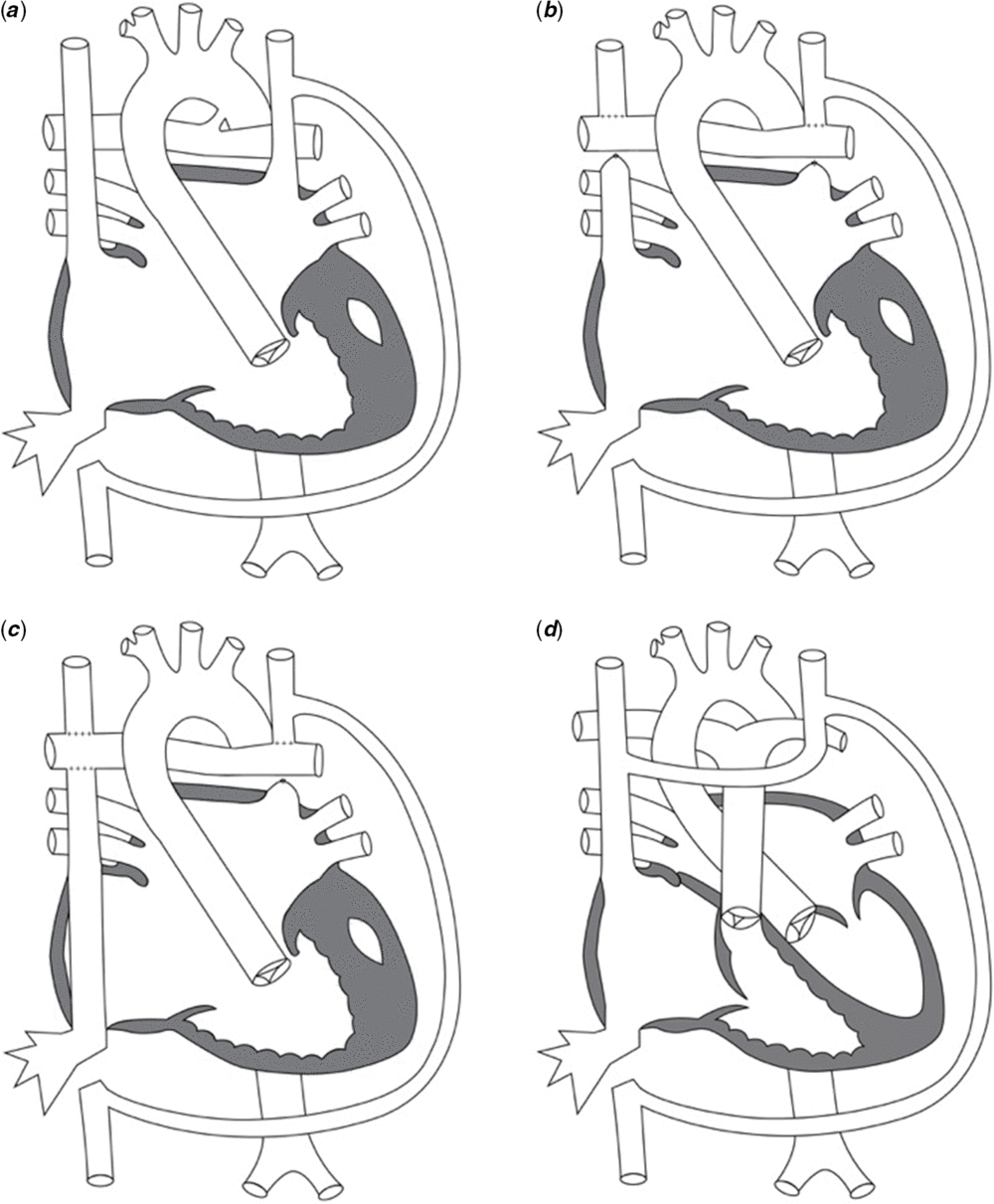
A female patient was born with heterotaxy-asplenia and complex CHD consisting of double-outlet right ventricle, pulmonary atresia, unbalanced atrioventricular canal towards the right ventricle, a common atrium, interrupted left inferior caval vein with hemi-azygous continuation, and bilateral superior caval veins. Palliative surgeries performed were modified Blalock–Taussig shunt placement at 3 days of age, bilateral bidirectional Glenn and Kawashima procedure, with repair of the atrioventricular valve as well as ligation of the Blalock–Taussig shunt at 10 months of age, and extra-cardiac autologous pericardial lateral tunnel Fontan procedure at 3 years of age (Fig 1). Unfortunately, the patient developed progressive cyanosis due to the development of left lung arteriovenous malformations by the age of 10. Physical examination revealed cyanosis and significant clubbing with an oxygen saturation of 80%, despite receiving oxygen at 2 L/minute. Angiography revealed significant streaming of the right superior caval vein and hepatic flow to the right lung, while the left superior and inferior caval veins supplied the left lung with associated pulmonary arteriovenous malformations.
Illustration of the patient’s anatomy. Diagrammatic illustration by Clara Awad, adapted from Mullins diagrams. (a) Anatomy at birth, consisting of left superior caval vein, single atrium, common atrioventricular valve, single ventricle, and pulmonary atresia. (b) Anatomy after bilateral bidirectional Glenn and Kawashima connection. (c) Anatomy after Fontan completion. (d) Final anatomy after heart transplantation.

At 13 years of age, she underwent orthotopic heart transplantation with tri-caval anastomosis, reconstruction of the left superior caval vein, patch arterioplasty of the left pulmonary artery, and takedown of Glenn and Fontan connections. One-year post-transplant and without improvement in oxygen saturation, the patient underwent CT angiogram of the chest, which showed a hypoplastic left lung and left pulmonary artery. The study showed enlargement of left pulmonary vessels in the anterior segment of the upper lobe and basilar segments of the lower lobe, suggesting presence of persistent pulmonary arteriovenous malformations, which was confirmed on cardiac catheterisation.
The patient’s oxygen saturation continued to decrease and she presented with an episode of acute cellular rejection with severe cyanosis. Her resting saturation was in the 60s and increased to the 70s with supplemental oxygen. Following treatment, she underwent a bedside nitric oxide challenge with 100% oxygen and 60 ppm of nitric oxide, which improved her saturation to the low 90s. She was placed on oral sildenafil and continuous supplemental oxygen.
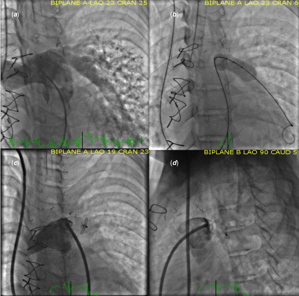
After weighing the advantages and disadvantages of other treatment options including lung transplantation and embolisation of the pulmonary arteriovenous malformations, the patient underwent repeat cardiac catheterisation with test occlusion of her left pulmonary artery and simultaneous selective right mainstem intubation. This resulted in improved oxygen saturation from 79 to 94%, without a significant increase in mean pulmonary artery pressure (Fig 2a and b). Encouraged by these findings, the patient underwent elective embolisation of the left pulmonary artery with a 16 mm AmplatzerTM vascular plug II at age 16 (Fig 2c and d). The procedure was well-tolerated and she showed immediate improvement in oxygen saturation to 97% in room air without significant changes in haemodynamics. In the 18 months of follow-up, her oxygen saturation remains 98% in room air, with a mean pulmonary artery pressure of 17 mmHg.
Heart catheterisation with left pulmonary artery test occlusion and left pulmonary artery device deployment: (a) Left anterior oblique/cranial injection of main pulmonary artery shows diffuse left pulmonary arteriovenous malformations. (b) Left anterior oblique/cranial, test occlusion of left pulmonary artery with sizing balloon. Note, right main stem intubation. (c) Left anterior oblique/cranial and (d) lateral projection of main pulmonary artery angiogram after embolisation of left pulmonary artery with a 16 mm AmplatzerTM vascular plug II.

Discussion
We present the first reported case of therapeutic embolisation of the left pulmonary artery to correct severe, persistent cyanosis in a patient with diffuse microvascular arteriovenous malformations in the left lung following heart transplantation for a failed Fontan. The procedure normalised her oxygen saturation and enhanced her quality of life. To our knowledge, all similar published cases showed resolution of pulmonary arteriovenous malformations and cyanosis after heart transplant alone in Fontan patients.
Pulmonary arteriovenous malformations are abnormal anastomoses between pulmonary arteries and veins causing insufficient oxygenation, dyspnoea, and cyanosis.Reference Sharma, Kochar and Sharma1 Pulmonary arteriovenous malformations can develop secondary to cavopulmonary anastomoses. The exact cause of their development is unknown, but multiple theories exist including lack of a “hepatic factor” to the affected lungReference Marianeschi, McElhinney and Reddy2 and lack of pulsatile flow through pulmonary arteries.Reference Srivastava, Preminger and Lock3 Importantly, not one single theory can account for all aspects of their development.Reference Kwon, Bae, Kim, Noh, Choi and Yun4
Treatment depends on the severity; studies report resolution of cyanosis after isolated heartReference Lamour, Hsu, Kichuk, Galantowicz, Quaegebeur and Addonizio5 or combined heart–lung transplant.Reference Mott, Spray and Bridges6 Embolisation of pulmonary arteriovenous malformations is the first-line therapyReference Hsu, Kwan, Thompson, Evans-Barns and Van Driel7; however, it is less reliable in cases of diffuse, or microvascular disease, where surgical lobectomy is the recommended treatment.Reference Park, Kim and Park8 Implantation of covered stents to redirect hepatic venous return and allow for its even distribution to both lungs has also been reported, but this could potentially increase the already high risk of thrombosis in Fontan patients.Reference Dori, Sathanandam, Glatz, Gillespie and Rome9 Lung transplantation is another treatment option for isolated diffuse pulmonary arteriovenous malformations; however, it is rarely performed due to the potential complications and low survival rate.
In our case, we hypothesise that the patient’s complex venous anatomy led to streaming of venous blood from the liver predominantly to the right lung. This led to the development of diffuse left-sided pulmonary arteriovenous malformations. Elective left pulmonary artery embolisation normalised her oxygen saturation without an associated increase in her mean pulmonary artery pressure. Importantly, she did not develop pulmonary hypertension secondary to occlusion of lower vascular resistance vessels and increased flow to a single lung. This was the first case of therapeutic left pulmonary artery embolisation in this setting, corroborating studies showing that embolisation of pulmonary arteriovenous malformations does not lead to pulmonary hypertension.Reference Shovlin, Tighe, Davies, Gibbs and Jackson10
We demonstrate that treatment of unilateral diffuse microvascular unilateral pulmonary arteriovenous malformations with embolisation of the affected pulmonary artery is a viable alternative to lung resection or transplant. In expert hands, the procedure can be performed safely and with normalisation of saturations and pulmonary artery pressures.
Acknowledgements
We would like to thank the patient and her family, as well as the artist, Clara Awad, for drawing an illustration of the patient’s anatomy (Fig 1).
Financial Support
This research received no specific grant from any funding agency, commercial, or not-for profit sectors.
Conflicts of Interest
None
Ethical Standards
This case report did not involve human experimentation. Written consent obtained from patient and patient’s mother (6/18/2018). This research was exempt from review by the Institutional Review Board.