Implications
In bovine, in vitro embryo production (IVEP) is especially interesting when applying the ovum pick-up technique (OPU). However, a small number of oocytes per female are usually recovered after OPU, and when embryos are cultured individually or in a reduced group blastocyst yield efficiency and quality are generally reduced. The improvement of IVEP efficiency and quality with low numbers of embryos would be of great interest for applying this technique in the dairy industry.
Introduction
In vitro embryo production (IVEP) has numerous applications in various scientific areas such as animal production or biomedicine. IVEP-related studies provide us with basic knowledge to better understand the mechanisms of oogenesis, fertilisation and embryo development. Moreover, IVEP from slaughterhouse material provides a high number of oocytes or embryos (after in vitro maturation and fertilisation) at a low cost to carry out other associated embryo technologies such as cryobiology. In bovine, IVEP is especially interesting to mitigate the decrease in fertility suffered by dairy cattle during environmental heat stress conditions (Ambrose et al., Reference Ambrose, Drost, Monson, Rutledge, Leibfried-Rutledge, Thatcher, Kassa, Binelli, Hansen, Chenoweth and Thatcher1999; Al-Katanani et al., Reference Al-Katanani, Drost, Monson, Rutledge, Krininger, Block, Thatcher and Hanse2002) or to take advantage of the ovum pick-up technique (OPU). In the latter case, a small number of oocytes per female are usually recovered after OPU (approximately three to six oocytes recovered per non-stimulated cow and session; Rizos et al., Reference Rizos, Burke, Duffy, Wade, Mee, O'Farrell, Macsiurtain, Boland and Lonergan2005; Chaubal et al., Reference Chaubal, Molina, Ohlrichs, Ferre, Faber, Bols, Riesen, Tian and Yang2006). IVEP efficiency has increased in recent years, although the quality of these embryos remains lower than in embryos produced in vivo (reviewed by Rizos et al., Reference Rizos, Clemente, Bermejo-Alvarez, De La Fuente, Lonergan and Gutiérrez-Adán2008). In particular, when embryos were cultured individually or in reduced groups, in addition to a lower efficiency of embryo production, the obtained embryos usually presented a lower number of cells per blastocyst (Paria and Dey, Reference Paria and Dey1990; O'Doherty et al., Reference O'Doherty, Wade, Hill and Boland1997; Nagao et al., Reference Nagao, Iijima and Saeki2008: 5 v. 25 embryos). Numerous authors have previously indicated, in several mammalian species, that IVEP efficiency is negatively affected when a single or a low number of oocytes/embryos were in vitro cultured in comparison with a higher number of the same (Paria and Dey, Reference Paria and Dey1990; Lane and Gardner, Reference Lane and Gardner1992; Carolan et al., Reference Carolan, Lonergan, Khatir and Mermillod1996; O'Doherty et al., Reference O'Doherty, Wade, Hill and Boland1997; Fujita et al., Reference Fujita, Umeki, Shimura, Kugumiya and Shiga2006). Moreover, we have observed that post-fertilisation culture, especially the last part of the culture period, seems to be the most important period for bovine embryo development when single and/or low numbers (5 to 10) of embryos were cultured (Salvador et al., Reference Salvador, Cebrian-Serrano, Salamone and Silvestre2011). Several factors could be involved in this negative effect. One of them would be the lack of growth factors (GFs) in the culture medium (CM), which could exert a beneficial effect stimulating embryo development, in a paracrine and/or autocrine manner, when embryos were in vitro cultured in groups (Paria and Dey, Reference Paria and Dey1990; Lim and Hansel, Reference Lim and Hansel1996). Previously, Bavister (Reference Bavister1995) had indicated that a rise in embryo number in culture could deplete embryotoxic substances or diminish the inhibitor concentration.
To improve the production and quality of embryos cultured in low numbers or singly, several physical modifications to optimise embryo environment have been proposed, the most simple being reducing the ratio embryo/volume in microdrops (Carolan et al., Reference Carolan, Lonergan, Khatir and Mermillod1996; Fujita et al., Reference Fujita, Umeki, Shimura, Kugumiya and Shiga2006; Nagao et al., Reference Nagao, Iijima and Saeki2008); other options are ‘Well of Well’ system (Vajta et al., Reference Vajta, Peura, Holm, Páldi, Greve, Trounson and Callesen2000), glass oviduct (Thouas et al., Reference Thouas, Jones and Trounson2003), woven polyester mesh (Booth et al., Reference Booth, Watson and Leese2007), Cell-Tak grids (Gopichandran and Leese, Reference Gopichandran and Leese2006) or the use of microfluidics (Krisher and Wheeler, Reference Krisher and Wheeler2010). Another possibility could be to modify directly the composition of culture media with the use of cell co-cultures (bovine oviduct epithelial cell, Lim and Hansel, Reference Lim and Hansel1996; granulosa cells, O'Doherty et al., Reference O'Doherty, Wade, Hill and Boland1997; cumulus cells, Goovaerts et al., Reference Goovaerts, Leroy, Van Soom, De Clercq, Andries and Bols2009), conditioned media (Fujita et al., Reference Fujita, Umeki, Shimura, Kugumiya and Shiga2006) or addition of GF (Paria and Dey, Reference Paria and Dey1990; Carolan et al., Reference Carolan, Lonergan, Khatir and Mermillod1996; Lim and Hansel, Reference Lim and Hansel1996; Brison and Schultz, Reference Brison and Schultz1997). The review by Hardy and Spanos (Reference Hardy and Spanos2002) clearly reflects the importance of a number of GF affecting preimplantation embryo development. These authors suggested that on in vivo conditions, endocrine, paracrine and autocrine pathways take place during human embryo development (Hardy and Spanos, Reference Hardy and Spanos2002). The supplementation of embryo culture media with factors such as epidermal GF (EGF), insulin GF-I and -II (IGF-I and -II, respectively), transforming GF-α (TGF-α), TGF-β1, fibroblast GF, platelet-derived GF-AB, granulocyte-macrophage colony-stimulating factor and stem cell factor has been used to try to increase in vitro embryo development (mouse: Paria and Dey, Reference Paria and Dey1990; bovine: Lim and Hansel, Reference Lim and Hansel1996; De Moraes and Hansen, Reference De Moraes and Hansen1997; Sirisathien et al., Reference Sirisathien, Hernandez-Fonseca and Brackett2003). In addition, several studies indicated that the GF could protect the embryo against oxidative or thermal stress (mouse: Kurzawa et al., Reference Kurzawa, Glabowski, Baczkowski, Wiszniewska and Marchlewicz2004; bovine: Jousan and Hansen, Reference Jousan and Hansen2004).
Little information is found in the literature about the effect of culture of single or reduced number of bovine embryos on apoptosis rate. Brison and Schultz (Reference Brison and Schultz1997) observed that culturing in groups or adding 0.1 pM TGF-α to medium reduced the apoptosis rate, using terminal deoxynucleotidyl transferase (TdT)-mediated dUTP nick end-labelling (TUNEL) technique, when murine embryos were cultured singly. Other studies on bovine embryos cultured singly or in reduced groups did not show significant differences on apoptotic rate per embryo (Hoelker et al., Reference Hoelker, Rings, Lund, Ghanem, Phatsara, Griese, Schellander and Tesfaye2009; Goovaerts et al., Reference Goovaerts, Leroy, Rizos, Bermejo-Alvarez, Gutierrez-Adan, Jorssen and Bols2011). In the first of them, it is possible that the number of embryos maintained in reduced culture was too high (16 embryos) for observing a significant effect in apoptosis, although a reduction in the number of in vitro cultured bovine embryos, from 50 to 16 embryos, significantly decreased the blastocyst rate (Hoelker et al., Reference Hoelker, Rings, Lund, Ghanem, Phatsara, Griese, Schellander and Tesfaye2009). In the second one, singly cultured embryos were cultured in the presence of a cumulus cell monolayer (Goovaerts et al., Reference Goovaerts, Leroy, Rizos, Bermejo-Alvarez, Gutierrez-Adan, Jorssen and Bols2011) and it was observed that oviductal cell coculture diminished the superoxide anion level in embryo CM (Joo et al., Reference Joo, Kim, Na, Moon, Lee and Kim2001).
The aim of this work was to investigate the effect of supplementation in the CM of several GF on embryo development and quality (number of cells per blastocyst and apoptotic rate) when a reduced number of bovine embryos were in vitro cultured.
Material and methods
Unless otherwise stated, all chemical products were purchased from Sigma-Aldrich Quimica (Madrid, Spain).
In vitro maturation
Cumulus–oocyte complexes (COCs) were collected by aspiration of ∼2- to 8-mm diameter follicles of bovine ovaries obtained from heifers younger than one year of age in a local slaughterhouse. After aspiration, COCs were washed three times in handling medium (HM199) based on Hepes Medium 199 (M7528) supplemented with 7.4% (v/v) heat-inactivated foetal bovine serum (FBS; 10108-157, GIBCO®, Invitrogen, Life Technologies S.A., Madrid, Spain) and antibiotics. From aspirated COCs, those with several surrounding layers of cumulus cells were selected. After washing, selected COCs were in vitro matured in Medium 199 (M4530) supplemented with 10% FBS and 10 ng/ml of EGF (E1257). In addition, culture media were supplemented with 75 μg/ml potassium penicillin G (P3032) and 50 μg/ml streptomycin sulphate (S6051) as antibiotics. COCs were matured for 22 to 24 h at 38.5°C in an atmosphere of 5% CO2 in humidified air. All culture media containing bicarbonate were covered with mineral oil (M8410) and equilibrated overnight in the culture conditions.
Spermatozoa preparation and in vitro fertilisation (IVF)
Three straws (0.25 ml) of frozen semen from three bulls were thawed at 37°C in a water bath for 1 min and centrifuged for 20 min at 350 × g through a Bovipure® gradient (Nidacon International AB, Mölndal, Sweden) with 2 ml of Bovipure bottom layer, and 2 ml of Bovipure top layer, in 15 ml centrifuge tubes. The sperm pellet was separated and resuspended in 10 ml of HM199 and separated again after centrifugation at 300 × g for 5 min. Approximately 50 μl of semen pellet remained after the last centrifugation and this was diluted with ∼100 μl of HM199. For IVF, a final concentration of 1 × 10⁶ sperm/ml was used. After in vitro maturation, COCs were washed three times in fertilisation Fert-Talp medium (Parrish et al., Reference Parrish, Susko-Parrish, Winer and First1988) with 10 μg/ml of heparin (H9399) without epinephrine and hypotaurine and coincubated with the spermatozoa, in 500 μl using 4-Well Nunc® plates (Nunc, Fisher Scientific, Madrid, Spain), for 18 to 20 h in 5% CO2 at 38.5°C.
In vitro culture of embryos
After IVF, presumptive zygotes were denuded from surrounding cumulus cells in HM199 and washed three times in CM, which was based on synthetic oviductal fluid aaci (Holm et al., Reference Holm, Booth, Schmidt, Greve and Callesen1999) with some modifications (Salvador et al., Reference Salvador, Cebrian-Serrano, Salamone and Silvestre2011) and with 3 mg/ml bovine serum albumin (A-8806) instead of bovine serum. Then, presumptive zygotes were in vitro cultured in groups of ∼30 to 50 zygotes in 500 μl using 4-Well Nunc® plates, or in a number of five zygotes in 50 μl microdrops, depending on the experimental group (see below in Section ‘Experimental design’), at 38.5°C in a humidified atmosphere with 5% CO2 and 5% O2 in air. Cleavage and blastocyst formation rates were recorded at Day 2 (∼48 h post fertilisation), and at Days 7, 8 and 9 (D7, D8 and D9, respectively) of in vitro culture. Blastocysts were fixed and stained in ethanol with 25 μg/ml of bisbenzimide (B2261), and the total number of cells was counted under an epifluorescence microscope.
Assessment of cellular apoptosis of embryos
Apoptosis rate was evaluated by cellular DNA fragmentation using the TUNEL procedure. The TUNEL procedure described by Schwarz et al. (Reference Schwarz, Pires, Adona, Câmara de Bem and Leal2008) was followed, with some modifications. The In situ Cell Death Detection Kit Fluorescein (11684795910, Roche Diagnostics SL, Barcelona, Spain) was used. Blastocysts of D7 and D8 were fixed in 4% paraformaldehyde in phosphate buffered-saline solution (PBS) for at least 1 h at room temperature. After that, embryos were washed in PBS plus 1 mg/ml of polyvinyl alcohol (PVA) (P8136) (PBS–PVA) and either processed immediately (for D8 blastocysts) or kept at 4°C for 24 h in PBS–PVA (for D7 blastocysts). Blastocysts were treated in a solution consisting of 1 μl/ml of Triton X-100 (T9284) and 1 mg/ml sodium citrate (S4641) in PBS during 30 min at 37°C in humidified atmosphere. Some embryos were used for positive and negative control groups. Positive control blastocysts were incubated in PBS–PVA with 50 IU/ml of DNase I (04536282001, Roche Diagnostics SL, Barcelona, Spain) for 30 min at 37°C. After washing, embryos from positive control and experimental groups were incubated in 30 μl of TUNEL reagent from the kit, composed of 10% enzyme solution and 90% staining solution in 0.2 ml PCR Eppendorf tube (Bio-Rad Laboratories, S.A., Madrid, Spain), for 1 h at 37°C in humidified atmosphere. Negative control blastocysts were only treated with the staining solution in the absence of TdT (enzyme solution). After this treatment, embryos were washed and stained with ethanol and bisbenzimide, mounted on Polysine® slides (Thermo Fisher Scientific S.L.U., Alcobendas, Spain) and evaluated by epifluorescence microscopy (Nikon Eclipse E-400, Izasa Distribuciones Técnicas S.A., Barcelona, Spain) using different filters. Blastocyst cells with nuclei showing green fluorescence were considered as TUNEL positive (DNA fragmented), and those showing blue fluorescence (bisbenzimide) indicated the total number of cells (Schwarz et al., Reference Schwarz, Pires, Adona, Câmara de Bem and Leal2008).
Experimental design
Two experimental studies (ES) were carried out.
In ES 1, five different treatments were tested:
1. Control group (Control), ∼30 to 50 embryos were cultured in 500 μl of CM, without addition of any GF in the medium.
2. Microdrop control group (Control 5), five embryos were cultured in 50 μl microdrops of CM, without addition of any GF in the medium.
3. EGF group, five embryos were cultured in 50 μl microdrops of CM with 10 ng/ml EGF (G502A, Promega Biotech Ibérica, Madrid, Spain).
4. IGF-I group, five embryos were cultured in 50 μl microdrops of CM with 10 ng/ml IGF-I (I3769).
5. TGF-α group, five embryos were cultured in 50 μl microdrops of CM with 10 ng/ml TGF-α (T7924).
Assessments of blastocyst formation were done at D7 and D8 of culture.
In ES 2, following the results obtained in ES 1, four different treatments were tested: Control; Control 5; EGF group, all three similar to ES 1; and EGF + IGF-I group, where five embryos were cultured in 50 μl microdrops of CM with 10 ng/ml EGF and 10 ng/ml IGF-I. Assessments of blastocyst formation rate and number of cells were done at D7, D8 and D9 of culture, and cell apoptosis rate per blastocyst were done at D7 and D8 of culture.
Statistical analysis
Six and eight replicates were performed for ES 1 and 2, respectively. Results for cleavage and blastocyst rates were analysed using the χ 2-test. Results of blastocyst cell number (ES 1 and ES 2) and cellular apoptosis rate (ES 2) were analysed using a multifactor ANOVA. In ES 1, two factors and their interaction were analysed: experimental group (Control, Control 5, EGF, IGF-I, TGF-α) and days of culture (D7, D8). In ES 2, factors analysed were: experimental group (Control, Control 5, EGF, EGF + IGF-I) and days of culture (D7, D8, D9) and their interaction.
Results
In ES 1, we studied the effect of several GF in CM on IVEP efficiency when embryos were cultured in reduced groups. Results of this first experimental study are shown in Table 1. No differences in cleavage rates were detected between the different experimental groups. However, significant differences were observed between treatments in the ability of the cleaved embryos to reach blastocyst stage. A higher proportion of embryos cultured in larger groups achieved blastocyst stage at D7 post-fertilisation than embryos cultured in reduced groups (21.8% v. 13.6%, 11.6% and 4.6% for Control v. Control 5, IGF-I, TGF-α respectively; P < 0.05), except for embryos cultured in medium supplemented with EGF (18.4%). However, the EGF group did not present significant differences in blastocyst rates in comparison with Control 5 group. Similar effects were observed when total blastocyst rates (putting blastocyst at D7 and D8 together) were analysed relative to cleaved embryos or COCs.
Effect of EGF, IGF-I and TGF-α in culture medium on in vitro embryo production efficiency when embryos were cultured in reduced groups (Experimental study 1)

EGF = epidermal growth factor; TGF-α = transforming growth factor-α; COC = Cumulus–oocyte complexes.
Control: ∼30 to 50 embryos in 500 μl of culture medium; Control 5: five embryos in 50 μl of culture medium; EGF: five embryos in 50 μl of culture medium supplemented with EGF (10 ng/ml). IGF-I: five embryos in 50 μl of culture medium supplemented with IGF-I (10 ng/ml). TGF-α: five embryos in 50 μl of culture medium supplemented with TGF-α (10 ng/ml).
a,b,cDifferent superscripts in the same column indicate a statistical difference (P < 0.05).
*Percentage of blastocyst is calculated from cleaved embryos.
**Percentage of blastocyst is calculated from COCs.
ES 2 consisted of a more in-depth study of the effect of EGF and its combination with IGF-I in the CM on IVEP efficiency and apoptosis when embryos were cultured in reduced groups. Results of embryo development are shown in Table 2. No significant differences in cleavage rates were found between experimental groups. However, it was observed that a higher proportion of embryos cultured in large groups achieved blastocyst stage than embryos cultured in reduced groups (24.9% v. 17.1% and 19.0% for Control v. Control 5 and EGF, respectively; P < 0.05), except for embryos supplemented with EGF + IGF-I (23.5%). Moreover, EGF + IGF-I group presented significantly higher total blastocyst rates (blastocysts/oocytes) than Control 5 group (P < 0.05). With regard to quality of obtained blastocysts, the total number of cells per blastocyst was significantly affected by the factor ‘days of culture’ (P < 0.01), but not by the ‘experimental group’ factor or their interaction. The number of cells per blastocyst depending on different experimental groups ranged from 107.3 to 132.5. In this sense, blastocysts at D7 presented the highest number of cells, and blastocysts at D8 a higher number of cells than blastocysts at D9 (134.2 v. 118.9 v. 98.3 for blastocysts at D7, D8 and D9, respectively; P < 0.01, Table 3). Both ‘days of culture’ and ‘experimental group’ factors and their interaction significantly affected apoptosis rate of blastocysts (P < 0.05). In this way, blastocysts at D7 presented a lower apoptosis rate than blastocyst at D8 (12.5 v. 15.2% of cells from blastocysts were apoptotic for blastocyst at D7 and D8, respectively; P < 0.05, Table 3). Embryos cultured in larger groups (∼30 to 50 embryos in culture) presented a lower apoptosis rate than embryos cultured in reduced groups (five embryos in culture). Moreover, embryos cultured in reduced groups and supplemented with GF, EGF alone or combined with IGF-I, presented a lower apoptosis rate than embryos cultured in reduced group without GF supplementation (11.6 and 10.5 v. 21.9% for EGF, EGF + IGF-I and Control 5 groups, respectively; P < 0.05, Table 4).
Effect of EGF and its combination with IGF-I in culture medium on in vitro embryo production efficiency when embryos were cultured in reduced groups (Experimental study 2)

EGF = epidermal growth factor; COC = Cumulus–oocyte complexes.
Control: ∼30 to 50 embryos in 500 μl of culture medium; Control 5: five embryos in 50 μl of culture medium; EGF: five embryos in 50 μl of culture medium supplemented with EGF (10 ng/ml). EGF + IGF-I: five embryos in 50 μl of culture medium supplemented with EGF (10 ng/ml) + IGF-I (10 ng/ml).
a,b,cDifferent superscripts in the same column indicate a statistical difference (P < 0.05).
*Percentage of blastocyst is calculated from cleaved embryos.
**Percentage of blastocyst is calculated from COCs.
Average number of cells and apoptosis percentage per blastocyst depending on days of embryo culture (Experimental study 2)

s.e. = standard error; n = number of embryos studied; ne = non-evaluated.
a,b,cDifferent superscripts in the same row indicate a statistical difference (P < 0.05).
Average apoptosis percentage per blastocyst depending on experimental group (Experimental study 2)
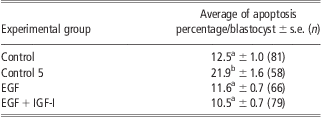
s.e. = standard error; n = number of embryos studied; EGF = epidermal growth factor.
a,bDifferent superscripts in the same column indicate a statistical difference (P < 0.05).
Discussion
In the present work, we studied the effect of embryo in vitro CM supplementation of different GF on embryo development and quality when a reduced number of embryos were cultured. Generally, it was observed that a greater proportion of embryos cultured in large groups achieved blastocyst stage than embryos cultured in reduced groups. In a previous work, in our conditions, we observed that culture of 10 embryos or less, maintaining the ratio 1/10 embryo/μL, impaired the efficiency of blastocyst yield (Salvador et al., Reference Salvador, Cebrian-Serrano, Salamone and Silvestre2011). In the ES 1 of the present work, we observed that the addition of 10 ng/ml EGF in the CM reduced the negative effect of the culture in reduced numbers, as IVEP efficiency was not significantly different compared with embryos cultured in larger groups. However, this slight improvement in the blastocyst rate was not observed in ES 2. This controversial result was also observed by Sirisathien et al. (Reference Sirisathien, Hernandez-Fonseca and Brackett2003); in their third experiment, the EGF group (5 ng/ml) significantly increased the rate of bovine blastocysts from oocytes, but this fact did not happen in their first experiment (20 embryos/50 μl). In this sense, some authors observed no beneficial effects of EGF in the embryo CM on the blastocyst rate either in individual (mouse: O'Neill, Reference O'Neill1997, 0.2 to 2000 ng/ml; bovine: Keefer et al., Reference Keefer, Stice, Paprocki and Golueke1994, 10 ng/ml) or in group culture (bovine: Flood et al., Reference Flood, Gage and Bunch1993, 10 ng/ml, Shamsuddin, Reference Shamsuddin1994, 10 to 50 ng/ml, Lee and Fukui, Reference Lee and Fukui1995, 10 ng/ml). However, other authors observed differences both in single (mouse: Paria and Dey, Reference Paria and Dey1990, 10 ng/ml; bovine: Lim et al., Reference Lim, Jang, Ko, Lee, Park, Kim, Lee, Hwang, Lee and Kanget2007, 10 ng/ml) and group culture (bovine: Lonergan et al., Reference Lonergan, Carolan, Van Langendonckt, Donnay, Khatir and Mermillod1996, 100 ng/ml; Mtango et al., Reference Mtango, Varisanga, Dong, Rajamahendran and Suzuki2003, 10 ng/ml; Sirisathien and Brackett, Reference Sirisathien and Brackett2003, 5 ng/ml).
On the other hand, the effect of TGF-α was also evaluated in the present work, and no beneficial effects, but rather negative, of this GF on embryo development were found, confirming results previously showed by other authors observing no beneficial effects (Brison and Schultz, Reference Brison and Schultz1997; Zhou et al., Reference Zhou, Liu, Cang, Ma, Yang, Li, Wang, Bou and Feng2008). Regarding IGF-I, in agreement with other studies (mouse: Paria and Dey, Reference Paria and Dey1990, 10 ng/ml; bovine: Flood et al., Reference Flood, Gage and Bunch1993, 10 ng/ml; mouse: Byrne et al., Reference Byrne, Southgate, Brison and Leese2002, 13 nM), we did not find a positive effect of this GF on embryo development when it was supplemented alone. In contrast with our results, other authors assaying with higher concentrations than our concentration (10 ng/ml) observed an increase in the blastocyst rate after IGF-I supplementation on embryo CM (O'Neill, Reference O'Neill1997; Block et al., Reference Block, Drost, Monson, Rutledge, Rivera, Paula-Lopes, Ocon, Krininger, Liu and Hansen2003). In this sense, both O'Neill (Reference O'Neill1997) and Sirisathien et al. (Reference Sirisathien, Hernandez-Fonseca and Brackett2003), in mouse and bovine, respectively, only found significant effects on embryo development when IGF-I concentration was higher than 30 ng/ml and 50 ng/ml, respectively. In particular, Sirisathien et al. (Reference Sirisathien, Hernandez-Fonseca and Brackett2003), in agreement with our results, did not find a significant effect of IGF-I with 10 ng/ml on embryo development (blastocyst from oocytes) when bovine embryos were cultured in larger groups (20 embryos/50 μl) in comparison with non-IGF-supplemented group. Recently, Sakagami et al. (Reference Sakagami, Umeki, Nishino, Uchiyama, Ichikawa, Takeshita, Kaneko, Akiyama, Kobayashi and Tamada2012) found similar results to Sirisathien et al. (Reference Sirisathien, Hernandez-Fonseca and Brackett2003). Despite this, even using a high concentration, Block et al. (Reference Block, Wrenzycki, Niemann, Herrmann and Hansen2008) did not detect any significant effect of the supplementation of 100 ng/ml IGF-I on the blastocyst rate (25 embryos/50 μl) in comparison with the non-supplemented group.
In ES 2, we studied the effect of culture of small numbers of embryos on embryo production and quality, in particular the number of cells and apoptosis rate per blastocyst. In bovine, in disagreement with our results, some authors observed that blastocysts obtained after culture individually contained a lower number of cells than embryos cultured in large groups (O'Doherty et al., Reference O'Doherty, Wade, Hill and Boland1997; Larson and Kubisch, Reference Larson and Kubisch1999). However, other authors did not find a significant decrease on blastocyst cell number of the embryos cultured singly (Goovaerts et al., Reference Goovaerts, Leroy, Van Soom, De Clercq, Andries and Bols2009 and Reference Goovaerts, Leroy, Rizos, Bermejo-Alvarez, Gutierrez-Adan, Jorssen and Bols2011) or in reduced groups (Hoelker et al., Reference Hoelker, Rings, Lund, Ghanem, Phatsara, Griese, Schellander and Tesfaye2009). However, in the experiments of Goovaerts et al. (Reference Goovaerts, Leroy, Van Soom, De Clercq, Andries and Bols2009 and Reference Goovaerts, Leroy, Rizos, Bermejo-Alvarez, Gutierrez-Adan, Jorssen and Bols2011), embryos were cultured with a cumulus cell coculture.
Apoptotic rates obtained in this work for IVEP blastocysts were similar to those obtained by other authors (∼7% to 14% embryo cells; Van Soom et al., Reference Van Soom, Yuan, Peelman, De Matos, Dewulf, Laevens and De Kruif2002; Vandaele et al., Reference Vandaele, Mateusen, Maes, de Kruif and Van Soom2006), but higher than others (around 2% to 3%; Goovaerts et al., Reference Goovaerts, Leroy, Rizos, Bermejo-Alvarez, Gutierrez-Adan, Jorssen and Bols2011). We observed that the in vitro culture of a reduced number of embryos impaired blastocyst production and also quality, in particular by increasing the apoptotic rate per blastocyst. In bovine, few works were found in the literature related with the effect of in vitro culture of individual or small groups of embryos on their quality, and information about apoptosis is particularly scarce. Programmed cell death or apoptosis may be necessary, as this mechanism would be involved in the elimination of injured embryo cells and also, to help to the preservation of a proper number of cells and inner cell mass (ICM)/trophectoderm (TE) ratio (reviewed by Leese et al., Reference Leese, Baumann, Brison, McEvoy and Sturmey2008). A non-adequate environment such as in vitro culture increases apoptotic rate of mouse embryos (Brison and Schultz, Reference Brison and Schultz1997). In the case of embryo culture in small groups, Nagao et al. (Reference Nagao, Iijima and Saeki2008) found that the effect of oxygen concentration of incubator atmosphere was related with the embryo number in culture, and that a reduction in oxygen concentration improved bovine embryo development mainly of the smallest groups. Although we do not have evidence, it might be possible that the surrounding microenvironment could be different when fewer embryos are cultured than in large groups. Exogenous reactive oxygen species are produced by many sources as high oxygen tension, exposure to visible light or excess of glucose in the medium among others (reviewed by Guérin et al., Reference Guérin, El Mouatassim and Ménézo2001) and it is known that oxidative stress induces DNA fragmentation in embryos (Kitagawa et al., Reference Kitagawa, Suzuki, Yoneda and Watanabe2004). In any case, the present work showed that supplementation of EGF plus IGF-I or EGF alone when embryos were cultured in small groups diminished apoptosis rate at similar levels to those of larger groups. In several works, it has been observed that GF reduced apoptosis rate in embryos in ‘standard’ in vitro conditions (IGF-I: Byrne et al., Reference Byrne, Southgate, Brison and Leese2002; Makarevich and Markkula, Reference Makarevich and Markkula2002) or artificially induced stressful conditions (hydrogen peroxide: EGF: Kurzawa et al., Reference Kurzawa, Glabowski, Baczkowski, Wiszniewska and Marchlewicz2004). However, this effect was not always evident; Sirisathien and Brackett (Reference Sirisathien and Brackett2003) observed a slight positive effect of IGF-I reducing apoptosis rates only on D8 bovine blastocyst but not on D7 blastocyst or after EGF treatment. In contrast, Block et al. (Reference Block, Wrenzycki, Niemann, Herrmann and Hansen2008) did not observe any effect of IGF-I on apoptosis.
In our case, the supplementation of EGF alone reduced the apoptotic rate of obtained blastocysts, although it had no effect on the total cells per blastocyst. Moreover, a significant effect of combination EGF + IGF-I on both embryo development and apoptotic rate was found, but without increasing the total cell number per blastocyst. Our results are consistent with several studies that did not show significant effects of EGF on total cells per blastocyst (Keefer et al., Reference Keefer, Stice, Paprocki and Golueke1994; Sirisathien and Brackett, Reference Sirisathien and Brackett2003) or on ICM and trophoblast cells (Sirisathien et al., Reference Sirisathien, Hernandez-Fonseca and Brackett2003). In contrast, other studies found a beneficial effect of EGF, increasing the number of cells per blastocyst cultured singly (Paria and Dey, Reference Paria and Dey1990) or in groups (Lee and Fukui, Reference Lee and Fukui1995; Mtango et al., Reference Mtango, Varisanga, Dong, Rajamahendran and Suzuki2003). In line with our results, Brice et al. (Reference Brice, Wu, Muraro, Adamson and Wiley1993) hypothesised that EGF receptor seemed to be more linked to the regulation of TE differentiation rather than cell proliferation in mouse preimplantation embryos. In agreement with our results but with larger groups of bovine embryos in culture (20 embryos/100 μl), a recent work found a significant effect of the combination IGF-I and EGF on embryo development in comparison with non-GF-supplemented (Sakagami et al., Reference Sakagami, Umeki, Nishino, Uchiyama, Ichikawa, Takeshita, Kaneko, Akiyama, Kobayashi and Tamada2012). With regard to total cells per embryo, supplementation of IGF-I and EGF did not increase total cell number per blastocyst, only the ICM cell number (Sakagami et al., Reference Sakagami, Umeki, Nishino, Uchiyama, Ichikawa, Takeshita, Kaneko, Akiyama, Kobayashi and Tamada2012).
On the other hand, the quality of embryos obtained at different days of culture was studied in the present work. We found that blastocyst at D7 had better quality, more cells per blastocyst and a lower apoptosis rate, than the rest of the blastocysts obtained later. In line with our results, Larson and Kubisch (Reference Larson and Kubisch1999) observed that blastocysts at D7 had higher numbers of cells than blastocysts at D8 or D9. Moreover, Van Soom et al. (Reference Van Soom, Ysebaert and De Kruif1997) observed that timing of blastocyst formation was linked with cell numbers per blastocyst, detecting that blastocysts at D8 had lower numbers of cells than blastocysts at D6 or D7. In turn, the apoptotic cell index increased significantly in blastocysts at D9 in comparison with the previous days (Van Soom et al., Reference Van Soom, Yuan, Peelman, De Matos, Dewulf, Laevens and De Kruif2002). Vandaele et al. (Reference Vandaele, Mateusen, Maes, de Kruif and Van Soom2006) also observed significantly higher apoptosis rates in blastocysts at D8 than at D7. Similar results were observed, but not in all experiments, by Sirisathien and Brackett (Reference Sirisathien and Brackett2003).
In conclusion, this study showed that supplementation of the CM with EGF and IGF could avoid partially the deleterious effect of in vitro culture of small groups of bovine embryos, increasing blastocyst rates and decreasing apoptosis rates of these blastocysts.
Acknowledgements
This work was partially supported by the INIA (RTA2007-0110) and Conselleria de Educación of the Generalitat Valenciana (ACOMP/2009/072), as well as FEDER and The European Social Fund. M.A.S. was supported by funds from INIA-CCAA, the Ramón y Cajal Subprogramme (RyC-2011-08750) and the European Social Fund. A.C-S. was supported by a research grant from the Conselleria de Agricultura, Pesca y Alimentación. C.J.A. and R.L. were supported by a grant from IAMZ-CIHEAM. Semen doses were donated by Euro Fomento Pecuario, S.A. The authors would like to thank the company Martinez Loriente S.A. (Buñol, Valencia, Spain) for their assistance in obtaining bovine ovaries.