Case report
A previously healthy 14-year-old Han Chinese male presented to a local hospital with dizziness following physical exertion and later developed right leg numbness. Lower extremity CT angiography identified an extensive thrombus extending from the right iliac to the femoral arteries, for which a thrombectomy was performed. Incidentally, transthoracic echocardiography revealed masses within the right ventricle, prompting transfer to our centre.
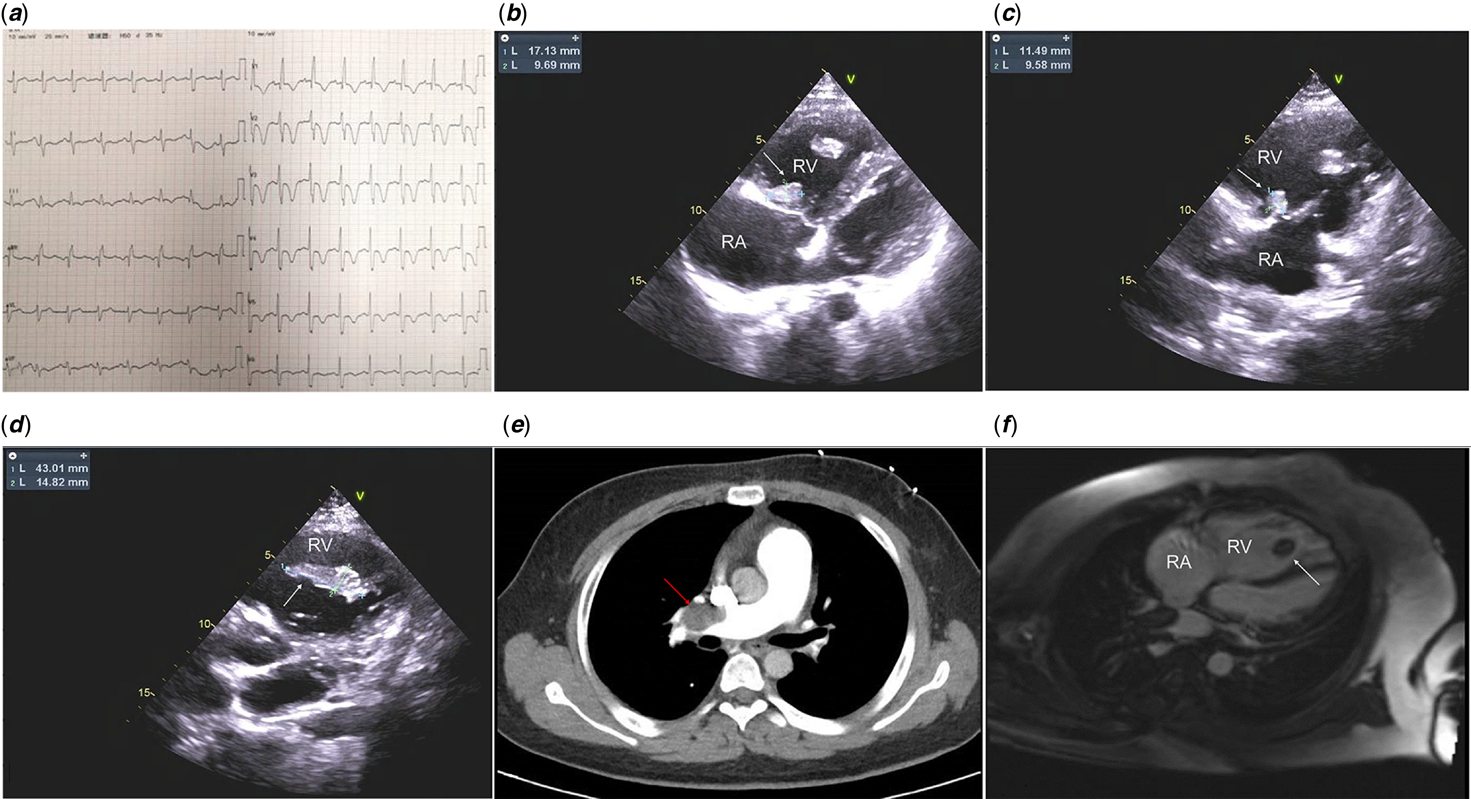
On admission, he was tachycardic (103 bpm), tachypnoeic (24 breaths/min), and hypoxic, with an oxygen saturation of 90% while on 5 L/min of oxygen via nasal cannula. Cardiac auscultation revealed a loud P2 and a systolic murmur. Laboratory studies were notable for elevated D-dimer (1.50 mg/L of fibrinogen equivalent units, normal 0–0.55 mg/L of fibrinogen equivalent units,) and BNP (761 pg/mL, normal 0–100 pg/mL), with decreased protein C (51.1%, normal 70.0–140.0%) and protein S (66.6%, normal 75.0–125.0%) activity. Tests for antiphospholipid antibodies were negative. An electrocardiogram revealed sinus rhythm, complete right bundle branch block, and T-wave inversion in V1–V5 (Figure 1 a ).
( a ) Electrocardiogram shows complete right bundle branch block and T-wave inversion in V1-V5. ( b – d ) Transthoracic echocardiography reveals three heterogeneous and irregular masses in the right ventricle, attached to the base of the right ventricle (17.1 mm × 9.6 mm), right ventricular apex (11.4 mm × 9.5 mm), and tricuspid valve chordae (43.0 mm × 14.8 mm), respectively. ( e ) CT pulmonary angiography reveals a large filling defect in the right main pulmonary arteries. ( f ) Late gadolinium enhancement on cardiac MRI in the apical long-axis view shows no contrast agent uptake in the mass, suggesting no vascularity within the mass. White arrow indicates right ventricular mass. Red arrow indicates thrombus.

Figure 1. Long description
Panel A: An electrocardiogram displaying multiple heartbeats with visible electrical activity. Panel B: An echocardiogram showing a cross-sectional view of the heart with labels indicating the right ventricle (RV) and right atrium (RA), and a mass measuring 17.13 millimeters by 9.69 millimeters. Panel C: Another echocardiogram with a different view, showing a mass measuring 11.49 millimeters by 9.58 millimeters. Panel D: A third echocardiogram view highlighting a mass measuring 43.01 millimeters by 14.82 millimeters. Panel E: A computed tomography (CT) scan of the chest showing a cross-sectional view of the pulmonary arteries with a large filling defect indicated by a red arrow. Panel F: A cardiac magnetic resonance imaging (MRI) scan showing a longitudinal view of the heart with labels indicating the right atrium (RA) and right ventricle (RV), and a mass indicated by a white arrow.
Multimodal imaging was pivotal (Figure 1 b – f ). Transthoracic echocardiography demonstrated three right ventricle masses: a large (43.0 × 14.8 mm) highly mobile mass attached to tricuspid valve chordae, an 11.4 mm × 9.5 mm highly mobile mass at the apex, and a 17.1 mm × 9.6 mm mass attached the base. This was accompanied by severe pulmonary hypertension (estimated systolic pulmonary artery pressure 105 mmHg), right ventricular dilation, and reduced function (tricuspid annular plane systolic excursion, 15 mm). Cardiac MRI with late gadolinium enhancement showed no contrast uptake within any of the right ventricular masses and revealed dilation of the main and right pulmonary arteries as well as right ventricular hypokinesis. CT pulmonary angiography showed multiple filling defects in both pulmonary arteries, suggestive of acute-on-chronic pulmonary thromboembolism with partial organization. Notably, the main and right pulmonary arteries were ectatic, whereas the left main pulmonary artery was attenuated. A small (0.8 mm) atrial septal defect was also identified.
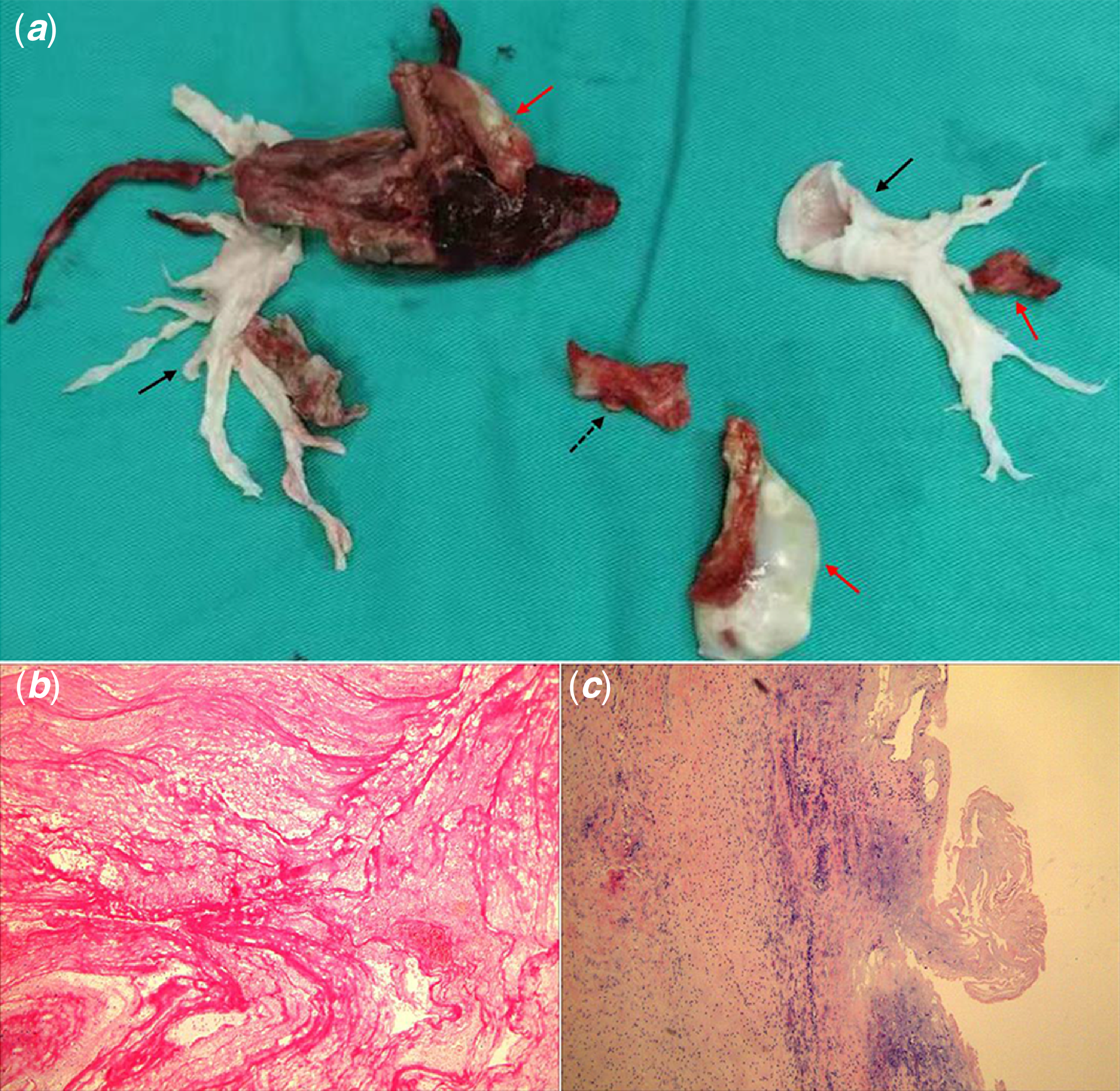
Following the diagnosis of acute-on-chronic pulmonary embolism and chronic thromboembolic pulmonary hypertension, therapeutic enoxaparin was initiated immediately. Despite this, the patient’s clinical condition deteriorated on the 4th day of hospitalization, with worsened dyspnoea, chest pain, and severe hypoxia with an oxygen saturation of 75% on unchanged supplemental oxygen support. Repeat transthoracic echocardiograph showed a reduction in right ventricular thrombus size, suggesting embolization. Given the high risk of recurrent, potentially fatal pulmonary embolism from the large, mobile residual thrombus compounded by chronic thromboembolic pulmonary hypertension, the multidisciplinary heart team opted for urgent surgery. Intraoperative findings included free-floating thrombi in the right ventricle, a large organized thrombus burden in the main and bilateral pulmonary arteries, along with fresh thrombi in the left pulmonary artery (Figure 2 a ). A small ostium secundum atrial septal defect was also confirmed. Surgical pulmonary embolectomy, right ventricular thrombectomy, and repair of the atrial septal defect were successfully performed. Postoperative transesophageal echocardiography showed complete thrombus removal. Concurrent right heart catheterization documented a dramatic reduction in mean pulmonary arterial pressure from 59 mmHg to 22 mmHg. Histopathological analysis confirmed organized thrombus and revealed fibrotic changes in the pulmonary artery wall (Figure 2 b , c ).
( a ) Thickened pulmonary artery intima (black arrow) and thrombi (red arrow) are observed in the bilateral artery lumen. Excised right ventricular mass (red arrow) and tricuspid valve chordae (black dashed arrow) are observed. ( b ) Right ventricular mass reveals mixed thrombus and mainly white thrombus. ( c ) Pulmonary arterial wall shows partial mucinous degeneration and hyalinosis, with infiltration of scattered lymphocytes and plasma cells.

Postoperatively, the patient was initiated on warfarin and bosentan. Extensive investigations, including whole-exome sequencing, revealed no definitive aetiology, and lifelong anticoagulation was recommended. At the three-month follow-up, he was asymptomatic with no recurrence of right ventricular thrombus.
Discussion
This complex case of a teenage boy with massive, mobile right ventricular thrombi culminating in life-threatening pulmonary embolism despite therapeutic anticoagulation highlights critical dilemmas in management. It underscores that in the presence of high-risk thrombus morphology, conventional first-line therapy may be inadequate, and a multidisciplinary strategy guided by advanced imaging is imperative.
The initial diagnostic challenge centred on the definitive characterization of the right ventricular masses. While echocardiography served as the cornerstone, our case exemplifies the complementary value of a multimodal approach. Transthoracic echocardiograph identified three distinct right ventricular masses, notably two highly mobile lesions. Although this morphology was highly suggestive of Type A (“in-transit”) thrombi and conferred an extremely high embolic risk, 1,Reference Dinesh Kumar, Nareppa and Shetty2 echocardiography alone proved insufficient to definitively characterize the masses. Crucially, cardiac MRI with late gadolinium enhancement provided definitive characterization by demonstrating complete avascularity, effectively ruling out neoplasm. Reference Motwani, Kidambi and Herzog3,Reference Barbagallo, Naef and Köpfli4 Both echocardiography and MRI revealed severe right ventricular dysfunction and pulmonary hypertension. Concurrently, CT pulmonary angiography detailed the massive, bilateral pulmonary embolism burden, and unveiled characteristic features of chronic thromboembolic pulmonary hypertension. This imaging triad did not merely diagnose right ventricular thrombus; it painted a complete picture of high embolic potential (morphology), definitive nature (avascularity), and severe physiological impact (massive pulmonary embolism, pulmonary hypertension, and right ventricular dysfunction).
The patient’s clinical deterioration despite therapeutic anticoagulation underscores the recognized limitation of anticoagulation for mobile, high-risk right ventricular thrombus. Reference Athappan, Sengodan and Chacko5,Reference Sakellariou, Efstathopoulos and Stamatis6 Given the type A morphology and the already-manifested large pulmonary embolism, the risk of further, potentially fatal embolization was deemed unacceptable. Our patient met the criteria for American Heart Association/American College of Cardiology pulmonary embolism category C3 (symptomatic, elevated severity score, abnormal right ventricular function, and positive biomarkers). As such, current guidelines provide a Class 2a recommendation for advanced therapies over anticoagulation alone to mitigate clinical deterioration. Reference Creager, Barnes and Giri7 While systemic thrombolysis is an option, the concern for fragmentation of such large, highly mobile thrombi and exacerbation of pulmonary hypertension favoured a definitive surgical approach. Specifically, underlying chronic thromboembolic pulmonary hypertension drove the decision to pursue a single-stage surgical approach encompassing both emergent thrombectomy and pulmonary endarterectomy—a procedure best achieved surgically. This strategy allowed for complete removal of the right heart thrombotic source, extensive pulmonary endarterectomy to address chronic changes, and incidental atrial septal defect repair in a single procedure. The immediate postoperative result—a dramatic drop in mean pulmonary arterial pressure from 59 mmHg to 22 mmHg—confirms the haemodynamic rationale for this aggressive intervention and aligns with outcomes sought in pulmonary endarterectomy for chronic thromboembolic pulmonary hypertension management. Reference Delcroix, Torbicki and Gopalan8
A comprehensive thrombophilia workup, extending to whole-exome sequencing, yielded no definitive cause. The transient depression of protein C and S levels was likely acute-phase/reactant, given the confounding effect of heparin therapy and the negative genetic screen. The lack of deep vein thrombosis argues against a simple “in-transit” phenomenon from the periphery, suggesting possible in situ formation in a predisposed heart, though no structural or arrhythmogenic cardiomyopathy was identified. This diagnostic ambiguity directly informed long-term management. Despite surgical clearance of the major thrombi, the presence of residual peripheral emboli and the idiopathic nature of the event justified the recommendation for lifelong anticoagulation, consistent with guidelines for similar high-risk scenarios. Reference Delcroix, Torbicki and Gopalan8 Bosentan was utilized in the context of the preoperative chronic thromboembolic pulmonary hypertension diagnosis.
In conclusion, this case highlights a refined approach in managing paediatric right ventricular thrombi. Multimodal imaging—specifically cardiac MRI—was pivotal for tissue characterization, allowing for definitive differentiation of the right ventricular mass and risk stratification. Crucially, the combination of a highly mobile right ventricular thrombus and underlying chronic thromboembolic pulmonary hypertension drove the decision to pursue a single-stage surgical approach encompassing both emergent thrombectomy and pulmonary endarterectomy—a procedure best achieved surgically. This case underscores the necessity of individualized, multidisciplinary evaluation to balance the risks of embolism against intervention, ultimately leading to successful salvage.
Data availability statement
The authors confirm that the data supporting the finding of this study are available within the article and its supplementary materials.
Acknowledgements
The authors would like to thank the family for consenting the publication of this case report.
Financial support
None.
Competing interests
The authors have no conflict of interest to declare.