Introduction
Understanding parasite diversity and distribution patterns, and what drives them, has long resonated with parasitologists and ecologists alike (Morand, Reference Morand2015). Despite this, our understanding of host–parasite associations remains limited for many vertebrates and, where described, this is often at low taxonomic resolution (Poulin & Leung, Reference Poulin and Leung2010; Poulin & Presswell, Reference Poulin and Presswell2016). In New Zealand's freshwater fish community, for example, host–parasite relationships have been studied in approximately half of the 40 recognized host species (Hine et al., Reference Hine, Jones and Diggles2000; https://www.niwa.co.nz/freshwater-and-estuaries/nzffd/NIWA-fish-atlas/fish-species), with parasite taxa often identified to genus at best. While such a low resolution may be partly attributed to the global loss of taxonomic classification expertise (Poulin & Leung, Reference Poulin and Leung2010), taxonomic classification prior to the widespread use of molecular techniques, or the fish species themselves only being identified relatively recently (McDowall & Waters, Reference McDowall and Waters2003), for many freshwater fish species, their parasite communities have yet to be examined. Advances in the number of host species examined continue to be made (e.g. Kelly et al., Reference Kelly, Paterson, Townsend, Poulin and Tompkins2009; Poulin et al., Reference Poulin, Closs, Lill, Hicks, Herrmann and Kelly2012), with molecular approaches often aiding the verification of cryptic parasite taxa (e.g. Blasco-Costa et al., Reference Blasco-Costa, Poulin and Presswell2017). Although considerable progress has been achieved in regard to trematodes (e.g. Blasco-Costa et al., Reference Blasco-Costa, Poulin and Presswell2016, Reference Blasco-Costa, Poulin and Presswell2017), other parasite groups, such as nematodes, remain poorly described (2/14 taxa described to species) (Moravec & Taraschewski, Reference Moravec and Taraschewski1988; Hine et al., Reference Hine, Jones and Diggles2000; Luque et al., Reference Luque, Vieira, Herrmann, King, Poulin and Lagrue2010).
One such under-studied fish species is the brown mudfish Neochanna apoda, an endemic New Zealand fish occupying small (~1.6 m3) pools in peat-swamp-forests, formed predominantly by sporadic tree-fall events (Eldon, Reference Eldon1978; White et al., Reference White, McHugh, Glover and McIntosh2015a, Reference White, McHugh, Glover and McIntoshb). Although nationally threatened, this fish species thrives (e.g. 250 fish/m3, White et al., Reference White, McHugh and McIntosh2016) in these isolated pools, which are characterized by frequent and unpredictable droughts, high acidity (~3.5 pH) and low oxygen (White et al., Reference White, McHugh, Glover and McIntosh2015a, Reference White, McHugh and McIntosh2016) – conditions lethal to most fish species (Alabaster & Lloyd, Reference Alabaster and Lloyd1982). Mudfish move between pools (up to 112 metres, White et al., Reference White, McHugh, Glover and McIntosh2015b) during high rainfall events that flood the forest floor and create connections among isolated pools. Typically, these pools contain isolated mudfish populations; however, sympatric populations with banded kokopu Galaxias fasciatus may occur in pools within flood-plain habitats, where environmental conditions are less stressful to other fish species (White et al., Reference White, McHugh, Glover and McIntosh2015a). The increasing environmental severity gradient from flood-plain to forest-pool habitats, which limits the distribution of banded kokopu in the peat-swamp-forest landscape, also has the potential to influence the parasite meta-community structure.
To date, our understanding of parasite infections of Neochanna fishes (five New Zealand and one Australian species) has been largely limited to external observations (O'Brien, Reference O'Brien2005) and incidental reports of parasites encountered during stomach or gonad examinations (Eldon, Reference Eldon1978). Of the five parasite taxa known from brown mudfish, two nematode taxa, Hedruris spinigera Baylis, 1931 (family Hedruridae) and an unidentified rhabditoid, have been detected in the stomach (Eldon, Reference Eldon1978). In the present pilot study, several nematode specimens were recovered from the swim bladder of brown mudfish from Westland, New Zealand. These specimens were morphologically distinct from previously described nematode taxa from this host species, and from the only nematode species known to infect the swim bladder of New Zealand freshwater fish, Anguillicola novaezelandiae Moravec & Taraschewski (1988). Instead, the new specimens were found to resemble more closely capillariid nematodes, of the subfamily Capillariinae Railliet, 1915 (Nematoda: Capillariidae, following Moravec, Reference Moravec2001; Nematoda: Trichuridae, following Gibbons, Reference Gibbons2010). The subfamily Capillariinae includes over 300 described species parasitizing a large number of hosts, including fishes, mammals and birds (Moravec, Reference Moravec2001). While the classification of Capillariinae is considered one of the most difficult among nematodes (Moravec, Reference Moravec1982; Anderson, Reference Anderson2000), Moravec (Reference Moravec1982) proposed what is now the most accepted systematic of the group. According to this classification, one important morphological feature showing interspecific differences in these nematodes is the structure of the posterior end of the male. Currently, there are 27 genera assigned to this subfamily according to morphological characteristics (Gibbons, Reference Gibbons2010), and only a few have been characterized genetically.
Here, using a combination of morphological and molecular approaches, we characterized the new nematodes parasitizing the swim bladder of brown mudfish. We also investigated the distribution of this parasite in relation to pool habitat types (i.e. flood-plain or forest) utilized by this host.
Materials and methods
Collection and nematode isolation
Forty-six brown mudfish were collected during November 2013 from the Saltwater Forest, Westland, New Zealand (43°07′S, 170°26′E) from four ‘flood-plain’ pools (occasionally flooded from adjacent streams; 18 fish) and three ‘forest’ pools (isolated from permanent water sources; 28 fish). Mudfish were captured using unbaited gee minnow traps set overnight, with up to 10 individual fish per pool retained for parasitological examination. Fish were euthanized with an overdose of 2-phenoxyethanol (approximately 1 g l−1), then either frozen (−20°C) or preserved in 10% buffered formalin until all internal organs were examined for parasites in the laboratory. Nematodes obtained from the swim bladder were placed in 95% ethanol. Fixed specimens were mounted on temporary slides with a glycerol:water (1:1) solution. Morphological study consisted of microscopic examination of the different specimens at different magnifications using a light microscope (Olympus CX41, Olympus Australia Pty Ltd, Notting Hill, Victoria, Australia). All specimens were photographed using a digital Olympus DP25 camera as means of morphological vouchering information, and measured in ImageJ (Schneider et al., Reference Schneider, Rasband and Eliceiri2012). Studied specimens are deposited at the parasite collection of the Evolutionary and Ecological Parasitology group at University of Otago (New Zealand). All measurements are in μm unless otherwise stated, and are given as the range with the average in parentheses.
DNA extraction and sequencing
The genomic DNA of the new capillariids was extracted from six specimens (two females and four males, representing the three male morphs, see below). DNA was isolated using the PureLink® Genomic DNA Kit (Invitrogen New Zealand Ltd, Auckland, New Zealand) according to the manufacturer's protocol. We amplified two nuclear fragments: a 873-bp fragment of the 18S ribosomal DNA (18S) and 455-bp fragment of the ribosomal DNA first internal transcribed spacer (ITS1). The 18S was amplified using the primers Nem 18S F and Nem 18S R from Floyd et al. (Reference Floyd, Rogers, Lambshead and Smith2005), while the ITS1 was amplified with the primers rDNA2 (Vrain et al., Reference Vrain, Wakarchuk, Levesque and Hamilton1992) and rDNA1.58s (Cherry et al., Reference Cherry, Szalanski, Todd and Powers1997). Polymerase chain reactions (PCRs) were performed in a total volume of 20 μl, comprising 4 μl of MyTaq™ Red reaction buffer (Bioline (Aust.) Pty Ltd, Alexandria, New South Wales, Australia), primers at 0.5 µm each, MyTaq™ Red DNA Polymerase (Bioline) at 0.025 units/μl and approximately 10 ng of DNA template. The PCRs consisted of 35 iterations of the following cycle: 30 s at 95°C, 30 s at 54–58°C (for 18S and ITS1, respectively) and 1 min at 72°C; beginning with an additional denaturation step of 3 min at 95°C and ending with a final extension at 72°C for 10 min. PCR amplicons were purified and sequenced by a commercial facility (Macrogen Corporation, http://www.macrogen.com/).
Phylogenetic analysis
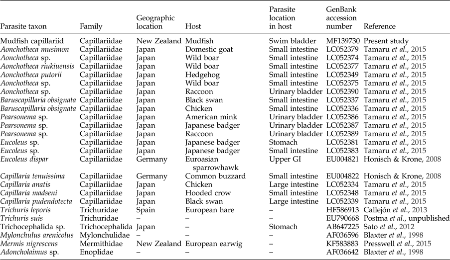
Sequence chromatograms of the New Zealand mudfish capillariid were edited and trimmed in Geneious v 8.1.4 (http://www.geneious.com/; Kearse et al., Reference Kearse, Moir, Wilson, Stones-Havas, Cheung, Sturrock, Buxton, Cooper, Markowitz, Duran, Thierer, Ashton, Mentjies and Drummond2012). Nematode 18S sequences comprising Capillaria, Aonchotheca, Baruscapillaria, Eucoleus, Trichuris and other non-identified Trichocephalida sequences were downloaded to Geneious, providing a total of 22 sequences for analysis (table 1). Since, for the ITS1 fragment, maximum query coverage with available sequences of other Capillariinae nematodes was 34%, phylogenetic analysis was only performed for the 18S fragment. Adoncholaimus, Mylonchulus and Mermis were used as outgroups (table 1). The 18S dataset was aligned using MAFFT (Katoh et al., Reference Katoh, Misawa, Kuma and Miyata2002) in Geneious, employing the E-INS-i algorithms (with default parameters). Final alignment consisted of 910 bp, including gaps.
Nematode sequence data included in the phylogenetic analysis, with respective family, geographic location, host identity, organ where it is located in the host and GenBank accession number.

GI, gastrointestinal tract.
To determine the phylogenetic position of the capillariid nematode studied, a Bayesian inference method was employed in MrBayes v. 3.2.2 (Ronquist et al., Reference Ronquist, Teslenko, van der Mark, Ayres, Darling, Höhna, Larget, Liu, Suchard and Huelsenbeck2012). A reversible-jump Markov chain Monte Carlo (MCMC) was implemented to integrate over the pool of all 203 possible reversible 4 × 4 nucleotide models. An a priori model was specified, allowing for the estimation of base frequencies, the proportion of invariable sites and rate-variation across sites with a gamma distribution. Ten million MCMC generations were sampled every 1000th step, with the first 25% discarded as burn-in. Two independent runs were carried out, each with one cold and three heated chains (T = 0.04) and the samples were pooled after burn-in was removed. Mixing and convergence of each run were monitored through the statistics provided in MrBayes (values of standard deviation of partition frequencies (<0.01), potential scale reduction factors (PSRF) (1.00), effective sample sizes (ESS) (>200)) and in Tracer v. 1.6 (Rambaut et al., Reference Rambaut, Suchard, Xie and Drummond2014). Estimates of evolutionary divergence for the 18S pairwise uncorrected differences (p-distance) between and within defined lineages were made in MEGA v. 6 (Tamura et al., Reference Tamura, Stecher, Peterson, Filipski and Kumar2013). Newly reported sequences from this study were submitted to GenBank under the accession numbers MF139730 (18S) and MF139729 (ITS1).
Results
Mudfish were found to be infected with nematode and acanthocephalan parasites in the swim bladder and intestinal regions. While acanthocephalans were found in mudfish populations from both ‘forest’ and ‘flood-plain’ pools (to be described elsewhere, Paterson et al., unpublished results), nematodes infecting the swim bladder were only found in ‘flood-plain’ pools. Twenty-two per cent of the mudfish from ‘flood-plain’ pools were infected with nematodes, with intensity ranging between 1 and 3 nematodes/fish. In total, 9 nematodes (6 males and 3 females) were found, although only 5 were complete specimens. The nematode specimens forming the present material were identified as belonging to the family Capillariidae (following Moravec, Reference Moravec2001), Trichuridae (following Gibbons, Reference Gibbons2010), subfamily Capillariinae based on the presence of an oesophagus consisting of a long glandular section, i.e. the stichosome (collection of a long chain of glandular cells, the stichocytes), and by the shape of the male posterior end. Morphological variability was observed at the male posterior end, namely in the thickness and extension of the lateral projections of the pseudobursa. Three different male morphotypes were provisionally defined. Male morph 1 (three specimens) was found infecting only one brown mudfish, whereas male morph 2 (one specimen) and morph 3 (two specimens) were found in the same host specimen.
Morphological description
General: Capillariinae Railliet, 1915
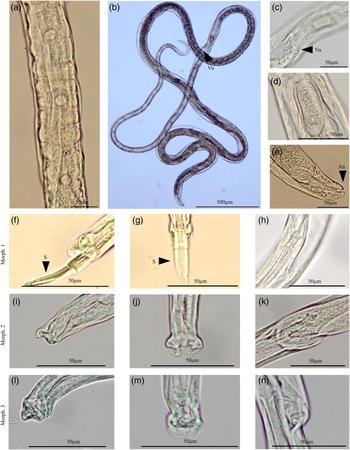
Based on five complete specimens and four incomplete specimens. Nematodes with rounded extremities, with anterior end narrower than posterior. Oral aperture terminal. Stichosome composed of one row of elongate stichocytes with small nuclei, subdivided into transverse annuli (fig. 1a).
Morphology of the new capillariid from the brown mudfish Neochanna apoda. (a) Stichocytes at middle part of the stichosome; (b) general view of adult female; (c) vulvar region, lateral view; (d) egg; (e) posterior end of female, lateral view. Male morph 1: (f) posterior end of male, lateral view; (g) posterior end of male, ventral view; (h) spicule proximal end. Male morph 2: (i) posterior end of male, lateral view; (j) posterior end of male, ventral view; (k) spicule proximal end. Male morph 3: (l) posterior end of male, lateral view; (m) posterior end of male, ventral view; (n) spicule proximal end. S, spicule; Vu, vulva; An, anus.

Male
Body 3.83–4.58 mm (4.05 mm, n = 4) long. Length of the entire oesophagus 1.38–1.61 mm (1.49 mm, n = 2), representing 37% of the body length. Muscular oesophagus 239.51–278.10 (258.80, n = 2) long, stichosome 1.1–1.37 mm (1.24 mm, n = 2) long, composed of 30 stichocytes (n = 1). Well-sclerotized spicule, 368.24–446.43 (409.43, n = 5) long, with a tronchanter-like shape at proximal end (fig. 1h, k) and with narrower and incised-like shape distal end (fig. 1g). Spicular sheath without spines. Male cloacal aperture subterminal. Excretory pore at level of the proximal end of spicule (when fully retracted). Caudal lateral alae absent. Membranous bursa of male supported on either side by one medium, round lobe, each lobe provided with a projection ventrally bent (fig. 1f, g, i, j, l, m). Male morph 1 (n = 3; fig. 1f–h) with a membranous bursa supported by two lateral lobes, each with one elongate papilla and a dorsal, ventrally bent projection extending beyond the papilla, but not reaching the posterior border of bursa (fig. 1g). Male morph 2 (n = 1, fig. 1i–k) with a small membranous bursa with a bell-like shape (fig. 1j), with two sessile, rounded papillae and two dorso-lateral projections (fig. 1i). Male morph 3 (n = 2; fig. 1l–n) with a membranous bursa, with two sessile papillae and two digitiform, ventrally bent projections.
Female (gravid)
Body 7.80 mm (n = 1) long. Length of the entire oesophagus 2.43 mm (n = 1), representing 31.15% of the body length. Muscular oesophagus 242.75 long, stichosome 2.19 mm long, composed of 37 stichocytes. Vulva situated at the level of the oesophago-intestinal junction, with vulva lips not elevated (fig. 1b, c). Vulvar appendage not observed. Ovary extending posteriorly to the junction of the intestine and rectum, with anus located subterminally (fig. 1e). Uterus containing numerous eggs, arranged in one row near the vulva and 2–3 rows more posteriorly (fig. 1d). Eggs (n = 6) 58.35–62.94 (61.71) in length and 25.94–31.23 (27.73) in width. Barrel-shaped eggs appearing with two layers: a hyaline inner layer and a thicker outer layer with a fine reticular, engraved structure (fig. 1d).
Remarks
Capillariid generic identification is based on specific morphological elements, namely the structure of the posterior end of the male. We followed Moravec (Reference Moravec1982) and Gibbons (Reference Gibbons2010), and restricted the taxonomic discussion to those found in fish only. Several genera were easily excluded based on the features of stichosome, characters of the male caudal region and female features, i.e. Capillaria Zeder, 1800, Gessyella Freitas, 1959, Schulmanela Ivashkin, 1964, Freitascapillaria Moravec, 1982 and Piscicapillaria Moravec, 1987. At a general level of the male pseudobursa, the studied specimens share some features with Paracapillaria Medonça, 1963, but the latter presents large sessile papillae. Paracapillaroides Moravec, Salgado-Maldonado and Caspeta-Mandujano, 1999 also presents some resemblance at the level of the pseudobursa, but it presents a spinose spicule sheath, which is not observed on the mudfish capillariid specimens. While, on the basis of shape of the spicule distal end and location in host, the new specimens may represent a new species or even a new genus, we refrain from providing a formal description due to the low number of specimens examined, together with the uncertainties surrounding the male morphs. In particular, it was uncertain whether the observed morphological variability of the three male morphs also represents genetic variability and, if so, if those specimens are closely related or not. In the latter case, we would need to determine to which morph the female morphology described here corresponds. However, the national conservation status of the host species of the studied nematode restricted the collection of additional specimens in order to clarify such uncertainties.
Phylogenetic analysis
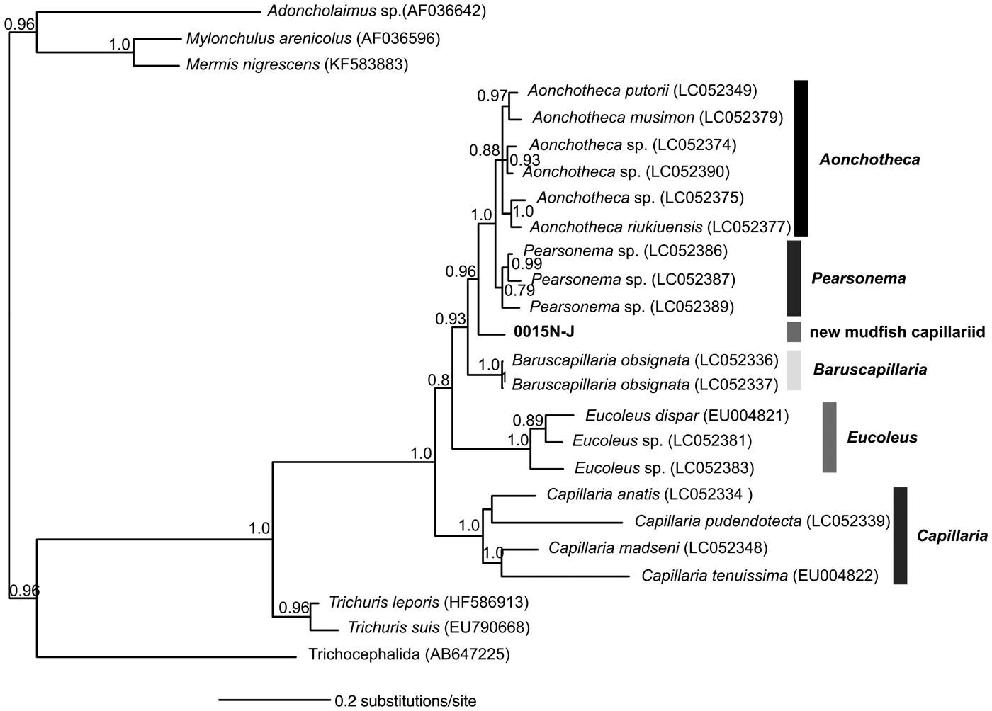
The DNA of a single male (morph 1) was successfully amplified and used for phylogenetic inference of the capillariid found in New Zealand mudfish. A phylogenetic inference tree based on the 18S nucleotide sequences is shown in fig. 2. The molecular analysis supported the morphological identification, placing the 18S sequence obtained in this study together with other sequences identified as belonging to the subfamily Capillariinae, but forming a separate clade. Among the available Capillariinae taxa, the new capillariid seems genetically more closely related to the Pearsonema clade (3.4% uncorrected p-distance). This observed genetic divergence is higher than that observed among other genera, namely between Pearsonema and Aonchotheca clades (2.6% uncorrected p-distance).
Phylogenetic relationships of the new capillariid from the brown mudfish Neochanna apoda based on Bayesian analysis of 18S gene sequences. Values represent Bayesian posterior probabilities (values below 0.75 are not represented). The sequence newly reported in this study is in bold and other sequences are presented with the respective GenBank accession numbers in parentheses (see table 1 for additional details).

Discussion
On the basis of both morphological and phylogenetic assessments, the nematode specimens found infecting the swim bladder of the brown mudfish were identified as belonging to the subfamily Capillariinae. At the morphological level, the new nematodes are distinct from other Capillariinae taxa by the shape of spicule distal end and the shape of the bursa lobes in males. Morphological variation at the male posterior end was observed; however, we were unable to determine if such variation was an artefact of specimen preservation, or reflects genetic variability within these nematodes, since only male morph 1 was successfully characterized molecularly. While presenting unique morphological features, we found some similarities between the overall morphological features of the specimens reported here and other capillariid parasites of fish, i.e. Paracapillaria and Paracapillaroides. Only a few Capillariinae taxa have been molecularly characterized, so we were unable properly to assess the level to which morphological similarities between the mudfish capillariid and other taxa also match phylogenetic relatedness.
According to Moravec & Justine (Reference Moravec and Justine2010), eight Capillariinae genera are known to parasitize fish, but none are documented in the swim bladder as observed in the present study. The only recorded trichinelloid nematode found in the swim bladder (Gibbons, Reference Gibbons2010), genus Huffmanela Moravec, 1987, belongs to a different family (Trichosomoididae Hall, 1916) and is also morphologically distinct from the specimens reported here. Following Moravec's (Reference Moravec1982) hypothetical evolution of capillariids based on the structure of the male caudal end and spicule sheath, Capillaria was one of the most basal divergences, and Pearsonema was more closely related to Baruscapillaria than to Aonchotheca. In the reported inferred phylogeny, and similar to the one estimated by Tamaru et al. (Reference Tamaru, Yamaki, Jimenez and Sato2015), Aonchotheca is more closely related to Pearsonema. While our results and those of Tamaru et al. (Reference Tamaru, Yamaki, Jimenez and Sato2015) seem to support to some extent the validity of Moravec's (Reference Moravec1982) taxonomic classification, we highlight the need for thorough analysis of the morphological evolution of this nematode subfamily. We highly recommend that new species' descriptions provide a molecular characterization together with the detailed morphological assessment. As several unreported species may await discovery, inferences regarding the evolutionary history of capillariid nematodes will remain challenging if such efforts are not undertaken.
Although the life cycle of the studied capillariid remains unknown (i.e. whether it requires a facultative intermediate invertebrate host), its apparent restricted distribution to ‘flood-plain’ pools, despite the considerable mobility of its mudfish host (White et al., Reference White, McHugh, Glover and McIntosh2015b), suggests that this nematode may be less tolerant of the environmental extremes (e.g. drought, high acidity, hypoxia) of the ‘forest’ pools. While mudfish are capable of surviving periods of drought (over 30 days without water; White et al., Reference White, McHugh and McIntosh2016) by burrowing into the soft peat-pool substrate and maintaining a low metabolic rate via cutaneous respiration (Urbina et al., Reference Urbina, Meredith, Glover and Forster2014a, Reference Urbina, Walsh, Hill and Gloverb), the environmental extremes of ‘forest’ pools may be lethal to infective stages of this capillariid. Thus adaptations to the harsh environmental conditions in addition to pool isolation may enable brown mudfish to escape some of their enemies, both in the form of parasites and their predators (e.g. banded kokopu).
It remains to be determined whether other mudfish species also harbour this, or related, capillariid species, given the paucity of parasitological knowledge currently available for the Neochanna genus in general. However, the unique habitat niche occupied by brown mudfish, in combination with the non-overlapping distribution of mudfish species, suggest that this capillariid is likely to have evolved in isolation together with its host. Furthermore, recent parasitological investigations suggested that other parasite taxa also found in mudfish may be genetically distinct from those infecting other New Zealand freshwater fish species (Paterson & Hernadez-Ortez, pers. comm.). Regarding the capillariid reported herein, future studies should assess host specificity of the new taxon, i.e. whether other fish species (e.g. banded kokopu) in the flood-plain habitat are also hosts of this nematode, and also assess the diversity of other capillariids present in New Zealand to assess their phylogenetic relatedness. Only then can a proper inference of this parasite's evolutionary history be made.
In this study we described a new capillariid nematode parasitizing the swim bladder of the brown mudfish in New Zealand. Our findings highlight the potential diversity of parasite assemblages that has likely lain hidden in this very unique ecosystem, and establishes a baseline for future studies. The findings of the study also highlight the necessity of a more collaborative approach between parasitologists and researchers from other fields, in our case fish biologists, which could promote the discovery of host–parasite relationships in New Zealand.
Acknowledgements
The authors would like to thank Professor Angus McIntosh and Professor Robert Poulin for field and laboratory support, and in-kind support from Glendevon Research.
Financial support
This manuscript was put together in the space between other funded PhD and postdoctoral opportunities. Nevertheless, we would like to thank the funding agencies and grants that have supported us during this time: F.J. was funded through a doctoral grant (SFRH/BD/77332/2011), and by a 2017 University of Otago Research Grant (UORG) to Professor R. Poulin; R.S.A.W. and R.A.P. were supported by NIWA (National Institute of Water and Atmospheric Research) Sustainable Water Allocation Program subcontract (E6233); R.A.P. was also supported by funding from the European Union's Horizon 2020 research and innovation programme under the Marie Skłodowska-Curie grant agreement no. 663830.
Conflict of interest
None.
Ethical standards
All procedures described were approved by the University of Canterbury Animal Ethics Committee, permit 2013/27R.