Introduction
First branchial cleft anomalies are very rare, the annual incidence being approximately one per 1 000 000.Reference Arndal and Bonding1 Such anomalies are the result of incomplete obliteration of the clefts that arise between the branchial arches during embryogenesis, producing persistent anomalous structures with considerable variation in their anatomical course.Reference D'Souza, Uppal, De and Zeitoun2 Due to this high degree of variability, several different classifications have been published, based on the anatomical position, number of openings and type of tissue lining the tract.Reference Whetstone, Branstetter and Hirsch3–Reference Work5 In the present case, a tortuous fistulous course was found between the infra-auricular region and the fossa of Rosenmuller, diagnosed by computed tomography fistulography. An extensive literature search revealed that this variant of first branchial cleft anomaly has not previously been reported.
Case report
A 15-year-old girl presented with the chief complaint of recurrent mucopurulent discharge from an opening in the left infra-auricular region, persisting since early infancy.
On examination, a 1-mm opening was present at the infra-auricular region, about 3 mm below the attachment of the lobule. Application of pressure resulted in extrusion of thick, white discharge from the opening.
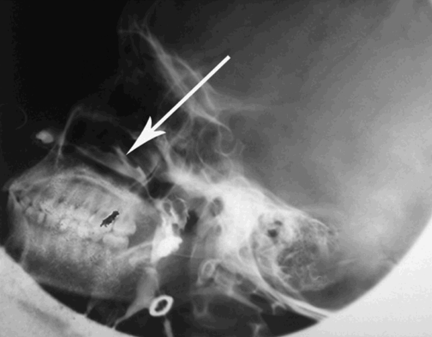
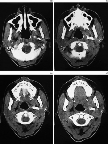
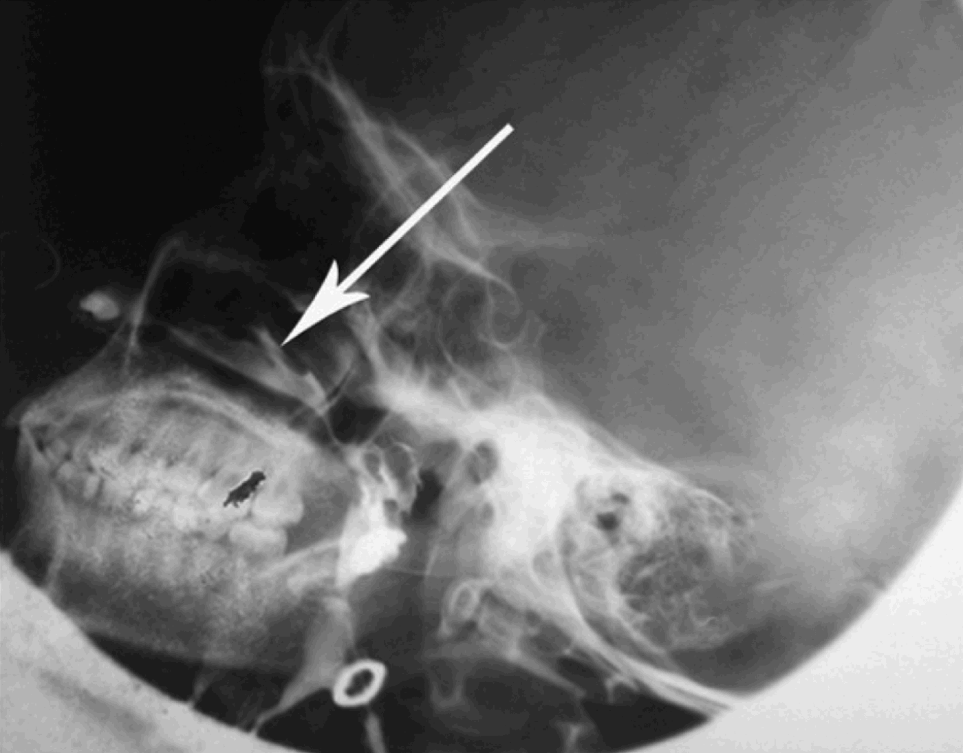
A sinogram of the left infra-auricular opening (Figure 1) showed spillage of contrast in the left nasal cavity, suggesting that the tract lay between the infra-auricular area and the nasopharynx. Computed tomography fistulography using diatrizoate meglumine dye (Figure 2) confirmed the presence of a tortuous tract approximately 4.61 cm in length, extending anteroinferiorly and medially from the external inframeatal opening to the lateral nasopharyngeal wall (anterior to the fossa of Rosenmuller). This tract was connected to the deep lobe of the parotid gland, and lay 0.67 cm anterior to the carotid artery and posterior to the medial pterygoid muscle.
Sinogram of left infra-auricular opening showing spillage of contrast in the left nasal cavity (arrow), suggesting that the tract lies between the infra-auricular area and the nasopharynx.
Axial, computed tomography fistulography images with diatrizoate meglumine dye, confirming the presence of a tortuous tract approximately 4.61 cm in length, extending anteroinferiorly and medially from the external inframeatal opening to the lateral nasopharyngeal wall (anterior to the fossa of Rosenmuller). The tract was connected to the deep lobe of the parotid gland and lay 0.67 cm anterior to the carotid artery and posterior to the medial pterygoid muscle.

Mandibulotomy was planned for complete resection of the tract. However, after the patient's parents were counselled regarding the nature of the problem, its surgical treatment, and the likely results of surgery versus conservative treatment, they opted to defer surgery until it became unavoidable.
Discussion
First branchial cleft anomalies are rare congenital malformations which are often misdiagnosed and mismanaged.Reference Lanisnik, Didanovic and Cizmarevic6 Their origin is uncertain; the presence of ectodermal and sometimes mesodermal elements has led some authors to conclude that they represent buried nests of cells which originally formed the first branchial cleft and its underlying mesoderm.Reference Lanisnik, Didanovic and Cizmarevic6
First branchial cleft anomaly can present as a cystic lesion, fistula or sinus extending towards the membranous external ear canal.Reference Lanisnik, Didanovic and Cizmarevic6 The sinus tract runs through the parotid gland in close association with the facial nerve. At present, there is no imaging method capable of identifying a first branchial cleft anomaly with certainty.Reference Arndal and Bonding1
During embryonic development, closure of the first branchial clefts is contemporaneous with the emergence of the developing parotid gland and migration of the facial nerve, originating from the second branchial arch; thus, first branchial cleft anomalies are typically associated with these structures.Reference Benson, Dalen, Mancuso, Kerr, Cacciarelli and Mafee7
• First branchial cleft anomalies are rare congenital malformations which are often misdiagnosed and mismanaged
• This paper describes a rare, variant branchial fistula passing from the external auditory canal to the nasopharynx
• Computed tomography fistulography is the imaging modality of choice for the diagnosis of branchial cleft fistula, and will also assist surgical planning
First branchial cleft anomalies are seen approximately twice as often in women (69 per cent) as in men (31 per cent).Reference D'Souza, Uppal, De and Zeitoun2 Fistulae occur more frequently on the left side (64 per cent), while sinuses show no side preference.Reference D'Souza, Uppal, De and Zeitoun2
Numerous schemes for classification of first branchial cleft lesions have been suggested. Classification systems rely on the location of the anomaly: type I lesions occur adjacent to the external auditory canal, whereas type II lesions occur at the angle of the mandible.Reference Whetstone, Branstetter and Hirsch3
Our patient was female and had her fistula opening in the left infra-auricular area. Computed tomography fistulography showed a tortuous tract between the infra-auricular area and the nasopharynx. As the tract course was in the area formed by the first branchial arch, the lesion appeared to be a rare variant of first branchial arch anomaly. The great variability in the course of these lesions, and the necessity of complete excision during treatment, makes radiographic visualisation crucial for definitive treatment.Reference Whetstone, Branstetter and Hirsch3 Complete excision of our patient's tract would require mandibulotomy with careful handling of cranial nerves, the facial nerve in particular, along with other vital structures.