Protein-losing enteropathy has been described in animal models and humans following ligation of the thoracic duct, or with elevated pressure in the brachiocephalic vein.Reference Drake, Anwar, Kee and Gabel1–Reference Blalock, Robinson, Cunningham and Gray8 In humans, this problem has been associated with constrictive pericarditis, restrictive cardiomyopathy, severe tricuspid insufficiency or stenosis, the Fontan operationReference Feldt, Driscoll and Offord9–Reference Ostrow, Freeze and Rychik12 and, obstruction of the superior caval vein, the latter occurring with obstruction of the superior caval venous limb of the interatrial rerouting that is part of the Mustard or Senning procedures.Reference Gleason, Roodman and Laks13 We describe our experience with 3 patients having protein-losing enteropathy, 2 with superior caval venous obstruction, and a third with stenosis in the Fontan circuit. All of the patients were treated successfully by relieving the obstruction of the caval vein.
Patients and methods
Our first patient was a 4 year old male who had resection of a mediastinal myofibroma as a neonate at another institution. At 32 weeks of gestation, he had a heart rate between 60 and 70 beats per minute, and at 36 weeks of gestation had 2 to 1 atrioventricular block and a pericardial effusion. In order to resect the mass, it was necessary to remove a portion of the superior caval vein, brachiocephalic vein, and right atrium. Continuity of the remaining tributaries of the superior caval vein with the right atrium was not reestablished. The right atrium was reconstructed with pericardium and prosthetic material, and an atrial septal defect intentionally was not closed. Following operation, he developed chylothorax and signs of superior caval venous obstruction requiring hospitalization for 2 months. Over the subsequent two years, he developed progressive pedal oedema and ascites. At 3 years of age, he developed tetany. He was hypocalcaemic, hypoalbuminaemic, and hypokalaemic. His serum albumin was 1.8 mg/dl, and the fecal alpha 1 antitrypsin was 580 mg/dl, the normal being less that 54 mg/dl. Biopsy of the small bowel revealed evidence of lymphatic obstruction. There was no renal or hepatic disease. His echocardiogram showed trivial tricuspid regurgitation, normal right and left ventricular systolic function and pressures, and a small atrial septal defect. Over the next year, he was treated with infusions of albumin, diuretics, a diet of low fat and high protein, and steroids, but without any significant change in his protein-losing enteropathy. Since birth, he was in complete heart block, the aetiology of which was unclear. At 3½ years of age, an epicardial pacemaker was placed. The physicians caring for him thought that atrioventricular synchronization would improve his cardiac output and protein-losing enteropathy. The protein-losing enteropathy improved transiently for 2 months following placement of the pacemaker, but recurred soon after.
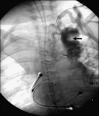
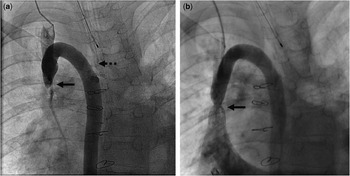
At 4 years of age, he was evaluated at the Mayo Clinic for the first time. During this initial evaluation, a venogram and lymphangiogram were done in order to ascertain the location of drainage of the thoracic duct and to determine the location of drainage of the tributaries of the superior caval vein. The axillary veins, brachial veins, and cephalic veins were patent bilaterally. The left arm veins drained into a tortuous and dilated plexus of veins in the left superior thorax (Fig. 1). There was no opacification of either subclavian or brachiocephalic veins. There was no identifiable internal jugular vein. There were collateral veins in the neck and the back that appeared to drain to the infradiaphragmatic area. The left sided axillary vein and a cephalic vein drained into a large collateral vein in the shoulder region, and to vessels in the mediastinum. There was drainage through the azygos system to the infradiaphragmatic area. The exact termination of the drainage of these veins was not apparent. The superior caval vein could not be visualized.
Pre-operative left subclavian venogram demonstrates its drainage into a tortuous, dilated and obstructed plexus of veins in the left superior thorax (arrow). This venous plexus eventually drained into mediastinal veins (dotted arrow).

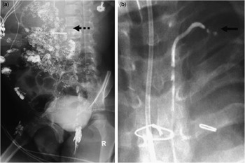
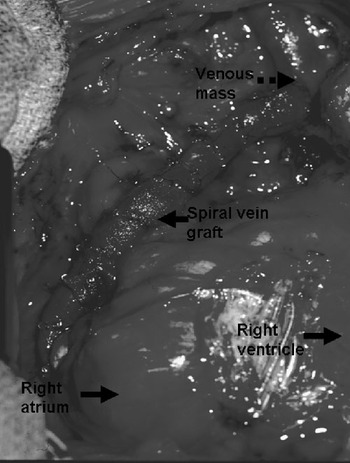
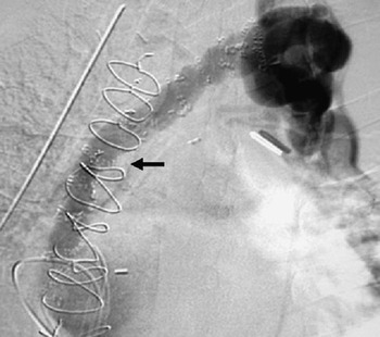
The lymphangiogram revealed severely dilated intestinal mucosa lymphatic channels, and drainage of the thoracic duct into the tortuous venous complex in the left hemithorax (Fig. 2). We inserted a spiral venous graft was used to connect this mediastinal venous plexus to the right atrium, and closed the atrial septal defect (Fig. 3). A postoperative venogram revealed effective decompression of the venous collaterals draining into to right atrium (Fig. 4). Over the next 2 months, the serum albumin normalized, increasing to 4.6 mg/dl and the fecal alpha 1 antitrypsin normalized at less than 4 mg/dl. At the age of 5, he was reevaluated, when it was noted that the spiral vein graft had become occluded. Despite that finding, he remains free of protein-losing enteropathy 6 years after operation.
Pre-operative lymphangiogram (a) revealed severely dilated intestinal mucosal lymphatic channels (dotted arrow). The pre-operative lymphangiogram (b) showed the termination of the thoracic duct into the region of the venous plexus in the left hemithorax (arrow).

Intra-operative photograph showing the spiral vein graft connecting the obstructed venous plexus to right atrium.

Post-operative venogram demonstrating effective decompression of the venous plexus by the spiral vein graft (arrow).

Our second patient is a 12-year-old girl with tetralogy of Fallot and pulmonary atresia who had closure of her ventricular septal defect and insertion of a homograft conduit from the right ventricle to the pulmonary arteries at 1 year of age at another institution. She had hypotension postoperatively, and suffered a stroke. She was in the hospital for 2½ months. She also developed the superior caval venous syndrome, and was treated with streptokinase, presumably, to dissolve a clot in the tributaries of the superior caval vein. She was noted to have Di-George syndrome and immunodeficiency requiring intermittent intravenous infusions of immunoglobulin.
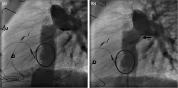
At 11 years of age, she developed progressive pedal oedema, ascites and hypo-albuminaemia. Her serum albumin was 1.5 g/dl, total protein 3.7 mg/dl, and fecal alpha 1-antitrypsin was 704 mg/dl. Biopsy of the small bowel was negative for sprue. There was no evidence of any coexisting renal or hepatic disease. Using echocardiography the estimated right ventricular pressure was 38 mmHg, the mean pressure gradient across the conduit from the right ventricle to pulmonary arteries was 10 mmHg, there was mild decrease in right ventricular systolic function, and there was moderate aortic regurgitation. A lymphangiogram was not done. At cardiac catheterization, the proximal superior caval venous mean pressure was 15 mmHg, and there was near occlusion of the vein itself (Fig. 5a). The right atrial pressure was 7 mmHg. She had balloon dilation of the stenosis using 8 mm×2 cm Z-Med II balloon, followed by placement of a 29 by 10 mm Palmaz-Genesis stent over a 10 mm by 2 cm Z-Med II balloon (Fig. 5b). At the end of the procedure, the previously stenotic segment of the superior caval vein was widely patent, albeit that the stent had a moderate waist. There was only 1 mmHg residual gradient between the proximal superior caval vein and the right atrium. Within one month, her symptoms of superior caval vein obstruction, pedal edema and ascites resolved. Her serum albumin increased to 3.8 g/dl, and the fecal alpha-1 antitrypsin decreased to 8 mg/dl. She has had no further symptoms or ongoing loss of enteric protein. Her serum albumin and total protein have remained normal 14 months after placement of the stent, and she no longer requires infusions of intravenous immunoglobulin to maintain normal levels.
The superior caval venous angiogram (a) demonstrates almost complete occlusion of the superior caval vein (solid arrow) and decompression of the superior caval vein by the azygous vein (dotted arrow). The inferior caval venous angiogram (b) shows restored patency of the superior caval vein with residual stenosis (arrow).

Our third patient is a 13-year-old boy born with an unbalanced atrioventricular septal defect, hypoplastic left ventricle, and dominant right ventricle. At 10 weeks of age, he underwent banding of the pulmonary trunk, followed at 9 months of age by a Damus-Kaye-Stansel procedure to relieve subaortic obstruction. He subsequently underwent placement of a bidirectional cavopulmonary anastomosis, followed by a nonfenestrated modified extracardiac Fontan procedure at the age of 5 years. Due to severe regurgitation across the common atrioventricular valve, he had insertion of a mechanical prosthesis at 9 years of age.
At the age of 11 years, he had progressive worsening pedal oedema, ascites, and scrotal oedema. Laboratory evaluation revealed hypoalbuminemia, and elevated fecal alpha-1 antitrypsin, at 460 mg/dl. Once the diagnosis of protein-losing enteropathy was established, he was treated with increasing doses of diuretics, low fat-high protein diet, inhibitors of angiotensin converting enzyme, and carvedilol, with no significant improvement. In December, 2006, cardiac catheterization revealed stenosis of the extracardiac conduit (Fig. 6a). The mean inferior caval venous pressure was 13 mmHg, with a mean gradient across the stenosis of 2 mmHg. The stenosis was dilated and a stent of 14 mm diameter was placed relieving the obstruction (Fig. 6b). Subsequent to the procedure, there was no gradient between the inferior caval vein and the right pulmonary artery. The mean inferior caval venous pressure remained at 13 mmHg. In January, 2007, his albumin and serum protein were normal, there was no evidence of anasarca, scrotal or pedal oedema, and he noted resolution of fatigue. In March, 2007, the stool alpha-1 antitrypsin was within normal levels, but the protein and albumin were 5.8 g/dl and 3.7 g/dl, respectively. In August, 2007, the alpha-1 antitrypsin, protein and albumin were 52 mg/dl, 4.8 g/dl, and 3.2 g/dl respectively. The patient had no oedema, and continues to feel well.
The angiogram prior to stenting (a), shown in the lateral projection, demonstrates the stenosis of the extracardiac Fontan conduit connecting the inferior caval vein to the right pulmonary artery (arrow). After stenting, a comparable angiogram (b) shows a widely patent anastomosis between the inferior caval vein and the right pulmonary artery (arrow).

Discussion
Cardiac conditions associated with protein-losing enteropathy include constrictive pericarditis, restrictive cardiomyopathy, severe tricuspid insufficiency or stenosis, the Fontan operation,Reference Feldt, Driscoll and Offord9–Reference Ostrow, Freeze and Rychik12 and obstruction of the superior caval vein, the latter often being encountered with obstruction of the superior caval limb of the baffle inserted as part of the Mustard or Senning procedures.Reference Gleason, Roodman and Laks13 Elevated systemic venous pressure in the tributaries of the superior caval vein is common to all of these conditions. In most, there also is elevation of systemic venous pressure in the tributaries of the inferior caval vein.
In all of these conditions, the thoracic duct drains into a vein in which the pressure is elevated, which may alter the dynamics of intestinal lypmphatic flow.Reference Blalock, Cunningham and Robinson4, Reference Neyazaki, Kupic, Marshall and Abrams6, Reference Blalock, Robinson, Cunningham and Gray8 This is a critical contributing factor to the development of protein-losing enteropathy. In dogs, occlusion of the left brachiocephalic vein results in reduced flow of lymph in the thoracic duct, accumulation of lymph in the lymphatic system, and perhaps in the intercellular spaces.Reference Wegria, Zekert and Walter2 Indeed, in dogs, ligation of the thoracic duct results in increased intestinal loss of protein.Reference Marshall, Neyazaki and Abrams3 This can occur even in the absence of elevated systemic venous pressure. In sheep, elevation of venous pressure in the neck caused by infusion of saline leads to increased lymphatic pressure in the gut.Reference Drake, Anwar, Kee and Gabel1 In rats, protein-losing enteropathy also occurs after ligation of the thoracic duct.Reference Kondo, Nakanishi, Bamba, Hosokawa and Masuda7 Blalock and his colleaguesReference Blalock, Robinson, Cunningham and Gray8 demonstrated that there were multiple routes of lymphatic drainage to the venous system other than just through the thoracic duct. Because of these numerous sites of drainage, he found it difficult completely to disrupt lymphatic drainage simply by ligating the thoracic duct in dogs. With sclerosis of the cisterna chili, in addition to ligation of the thoracic duct, complete disruption could be accomplished. Some of the dogs in whom this was accomplished died, but others developed collateral lympho-venous connections and survived.Reference Neyazaki, Kupic, Marshall and Abrams6, Reference Blalock, Robinson, Cunningham and Gray8 The presence of lympho-venous communications other than the thoracic duct, and the development of lympho-veno collaterals after ligation of the thoracic channels, may explain why, in some cases, protein-losing enteropathy does not occur only after ligation of the thoracic duct. This also may explain why protein-losing enteropathy does not always occur in the presence of increased pressure in the tributaries of the superior caval vein, and why protein-losing enteropathy can resolve spontaneously in some cases.
If there is elevated pressure in the tributaries of the inferior caval vein, this pressure could be transmitted to, and contribute to, elevated mesenteric venous pressure. Ludwig and Tomsa,Reference Ludwig and Tomsa14 in 1861, demonstrated in rats that venous congestion greatly increases lymph production in the gut. In the face of obstruction to the flow in the thoracic duct, increased production of lymph in the gut could lead to leakage of intersitial lymphatic fluid into the lumen of the gut and protein-losing enteropathy. Portal hypertension also can be associated with increased production of lymph by the gut. Flow of lymph in the thoracic duct is increased with cirrhosis, but protein-losing enteropathy is uncommon. If there is obstruction to the thoracic duct, the complication can occur in association with portal hypertension and cirrhosis.Reference Dousset, Legmann and Soubrane15
An important observation is that protein-losing enteropathy occurs in fewer than one-tenth of patients after the Fontan operation, even though all patients have elevated systemic venous pressure after that operation. Indeed, there are many patients after Fontan operation who do not have protein-losing enteropathy, but who have higher systemic venous pressures than patients who do develop this complication. In patients with systemic venous hypertension, there does not appear to be a correlation between its degree and the development of protein-losing enteropathy. Hence, elevated systemic venous pressure in itself is not the cause of protein-losing enteropathy, but is an important component of the cause. The aetiology of the complication is multifactorial, but likely, does not occur in the absence of elevated pressure in, at least, the tributaries of the superior caval or the hepatic veins. Rychik and Gui-YangReference Rychik and Gui-Yang16 demonstrated that patients with protein-losing enteropathy after the Fontan operation have increased mesenteric vascular resistance, and suggested that reduced cardiac output in these patients also contributes to the development of protein-losing enteropathy.
An intriguing question is why do some patients after the Fontan operation develop protein-losing enteropathy and others do not? It may be the relative degree of obstruction to drainage through the thoracic duct, reduced cardiac output, and elevated mesenteric vascular resistance. Clearly, there are additional contributing factors. It has been shown that natriuretic peptide is elevated in some patients after the Fontan operation. In cattle, natriuretic peptide inhibits transport, and attenuates flow, of lymph through a reduction of spontaneous contractions and marked relaxation of lymphatic smooth muscle.Reference Ohhashi, Watanabe and Kawai17, Reference Atchison and Johnston18
Inflammation also may have a role in the development of protein-losing enteropathy. Acute infections can precipitate its onset or recurrence. Steroids have been reported to ameliorate the condition in some patients.Reference Rychik, Piccoli and Barber19 There are two histologic patterns for the small bowel in patients with protein-losing enteropathy associated with the Fontan operation. One of those patterns is reminiscent of sprue, with mucosa replaced by granulation tissue. It is interesting to speculate that this pattern may result from local inflammation caused by leakage of chyle into interstitial space. Perhaps, it is for these patients that steroids are potentially helpful. It also is intriguing to speculate that those patients with congenitally malformed hearts who develop protein-losing enteropathy may have an unrecognized congenital malformation of the lymphatic systemic predisposing them to develop protein-losing enteropathy in the presence of elevated systemic venous pressure.
In two of our patients, protein-losing enteropathy occurred secondary to superior caval venous obstruction. In the first patient, we had documented that the thoracic duct emptied into a collection of venous structures that had an indirect connection to the heart. Once a direct connection was established, the protein-losing enteropathy resolved. Presumably, this occurred because the flow of lymph in the thoracic duct normalized. Curiously, even after the spiral vein graft eventually became obstructed, the protein-losing enteropathy did not return. Perhaps, because of the surgery, enough collateral venous channels formed to allow decompression of the thoracic duct. In our second case, we did not know to where the thoracic duct emptied, but did know that the left brachiocephalic vein did not enter the superior caval vein. We were surprised that simply reopening the superior caval vein eliminated the protein-losing enteropathy. We can only speculate that opening the superior caval vein resulted in enough decompression of the thoracic duct via collateral venous structures connecting to the proximal superior caval vein that the flow in the thoracic duct normalized.
We were disappointed that asymptomatic hypoproteinaemia recurred in the third patient months after dilation of the junction of the superior caval vein with the pulmonary arteries. However, this patient continues to be asymptomatic. This illustrates that protein-losing enteropathy may be more difficult to treat in patients who have had a Fontan operation than in those patients in whom protein-losing enteropathy occurs only because of obstruction to the tributaries of the superior caval vein.
These cases suggest that, of the various factors that contribute to the development of protein-losing enteropathy in patients with congenitally malformed hearts, elevated pressure in the venous structures into which the thoracic duct drains is critical. In such patients, aggressive techniques are warranted to lower the pressure in the tributaries of the superior caval vein. Successful treatment in patients who have had a Fontan operation remains a challenge.