Introduction
Approximately 6% of adolescent girls, aged 12–17 years, meet criteria for post-traumatic stress disorder (PTSD) (Kilpatrick et al. Reference Kilpatrick, Acierno, Saunders, Resnick, Best and Schnurr2000, Reference Kilpatrick, Ruggiero, Acierno, Saunders, Resnick and Best2003). The type of trauma associated with greatest risk for PTSD is assaultive violence exposure (Cisler et al. Reference Cisler, Begle, Amstadter, Resnick, Danielson, Saunders and Kilpatrick2012), which has a national prevalence rate of about 50% among adolescents and includes sexual assault, physical assault or witnessed violence (Kilpatrick et al. Reference Kilpatrick, Ruggiero, Acierno, Saunders, Resnick and Best2003). Trauma-focused cognitive–behavioral therapy (TF-CBT) is the ‘gold standard’ psychological treatment for pediatric PTSD with numerous randomized clinical trials demonstrating clear efficacy for PTSD symptoms, depression and behavioral problems (Cohen et al. Reference Cohen, Deblinger, Mannarino and Steer2004, Reference Cohen, Bukstein, Walter, Benson, Chrisman, Farchione, Hamilton, Keable, Kinlan, Schoettle, Siegel, Stock and Medicus2010, Reference Cohen, Mannarino and Iyengar2011; Deblinger et al. Reference Deblinger, Mannarino, Cohen and Steer2006, Reference Deblinger, Mannarino, Cohen, Runyon and Steer2011; Cary & McMillen, Reference Cary and McMillen2012). TF-CBT typically includes 12–16 weekly sessions organized into specific modules targeting: psychoeducation about trauma and PTSD; parenting skills; affect regulation and coping skills; and developing a narrative of the traumatic event and cognitive processing of associated thoughts and feelings.
While TF-CBT has demonstrated clear efficacy in improving traumatic stress symptoms among traumatized youth, the neural information processing mechanisms by which TF-CBT enacts clinical benefits remain unknown. Clearer delineation of the specific mechanisms that produce clinical change in TF-CBT could potentially inform efforts to enhance the speed, efficacy and/or maintenance of clinical response to TF-CBT. For example, if evidence suggested that one mechanism by which TF-CBT enabled PTSD symptom reduction was through the functional reorganization of specific neural circuits involved in emotion regulation, then subsequent research might test whether pharmacological or behavioral adjuncts to TF-CBT that specifically target this mechanism enhances overall efficacy of TF-CBT. The critical role of understanding the underlying mechanisms of treatment is echoed by the recent shift of the National Institute of Mental Health (NIMH) towards funding mechanisms for clinical trials that focus on establishing target engagement (i.e. intermediate mechanisms, such as brain function) rather than symptom reduction (Insel & Gogtay, Reference Insel and Gogtay2014). Within such a framework, the focus of inquiry with respect to TF-CBT would be on what intermediate mechanisms (e.g. altered neurocircuitry) are engaged by TF-CBT that enable its clinical outcomes. Subsequent efforts would then focus on identifying new ways to more effectively engage these key mechanisms during TF-CBT. Here, we report results from an exploratory study designed to provide initial data regarding the neural mechanisms engaged by TF-CBT that enable PTSD symptom reduction.
There is a significant literature supporting emotion regulation deficits in PTSD. Behaviorally, trauma-exposed youth with PTSD symptoms report greater difficulty in regulating emotions (Goldsmith et al. Reference Goldsmith, Chesney, Heath and Barlow2013; Sundermann & DePrince, Reference Sundermann and DePrince2015). For example, among adolescent girls with a history of maltreatment, self-reported emotion regulation difficulties were positively correlated with PTSD symptom severity when controlling for maltreatment characteristics (Sundermann & DePrince, Reference Sundermann and DePrince2015). With respect to neurocognitive mechanisms, neurocircuitry models of PTSD (Rauch et al. Reference Rauch, Shin and Phelps2006; Patel et al. Reference Patel, Spreng, Shin and Girard2012; Pitman et al. Reference Pitman, Rasmusson, Koenen, Shin, Orr, Gilbertson, Milad and Liberzon2012; Admon et al. Reference Admon, Milad and Hendler2013) consistently emphasize disruption within networks implicated in emotion regulation. Specifically, PTSD is often associated with hyper-reactivity of the amygdala, dorsal anterior cingulate cortex (dACC) and insula, and hypo-reactivity of the medial and lateral prefrontal cortex and hippocampus (Rauch et al. Reference Rauch, Whalen, Shin, McInerney, Macklin, Lasko, Orr and Pitman2000; Shin et al. Reference Shin, Whalen, Pitman, Bush, Macklin, Lasko, Orr, McInerney and Rauch2001, Reference Shin, Bush, Milad, Lasko, Brohawn, Hughes, Macklin, Gold, Karpf, Orr, Rauch and Pitman2011; Etkin & Wager, Reference Etkin and Wager2007; New et al. Reference New, Fan, Murrough, Liu, Liebman, Guise, Tang and Charney2009; Patel et al. Reference Patel, Spreng, Shin and Girard2012). While the neurocircuitry of pediatric PTSD has been less studied, emerging data similarly suggest altered function within the neurocircuitry implicated in adult PTSD, including hyper-reactivity of the amygdala (Garrett et al. Reference Garrett, Carrion, Kletter, Karchemskiy, Weems and Reiss2012) and dACC (Wolf & Herringa, Reference Wolf and Herringa2016) during emotion processing, altered functional connectivity (FC) of the amygdala and medial prefrontal cortex (PFC) during emotion processing (Cisler et al. Reference Cisler, Scott, Smitherman, Lenow and Kilts2013; Wolf & Herringa, Reference Wolf and Herringa2016), and smaller ventromedial PFC volumes (Keding & Herringa, Reference Keding and Herringa2015; Morey et al. Reference Morey, Haswell, Hooper and De Bellis2016). Disruptions in the function and structure of these neural regions in pediatric PTSD are consistent with the behavioral data demonstrating emotion regulation deficits.
Given the behavioral and neuroimaging data regarding emotion regulation deficits in PTSD, a plausible hypothesis regarding the intermediate mechanisms of symptom reduction in TF-CBT is that TF-CBT promotes functional reorganization of the neurocircuitry of amygdala-based neural networks mediating emotion regulation, subsequently improving the child or adolescent's ability to regulate strong negative emotions, which in turn leads to reduced PTSD symptoms. The current study provides an initial test of this specific hypothesis. Adolescent girls with PTSD related to assaultive violence exposure were engaged in a cognitive reappraisal task during 3 T functional magnetic resonance imaging (fMRI) before and after receiving 12 sessions of TF-CBT. PTSD symptoms and emotion regulation ability were additionally measured before and after treatment.
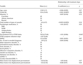
We focused the analyses of TF-CBT outcomes on changes in task-modulated FC for the bilateral amygdala. This was motivated by (1) the general conceptualization that emotion regulation is mediated neurally through the down-regulation of amygdala function (Etkin et al. Reference Etkin, Egner and Kalisch2011) and (2) PTSD neurocircuitry models that posit disrupted amygdala–prefrontal cortex FC (Rauch et al. Reference Rauch, Shin and Phelps2006). Focus on assault exposure was motivated by the greater risk for psychopathology conferred via assault exposure relative to other types of traumas (Cisler et al. Reference Cisler, Begle, Amstadter, Resnick, Danielson, Saunders and Kilpatrick2012) and the goal of increasing homogeneity of the sample by limiting variance in brain function due to type of trauma exposure. Focus on girls only was motivated by the increased risk for PTSD among girls (Kilpatrick et al. Reference Kilpatrick, Ruggiero, Acierno, Saunders, Resnick and Best2003) and the importance of increasing homogeneity of the sample by removing variability in brain function due to sex differences. A proportion of the treatment outcome data and clinical characteristics (see Table 1) were included in our prior report (Cisler et al. Reference Cisler, Sigel, Kramer, Smitherman, Vanderzee, Pemberton and Kilts2015), but all current imaging findings and relationships with treatment outcome are new.
Demographic, clinical characteristics and treatment response of the samples a

Data are given as mean (s.d.) unless otherwise indicated.
s.d., Standard deviation; s.e., standard error; IQ, intelligence quotient; SSRI, selective serotonin re-uptake inhibitors; NDRI, norepinephrine–dopamine reuptake inhibitors; UCLA, University of California at Los Angeles; PTSD, post-traumatic stress disorder; SMFQ, Short Mood and Feelings Questionnaire; ADHD, attention-deficit/hyperactivity disorder; ODD, oppositional defiant disorder.
a B coefficients come from robust regression models in which pre-treatment PTSD symptom severity was also controlled for, except in the model with only PTSD symptom severity as a predictor.
* p < 0.05.
Method
Participants and assessments
A total of 34 adolescent girls, aged 11–16 years, meeting Diagnostic and Statistical Manual of Mental Disorders, fourth edition criteria for PTSD, having a positive history of assaultive violence exposure, and having a consistent caregiver with whom to participate in treatment, were enrolled in the study and began TF-CBT. Participants were recruited through networking with local out-patient clinics, child advocacy centers, schools, juvenile justice, churches and community organizations. Exclusion criteria consisted of MRI contraindications (e.g. internal ferrous metal objects), psychotic symptoms, lack of a consistent caregiver and presence of a developmental disorder. Concurrent psychotropic medication was not exclusionary. Demographic and clinical characteristics of the sample are provided in Table 1. Adolescents provided assent and a caregiver/legal guardian provided consent. This study was conducted with University of Arkansas for Medical Sciences (UAMS) Institutional Review Board approval.
Of the 34 girls who began TF-CBT, 25 completed all TF-CBT modules, and 22 of these treatment completers also completed the post-treatment fMRI scan. Two were excluded from analyses due to excessive head motion (see below), leaving 20 adolescent girls completing TF-CBT and having usable pre- and post-treatment fMRI data. The 20 girls whose data are analysed here did not differ from the other 14 girls who did not complete all procedures in PTSD symptom severity, assault frequency, intelligence quotient (IQ), age or emotion regulation (all p’s > 0.22).
Participants’ pre- and post-treatment mental health status was assessed with the Mini International Neuropsychiatric Interview for Children and Adolescents (MINI-KID; Sheehan et al. Reference Sheehan, Sheehan, Shytle, Janavs, Bannon, Rogers, Milo, Stock and Wilkinson2010), a structured clinical interview for most Axis I disorders found in childhood and adolescence. Assaultive trauma histories were characterized using the trauma assessment section of the National Survey of Adolescents (NSA) (Kilpatrick et al. Reference Kilpatrick, Acierno, Saunders, Resnick, Best and Schnurr2000, Reference Kilpatrick, Ruggiero, Acierno, Saunders, Resnick and Best2003), a structured interview used in prior epidemiological studies of assault exposure and mental health functioning among adolescents that uses behaviorally specific dichotomous questions to assess sexual assault, physical assault, severe abuse from a caregiver and witnessed violence. A trained female research coordinator with several years of experience with structured clinical interviews completed the MINI-KID and NSA interviews with participants under the supervision of a licensed clinical psychologist.
The pre- and post-treatment assessment also included measures of verbal IQ [receptive one-word picture vocabulary test (Brownell, Reference Brownell2000), PTSD symptom severity (UCLA PTSD Reaction Index; Steinberg et al. Reference Steinberg, Brymer, Decker and Pynoos2004), depression (Short Mood and Feelings Questionnaire; SMFQ; Angold et al. Reference Angold, Costello, Messer, Pickles and Winder1995) and emotion regulation ability using the Difficulty in Emotion Regulation Scale (DERS; Gratz & Roemer, Reference Gratz and Roemer2004)]. Additionally, participants completed these measures of PTSD and depression symptom severity prior to each therapy visit.
TF-CBT
TF-CBT was delivered by two postdoctoral clinical psychology fellows and a doctoral-level graduate student. The therapists were trained in TF-CBT according to an established protocol approved by Anthony Mannarino, Ph.D., a co-developer of TF-CBT, which included completion of TF-CBTWeb (accessible at www.musc.edu/tfcbt), an online resource for TF-CBT training; 3 days of in-person TF-CBT training with Dr Mannarino; and 1 h of weekly supervision with a licensed clinical psychologist with expertise in supervising the model. TF-CBT in this study used a 12-week protocol of 60 to 90 min weekly sessions.
Fidelity to the TF-CBT model was assessed by recording a randomly selected 10% of therapy sessions and rating the session according to an a priori checklist of key therapy elements for each session (see online Supplementary material). Therapist fidelity in the current study was 100%.
MRI acquisition and image pre-processing
MRI acquisition
A Philips 3 T Achieva X-series MRI system with a 32-channel head coil (Philips Healthcare, USA) was used to acquire imaging data. Anatomic images were acquired with an MPRAGE sequence [matrix = 256 × 256, 160 sagittal slices, repetition time (TR)/echo time (TE)/flip angle = 2600 ms/3.02 ms/8°, final resolution = 1 × 1 × 1 mm3 resolution]. Echo planar imaging (EPI) sequences were used to collect the functional images using the following sequence parameters: TR/TE/flip angle = 2000 ms/30 ms/90°, field of view = 240 × 240 mm, matrix = 80 × 80, 37 oblique slices (parallel to anterior commissure–posterior commissure plane to minimize orbitofrontal cortex sinal artifact), slice thickness = 2.5 mm with a 0.5 mm gap between slices, resampled during pre-processing to a final resolution = 3 × 3 × 3 mm3.
Image pre-processing
Image pre-processing followed standard steps and was completed using AFNI18 software (https://afni.nimh.nih.gov/afni). In the following order, images underwent despiking, slice timing correction, deobliquing, motion correction using rigid body alignment, alignment to participant's normalized anatomical images, spatial smoothing using an 8 mm full width half maximum (FWHM) Gaussian filter (AFNI's 3dBlurToFWHM that estimates the amount of smoothing to add to each dataset to result in the desired level of final smoothing), and rescaling into percentage signal change. Images were normalized using the Montreal Neurological Institute (MNI) 152 template brain. Following recent work (Power et al. Reference Power, Mitra, Laumann, Snyder, Schlaggar and Petersen2014), we corrected for motion-related signal artifacts by using motion regressors derived from Volterra expansion, consisting of (R R 2 R t−1 R 2 t−1), where R refers to each of the six motion parameters, and separate regressors for the first principal component time courses (using AFNI's 3dmaskSVD) in the cerebrospinal fluid (CSF) and white matter (WM). This step was implemented directly after motion correction and normalization of the EPI images in the pre-processing stream. We used FSL (www.fmrib.ox.ac.uk/fsl) to segment the anatomical file into CSF and WM, transformed these masks into the size and shape of the functional images, and eroded them by a voxel to make them non-overlapping with gray matter. Additionally, we censored TRs from the first-level general linear models based on a previously used threshold of frame-wise displacement (FD) > 0.5. FD refers to the sum of the absolute value of temporal differences across the six motion parameters; thus, a cut-off of 0.5 results in censoring TRs where the participant moved, in total across the six parameters, more than about 0.5 mm plus the immediately following TR (to account for delayed effects of motion artifact). Additionally, we censored isolated TRs where the preceding and following TRs were censored, and we censored entire runs if more than 50% of TRs within that run were censored. Based on this recommended procedure (Power et al. Reference Power, Mitra, Laumann, Snyder, Schlaggar and Petersen2014), two participants were removed from all analyses due to not having any usable runs after censoring. Online Supplementary Figs S1 and S2 display the mean FD across all TRs of the task at pre- and post-treatment and the relationship between mean FD and PTSD symptom slopes.
fMRI tasks
Cognitive reappraisal task
Participants completed a cognitive reappraisal task (Ochsner et al. Reference Ochsner, Bunge, Gross and Gabrieli2002, Reference Ochsner, Ray, Cooper, Robertson, Chopra, Gabrieli and Gross2004), in which they were presented with images selected from the International Affective Picture System (IAPS) depicting scenes with either neutral or negative valence. Prior to the image appearing, participants were given an instruction (3 s duration followed by jittered inter-trial interval; ITI) indicating either to pay attention to their feelings about the pictures without attempting to alter them (i.e. ‘view’ instructions) or to think about the picture in a way that made them feel better about the picture (i.e. ‘reappraise’ instructions). All neutral images and half of the negative images were preceded by the instruction to view; the other half of negative valence images were preceded by the instruction to reappraise. Each image was presented for 8 s followed by a jittered ITI. There were 60 trials (20 neutral, 20 negative with view instructions, 20 negative with reappraise instructions), implemented in an event-related design across three runs of about 6 min each. Following each trial, participants were asked to indicate the degree to which they felt negatively about the image (Likert scale ranging from 1 to 4), and these rating trials lasted 3 s followed by a jittered ITI. See online Supplementary Fig. S3 for graphical depiction of the task.
Prior to administering the task, all participants were given a standardized tutorial for cognitive reappraisal, which consisted of a verbal description of cognitive reappraisal, observing the research coordinator engage aloud in cognitive reappraisal of example negative images, and having the adolescent practise cognitive reappraisal aloud of practice negative images.
Data analysis
Task-modulated FC with left and right amygdala
Task-modulated FC during the cognitive reappraisal task was characterized using the Beta Series Method (BSM) approach (Rissman et al. Reference Rissman, Gazzaley and D'Esposito2004; Cisler et al. Reference Cisler, Bush and Steele2014a ). In the BSM, each unique event during the cognitive reappraisal task is treated as an individual regressor [using an iterative regression approach (Mumford et al. Reference Mumford, Turner, Ashby and Poldrack2012) implemented with AFNI's 3dLSS], resulting in β coefficients unique to each trial for each voxel. The remainder of the BSM steps were conducted in Matlab R2015b (www.mathworks.com). The mean time course from the seed is then calculated for each stimulus condition separately using the trial-specific β coefficients unique to each task condition (e.g. 20 β coefficients for the 20 trials of the reappraise negative images condition). This seed time course of β coefficients, unique to each task condition, is then correlated with the corresponding β coefficients for every other voxel during the corresponding task condition. This approach characterizes FC between the seed region and every other voxel unique to each stimulus condition, and following r-to-z transformation, we created contrast FC maps for reappraise negative images v. view negative images and view negative images v. view neutral images. The seed regions were defined by 6 mm radius spherical volumes in the right (MNI center-of-mass xyz coordinates: 19, −5, −14) and left (xyz coordinates: −19, −5 −14) amygdala regions of interest (ROIs) used in our prior studies (Cisler et al. Reference Cisler, Steele, Lenow, Smitherman, Everett, Messias and Kilts2014b , Reference Cisler, Sigel, Kramer, Smitherman, Vanderzee, Pemberton and Kilts2015). The amygdala ROIs are graphically depicted in online Supplementary Fig. S4.
TF-CBT-related symptom change
Following our previous study and another prior study linking fMRI data to symptom change in depression (Heller et al. Reference Heller, Johnstone, Peterson, Kolden, Kalin and Davidson2013; Cisler et al. Reference Cisler, Sigel, Kramer, Smitherman, Vanderzee, Pemberton and Kilts2015), our primary measure of clinical response consisted of slope estimates representing trajectories of PTSD symptom change across treatment sessions calculated within an autoregressive general linear model implemented in Matlab (for further information, see online Supplementary material). We used three additional convergent measures of symptom change: slopes of changes in depression symptoms across treatment as measured by the SMFQ, magnitude of pre- to post-treatment change in emotion regulation ability, as measured by the DERS, and magnitude of pre- to post-treatment changes in mean valence ratings on the cognitive reappraisal task for negative view and negative reappraise images. Given that the DERS and cognitive reappraisal task were only collected twice (pre- and post-treatment), we could not calculate trajectory slopes comparable with the PTSD or depression symptom slopes.
Identifying changes in amygdala FC that track symptom change
Second-level analysis was conducted in Matlab and consisted of whole-brain, voxel-wise, robust regression (Wager et al. Reference Wager, Keller, Lacey and Jonides2005) analysis, in which the β coefficient representing slope of PTSD symptom change across time for each participant was regressed simultaneously onto (1) the intercept from the within-subject regression models representing severity of pre-treatment PTSD symptoms (i.e. controlling for any confounding effects of pre-treatment symptom severity), (2) the voxel's pre-treatment FC contrast value (i.e. controlling for individual differences in FC at pre-treatment), and (3) the voxel's post-treatment FC contrast value. Analyses were repeated for FC with both the right and left amygdala. To correct for multiple comparisons (voxel-wise, two ROIs, two contrasts), we maintained a corrected p < 0.0125 (p = 0.05/4 comparisons) using cluster-level thresholding (Forman et al. Reference Forman, Cohen, Fitzgerald, Eddy, Mintun and Noll1995) defined with Monte Carlo simulation (AFNI's 3dClustSim) using the spatial autocorrelation function method, in which a significant cluster (corrected p < 0.0125) is defined as a minimum of 33 contiguous voxels that survive a primary (uncorrected) threshold of p < 0.001. The mean effective FWHM across participants, runs and time points estimated using the spatial autocorrelation function (ACF) method (AFNI's 3dFWHMx) was 11.79 (s.d. = 0.14).
Results
Treatment outcome
Demographic, clinical and treatment outcome variables are provided in Table 1. The mean slope of symptom change across treatment was −0.97 (s.d. = 0.68), which was significantly different from zero (t 1,19 = −6.44, p < 0.001). Demographic and clinical variables were generally unrelated to magnitude of treatment outcome slopes, except for pre-treatment PTSD symptom severity, which was negatively related (t 1,18 = −2.2; B = −0.01, p = 0.041) to outcome slopes (i.e. more severe initial PTSD symptoms predicted steeper subsequent slopes of symptom change).
Improvements in emotion regulation predict PTSD symptom reduction
We tested whether improvements in emotion regulation ability scaled with PTSD symptom reduction with robust regression models in which pre-treatment PTSD symptom severity, pre-treatment DERS score and post-treatment DERS score were entered as simultaneous predictors of PTSD symptom slopes. These analyses demonstrated that, when controlling for both pre-treatment symptom severity and emotion regulation difficulties, greater post-treatment emotion regulation ability was significantly related to greater improvements in PTSD symptoms (t 1,16 = 2.42, p = 0.028).
Regarding cognitive reappraisal performance on the task, at both pre- and post-treatment, participants rated the negative images during negative view instructions significantly more negative compared with neutral images (t’s19 > 12.33, p’s < 0.001). Participants rated negative images during the negative view conditions significantly more negative than during the reappraise conditions (t’s19 > 4.5, p’s < 0.001). However, changes in cognitive reappraisal performance on the task were not related to symptom reduction (t’s1,16 < 0.41) nor were they related to self-reported change in emotion regulation (DERS scores) (t’s1,16 < 0.63).
PTSD symptom reduction and changes in task-modulated amygdala FC during cognitive reappraisal
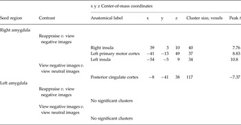
Full results of analyses of FC are depicted in Table 2.
Significant clusters where post-treatment task-modulated functional connectivity with the amygdala predicted post-traumatic stress disorder symptom slopes

Reappraising negative images v. viewing negative images contrast
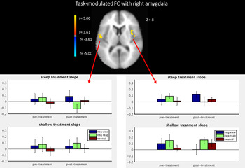
For the right amygdala, we observed that post-treatment task-modulated FC with both the right and left mid-insular cortex was positively correlated with PTSD symptom slope (Fig. 1; scatterplots in Supplementary Fig. S5). We further probed this contrast effect by entering the mean FC values within the ROI for the negative reappraise and negative view task conditions, at pre- and post-treatment, as predictors of symptom slopes (controlling for pre-treatment symptom severity) separately for the right and left insula clusters. For each of the insula clusters, greater post-treatment FC with the right amygdala during negative image viewing was associated with better PTSD symptom slopes (t’s1,15 < −4.5, p’s < 0.001), while greater FC during the reappraise negative images condition was associated with worse PTSD symptom slopes (t’s1,15 > 4.66, p’s < 0.001); there was no significant relationship of symptom change with insula FC during the neutral image conditions. The direction of the FC between the amygdala and each of the target regions (positive v. anti-correlation) is depicted in the relevant figures.
Top: clusters in the bilateral insular cortex where task-modulated functional connectivity (FC) with the right amygdala for the contrast of ‘reappraise negative images v. view negative images’ (neg reap v. neg view) predicts post-traumatic stress disorder symptom slopes. Bottom: bar graphs for girls with steep and shallow slopes (based on a median split) of mean FC values within each cluster for each of the task conditions at pre- and post-treatment. Values are means, with standard errors represented by vertical lines. neutral, View neutral images.

We also observed that post-treatment right amygdala FCs with the left primary motor cortex were positively correlated with PTSD symptom slopes (Table 2; scatterplot in online Supplementary Fig. S6). Comparable follow-up tests demonstrated that greater FC (greater positive correlation) with the left primary motor cortex while viewing negative images was associated with better PTSD symptom slopes (t 1,15 < −3.72, p’s < 0.01), while greater FC (less anti-correlation) while reappraising negative images was associated with worse PTSD symptom slopes (t 1,15 = 3.23, p < 0.01).
We did not observe any significant results for FC with the left amygdala.
Viewing negative images v. viewing neutral images contrast
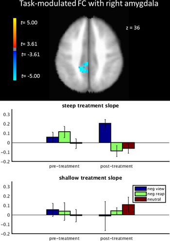
For the right amygdala, we observed one cluster in the posterior cingulate cortex where FC for this contrast at post-treatment was negatively correlated with PTSD symptom slopes (Fig. 2; scatterplot in online Supplementary Fig. S7). Follow-up analyses testing which specific task condition (negative images or neutral images) was driving this effect demonstrated that greater post-treatment FC while viewing negative images was associated with better PTSD slopes (t 1,15 = −3.06, p < 0.01), while lesser FC during neutral image viewing was associated with better PTSD slopes (t 1,15 = 3.30, p < 0.01).
Top: cluster in the posterior cingulate cortex where task-modulated functional connectivity (FC) with the right amygdala for the contrast of ‘view negative images v. view neutral images’ (neg view v. neutral) predicts post-traumatic stress disorder symptom slopes. Bottom: bar graphs for girls with steep and shallow slopes (based on a median split) of mean FC values within each cluster for each of the task conditions at pre- and post-treatment. Values are means, with standard errors represented by vertical lines. neg reap, Reappraise negative images.

We again did not observe any significant results for FC with the left amygdala.
To aid in interpretability, we provide the task activation of each cluster where we identified significant task-modulated FC with the amygdala in online Supplementary Figs S8 and S9.
Convergent validity for amygdala FC relationships with symptom reduction
We then tested whether the voxel clusters where post-treatment task-modulated FC with the amygdala were significantly related to PTSD symptom reduction were also related to depression symptom reductions during TF-CBT, pre- to post-treatment improvements in emotion regulation (measured with the DERS), and pre- to post-treatment changes in cognitive reappraisal ratings on the fMRI task. Full results for all clusters and convergent measures are provided in online Supplementary Table S1. Controlling for family-wise multiple comparisons with Bonferroni correction, we observed that post-treatment FC between the right amygdala and left insula for the contrast of reappraise negative images v. view negative images contrast was also predictive of improvements in self-reported emotion regulation (p < 0.01). No other relationships survived correction for multiple comparisons.
Addressing effects of potential pre-treatment confounding factors
When the primary analyses were repeated when including the possible pre-treatment confounding factors of age, verbal IQ, concurrent psychotropic medication (dichotomized into ‘yes’ or ‘no’), total number of co-morbid diagnoses and assault exposure severity (total number of assaultive event exposures), the observed relationships between post-treatment FC with the right and left amygdala for all voxel clusters identified in the primary analyses remained significant (all p’s < 0.0015). B coefficients and p values for all of these covariate analyses are listed in Supplementary Table S2, which demonstrates that the magnitude and significance of the identified relationships are essentially unchanged by the inclusion of these potentially confounding variables.
Discussion
The purpose of this study was to provide an exploratory test of the hypothesis that TF-CBT associates with PTSD symptom change through the functional reorganization of the neurocircuitry mediating emotion regulation and subsequent downstream effects on emotion regulation ability. Behaviorally, we observed that pre- to post-treatment improvements in self-reported emotion regulation ability positively correlated with improvements in PTSD symptoms. Though temporal precedence of the improvements in emotion regulation with respect to PTSD symptom reduction cannot be established from the current design, this result is nonetheless consistent with the hypothesis that TF-CBT produces PTSD symptom reduction through improvements in specific domains of emotion regulation. It should also be noted that the results pertaining to self-reported emotion regulation (DERS score) did not extend to the valence ratings on the cognitive reappraisal task, and caution should accordingly be used in interpreting this result.
The results of the analyses of task-modulated FC with the amygdala during cognitive reappraisal are similarly consistent with this hypothesis. We observed that greater PTSD symptom reduction was associated with greater suppression of amygdala–insula FC during reappraisal of negative images. As indicated in Fig. 1, those girls who improved the most demonstrated decreased FC post-treatment, whereas those girls who improved less failed to decrease FC at post-treatment. The specific site of the amygdala FC cluster in the insula was in the mid- to posterior insular cortex. The insular cortex is functionally heterogeneous (Craig, Reference Craig2002; Deen et al. Reference Deen, Pitskel and Pelphrey2011), with the posterior insular cortex more strongly linked with the representation of interoceptive/bodily state changes, such as sympathetic arousal during negative affective states as would be suggested by FC with the amygdala. Diminished amygdala–insula FC during reappraisal among girls with greater symptom reduction might indicate a lessened interoceptive representation of negative affective states at post-treatment compared with girls who respond less well. Accordingly, this suggests a plausible neural mechanism by which TF-CBT produces symptom reduction: specific skills training in affect regulation and cognitive reprocessing of the traumatic memory may stimulate functional reorganization of the neurocircuitry of emotion regulation to suppress the interoceptive representation of negative affective states. This mechanism might lead to a greater perceived ability to regulate emotions and subsequently fewer PTSD symptoms. Future research is clearly needed to provide corroborative evidence for this hypothesis.
The clinical translational significance of this work is rooted in its novel inferences for the development of approaches to increase the partial response of adolescent PTSD to TF-CBT. With respect to the identification of mechanisms that might be pharmacologically or behaviorally augmented to enhance TF-CBT outcomes, the current results suggest the possibility of targeting the behavioral and neural correlates of emotion regulation. The behavioral data and certain imaging findings suggest that suppression of amygdala–insula FC during reappraisal, and its putative downstream effect on emotion regulation ability, are viable targets for augmentation strategies for increasing clinical response to TF-CBT. For example, TF-CBT already includes modules addressing affect regulation and cognitive coping skills, and a prior dismantling study (Deblinger et al. Reference Deblinger, Mannarino, Cohen, Runyon and Steer2011) suggests that significant PTSD symptom reduction is attributable solely to these skill-building and parenting modules (i.e. in the absence of developing and processing the trauma narrative). As such, perhaps expanding the skills-training modules (e.g. doubling the amount of sessions, training to some objective criterion, generalization training, etc.) might engage the targeted neurocircuitry more robustly, leading to better subsequent clinical improvements. In a different vein, perhaps one of the family of pharmacological cognitive enhancers could enhance the consolidation of the skills-learning sessions, thereby more robustly the reorganization of the target neurocircuitry and boosting subsequent outcomes. Future research is clearly needed to continue testing these possible routes of boosting response to TF-CBT.
This study has limitations that temper its conclusions. Future research is clearly needed with larger participant samples, no-treatment control groups, expectancy effects resulting from the participants expecting their PTSD symptoms to decrease, and more repeated measures of brain function and emotion regulation to ascertain the temporal order of changes and better support causal inferences. Additional limitations of the current study include the relatively small sample, sole focus on adolescent girls, sole focus on assaultive violence exposure, and concurrent psychotropic medication usage among half of the sample, which limits generalizability and begs the question of sex differences. Additionally, we did not include a clinician-based measure of symptom change (e.g. Clinician-Administered PTSD Scale for Children; Ohan et al. Reference Ohan, Myers and Collett2002), which would be helpful in removing bias from self-report, nor did we have a control sample of non-PTSD adolescents that would be helpful in ascertaining whether observed changes in FC are linked to PTSD psychopathology. Finally, it also important to note that the amygdala seed time courses probably contain signals from surrounding structures, which limits specificity of inferences accordingly.
Supplementary material
The supplementary material for this article can be found at http://dx.doi.org/10.1017/10.1017/S0033291716001847
Acknowledgements
Portions of this work were supported through grant 1R21MH097784 from the NIMH and a NARSAD Young Investigator Award from the Brain and Behavioral Foundation. The content is solely the responsibility of the authors and does not necessarily represent the official views of the NIMH, National Institutes of Health or the Brain and Behavior Foundation.
Declaration of Interest
None.