Introduction
Among the numerous guano deposits along the northern coast of Chile, Pabellón de Pica is particularly noteworthy because here the guano is in contact with a chalcopyrite-bearing gabbro, and this has resulted in the formation of a remarkable array of unusual mineral species, several containing both Cu2+ and organic molecules. New minerals described previously from this deposit are: ammineite, Cu2+Cl2(NH3)2 (Bojar etal., Reference Bojar, Walter, Baumgartner and Farber2010), ammoniotinsleyite, (NH4)Al2(PO4)2(OH)⋅2H2O (Chukanov etal., Reference Chukanov, Möhn, Pekov, Zubkova, Ksenofontov, Belakovskiy, Vozchikova, Britvin and Desor2020a), antipinite, KNa3Cu2+2(C2O4)4 (Chukanov etal., Reference Chukanov, Aksenov, Rastsvetaeva, Lyssenko, Belakovskiy, Färber, Möhn and Van2015a), bojarite, Cu2+3(N3C2H2)3(OH)Cl2⋅6H2O (Chukanov etal., Reference Chukanov, Möhn, Zubkova, Ksenofontov, Pekov, Agakhanov, Britvin and Desor2020b), chanabayaite, Cu2+2Cl(N3C2H2)2(NH3,Cl,H2O,□)4 (Chukanov etal., Reference Chukanov, Zubkova, Möhn, Pekov, Pushcharovsky and Zadov2015d), joanneumite, Cu2+(C3N3O3H2)2(NH3)2 (Bojar etal., Reference Bojar, Walter and Baumgartner2017), möhnite, (NH4)K2Na(SO4)2 (Chukanov etal., Reference Chukanov, Aksenov, Rastsvetaeva, Pekov, Belakovskiy and Britvin2015b), shilovite, Cu2+(NH3)4(NO3)2 (Chukanov etal., Reference Chukanov, Britvin, Möhn, Pekov, Zubkova, Nestola, Kasatkin and Dini2015c) and triazolite, NaCu2+2(N3C2H2)2(NH3)2Cl3⋅4H2O (Chukanov etal., Reference Chukanov, Zubkova, Möhn, Pekov, Belakovskiy, Van, Britvin and Pushcharovsky2018). Pabellóndepicaite is the tenth new mineral and the fourth new 1,2,4-triazolate to be described from Pabellón de Pica.
The name pabellóndepicaite is for the name of the locality. The new mineral and the name have been approved by the International Mineralogical Association (IMA2023-104, Kampf etal., Reference Kampf, Möhn, Ma and Désor2024; Warr symbol: Pdp). The description is based upon one holotype specimen deposited in the collections of the Natural History Museum of Los Angeles County, 900 Exposition Boulevard, Los Angeles, CA 90007, USA, catalogue numbers 76305.
Occurrence
Specimens in which pabellóndepicaite was identified were collected by one of the authors (GM) in the guano deposit at Pabellón de Pica, 1.5 km south of Chanabaya village, Iquique Province, Tarapacá Region, Chile (20°54ʹ32ʹʹS 70°08ʹ17ʹʹW). Pabellóndepicaite occurs on salammoniac (with embedded fragments of augite, magnesio-hornblende and magnetite) in association with antipinite, bojarite, dittmarite, halite, joanneumite, sampleite, struvite, wheatleyite, whewellite and an unknown phase containing Cu, C, N, S, O and Cl. Pabellóndepicaite is a secondary mineral formed at the contact between the altered guano deposit and the chalcopyrite-bearing gabbro. The organic matter in the guano was the source of N and C, whereas Cu was sourced from the chalcopyrite in the gabbro.
Physical and optical properties
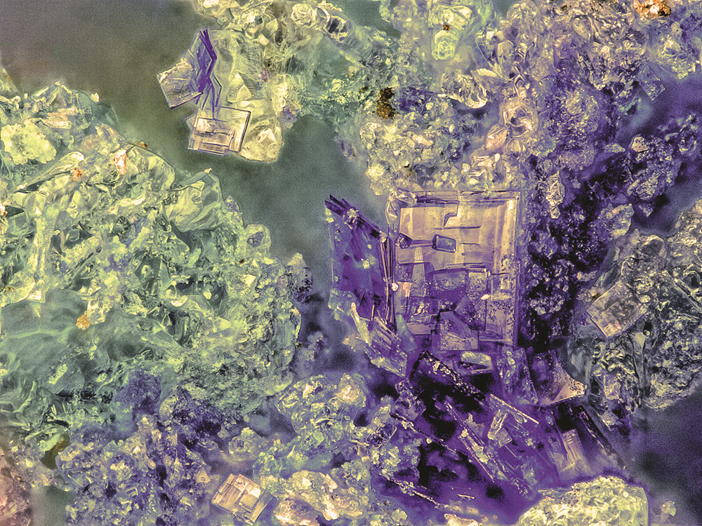
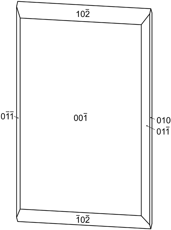
Pabellóndepicaite occurs as rectangular blades or tablets, up to ∼0.2 mm long(Fig. 1). Crystals are flattened on {001}, elongated along [100] and exhibit the forms {010}, {001}, {011} and {102} (Fig.2). The mineral is indigo blue and transparent with light indigo-blue streak. The mineral does not fluoresce in long- or short-wave ultraviolet light. The Mohs hardness is ∼2½ based on scratch tests. Crystals are brittle with curved and stepped fracture. Cleavage is perfect on {001} and good on {010}. The density measured by flotation in a mixture of methylene iodide and toluene is 1.95(2) g·cm–3. The calculated density is 1.980 g·cm–3 for the empirical formula and 1.959 g·cm–3 for the ideal formula, in both cases using the single-crystal cell. At room temperature, the mineral is insoluble in H2O and dissolves slowly in dilute HCl.
Indigo pabellóndepicaite crystals on green crystals of salammoniac (probably coloured by inclusions of antipinite) on holotype specimen 76305; the field of view is 0.68 mm across.

Crystal drawing of pabellóndepicaite; clinographic projection in nonstandard orientation, a vertical. The figure was created using SHAPE, version 7.4 (Shape Software, Kingsport, Tennessee, USA).

Optically, pabellóndepicaite is biaxial (+) with α = 1.583(3), β = 1.596(3) and γ = 1.644(3) measured in white light. The 2V measured directly on a spindle stage is 56(2)°; the calculated 2V is 56.3°. Dispersion is r > v distinct and the optical orientation is X = a, Y = b and Z = c. The mineral is pleochroic: X light indigo blue, Y indigo blue, Z pale indigo blue; Y > X > Z. The Gladstone–Dale compatibility could not be calculated because coefficients for NH3 and N3C2H2– groups are unknown.
Raman spectroscopy
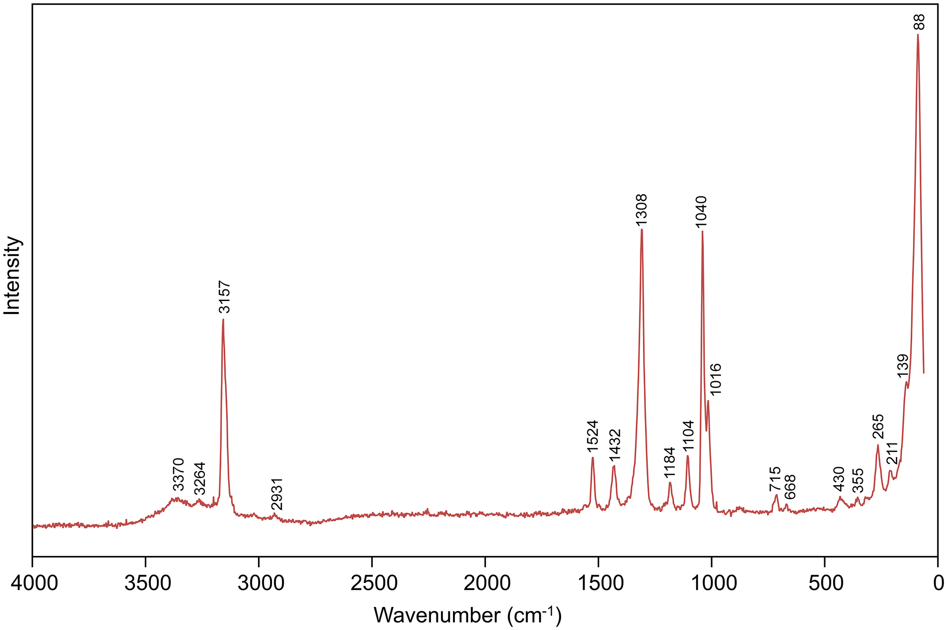
Raman spectroscopy was done on a Horiba XploRa PLUS micro-Raman spectrometer using an incident wavelength of 532 nm, 0.2mW power, a laser slit of 200 μm, 1200 gr/mm diffraction grating and a 100x (0.9 NA) objective. The spectrum from 4000 to 60cm–1 is shown inFig. 3.
Raman spectrum of pabellóndepicaite recorded with a 532 nm laser.

The spectrum exhibits similarities to that of bojarite, Cu3(N3C2H2)3(OH)Cl2·6H2O (Chukanov etal., Reference Chukanov, Möhn, Zubkova, Ksenofontov, Pekov, Agakhanov, Britvin and Desor2020b), except that bands assignable to nitrate are observed in the pabellóndepicaite spectrum. Bands between 3400 and 3200 cm–1 are attributed to O–H and N–H stretching. The band at 3157 cm–1 is due to C–H stretching with the C atom belonging to the system of conjugated bonds of the triazole ring. The very weak band at 2931 cm–1 may correspond to C–H stretching vibrations of an extraneous hydrocarbon impurity. Most of the bands between 1524 and 1016cm–1 are related to the in-plane stretching and mixed vibrations of the 1,2,4-triazolate ring; however, the band at 1040 cm–1 is assignable to the symmetric stretching of the nitrate group. The band at 715 cm–1 can be assigned to the out-of-plane bending of the nitrate group and that at 668 cm–1 to the out-of-plane bending vibrations of the 1,2,4-triazolate ring. The bands at wavenumbers below 500 cm–1 are assigned to lattice modes, largely related to Cu–O, Cu–N and Cu–Cl stretching vibrations.
Composition
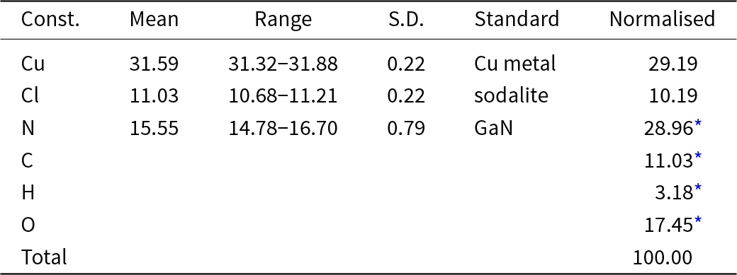
Analyses (five points) were performed at Caltech on a JEOL JXA-iHP200F field-emission electron microprobe in WDS mode. Analytical conditions were 15 kV accelerating voltage, 5 nA beam current and 10 μm beam diameter. A time-dependent intensity calibration was applied; however, significant instability of the mineral led to the loss of much of the volatile content under vacuum and in the beam. This is certainly the cause of much lower than expected N values. Analyses in EDS mode under milder conditions provided significantly higher N values (∼22 wt.%), but still below the amount predicted by the structure determination. It is worth noting that many of the phases found in the guano deposit at Pabellón de Pica are notorious for their instabilities. The very small amount of material available precluded the determination of N, C and H by CHN analysis or other techniques, so these elements are calculated based on the structure. The volatile loss resulted in higher-than-expected concentrations for Cu and Cl; therefore, they have been normalised to provide a total of 100% when combined with the calculated constituents. The cause of the excess Cl relative to Cu is unclear; however, a similar excess was reported by Chukanov etal. (Reference Chukanov, Möhn, Zubkova, Ksenofontov, Pekov, Agakhanov, Britvin and Desor2020b) in the analyses of bojarite. No other elements with an atomic number larger than 8 were detected. Analytical data are given inTable 1.
Analytical data (in wt.%) for pabellóndepicaite

* Based on the structure (Cu = 2).
S.D. – standard deviation.
The empirical formula based on the formula Cu2+2(N3C2H2)2(NH3)2(NO3)Cl·2H2O obtained from the structure refinement (see below) is Cu2+2Cl1.25N9C4H13.75O4.75. Note that it is not clear where the excess 0.25 Cl apfu is accommodated in the structure or even if it may be an artefact resulting from the deterioration of the sample during analysis. Assuming that the excess Cl replaces H2O, we have chosen to reduce the H2O by 0.25 and add back 0.25 H for charge balance. The ideal formula is Cu2+2(N3C2H2)2(NH3)2(NO3)Cl·2H2O, which requires Cu 29.50, Cl 8.23, C 11.15, N 29.26, H 3.28, O 18.57, total 100wt.%.
X-ray crystallography and structure refinement
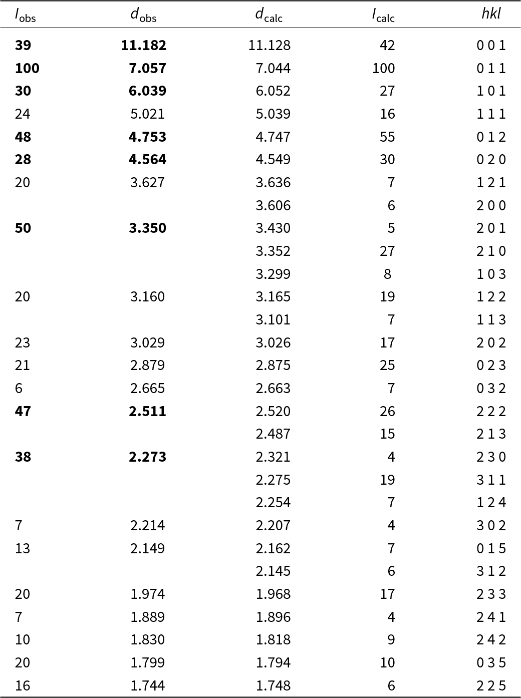
Powder X-ray diffraction data for the holotype were recorded using a Rigaku R-Axis Rapid II curved imaging plate microdiffractometer with monochromatised MoKα radiation. A Gandolfi-like motion on the φ and ω axes was used to randomise the sample. Observed d values and intensities were derived by profile fitting using JADE Pro software (Materials Data, Inc.). The powder data are presented in Table2. The unit-cell parameters refined from the powder data using JADE Pro with whole-pattern fitting (space group Pmma) are a = 7.192(6), b = 9.095(9), c = 11.111(10) Å and V = 726.8(14) Å3.
Powder X-ray diffraction data (d in Å) for pabellóndepicaite

Note: Only calculated lines with intensities > 3.5 are listed. The strongest lines are in bold.
Single-crystal X-ray studies were done on the same diffractometer and radiation noted above. Pabellóndepicaite crystals do not diffract strongly and suffer from somewhat high mosaicity (including some satellite reflections along the c axis that were included in the integration). This severely limited the data collection range and consequently provided a relatively low data to parameter ratio.
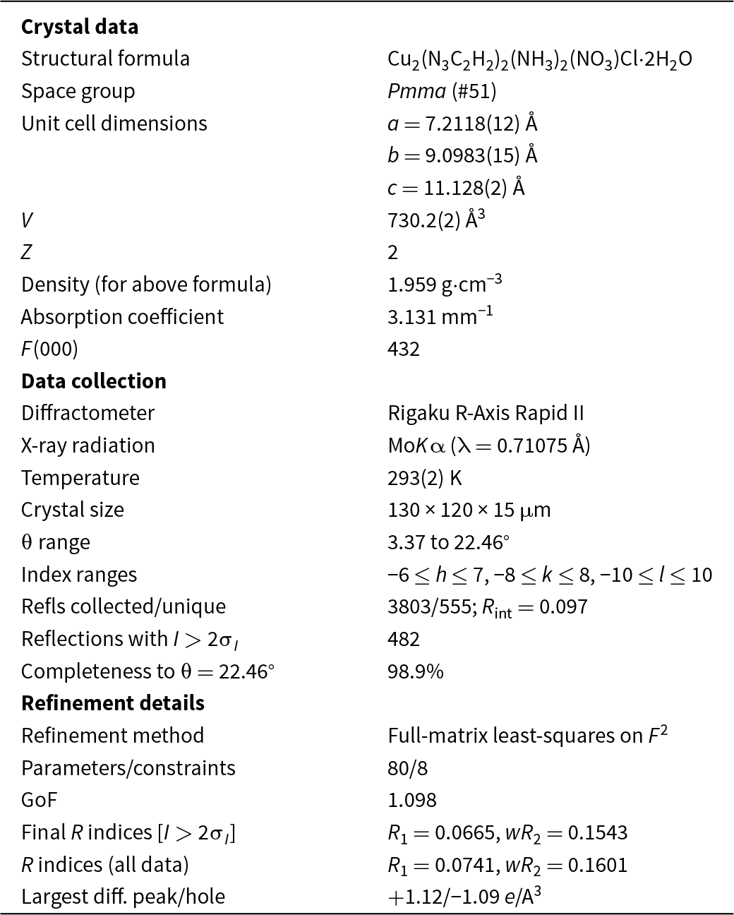
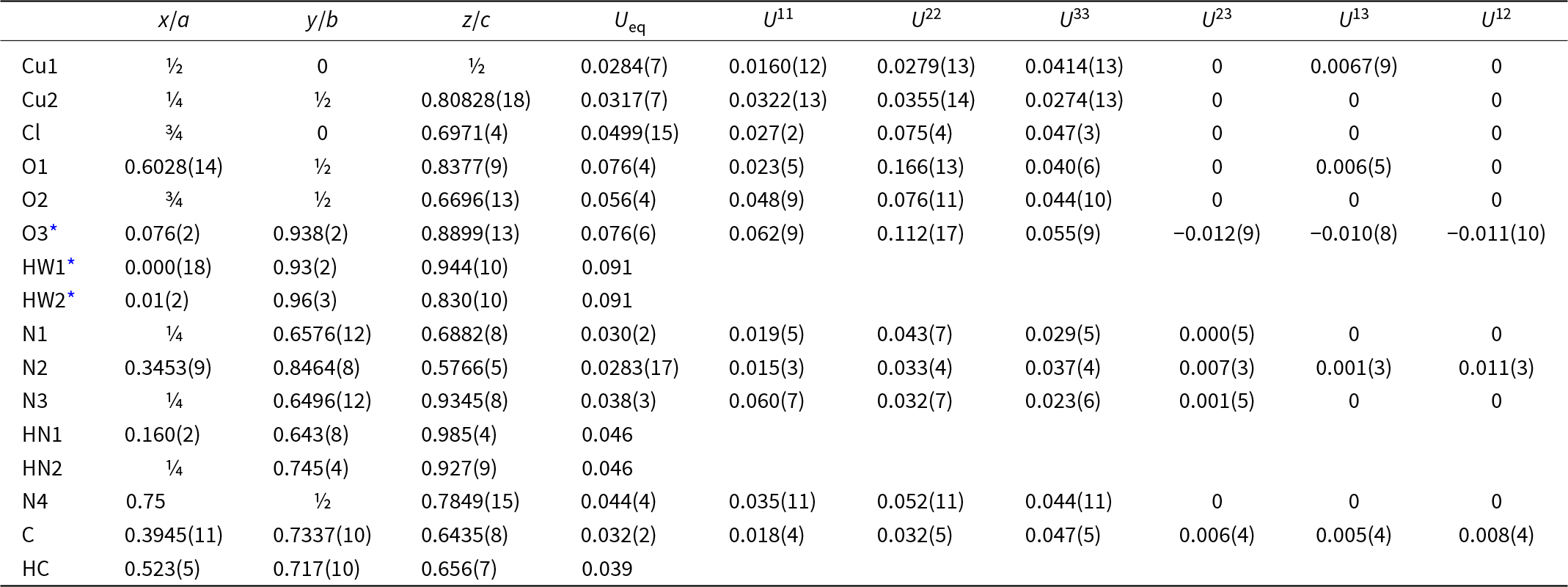
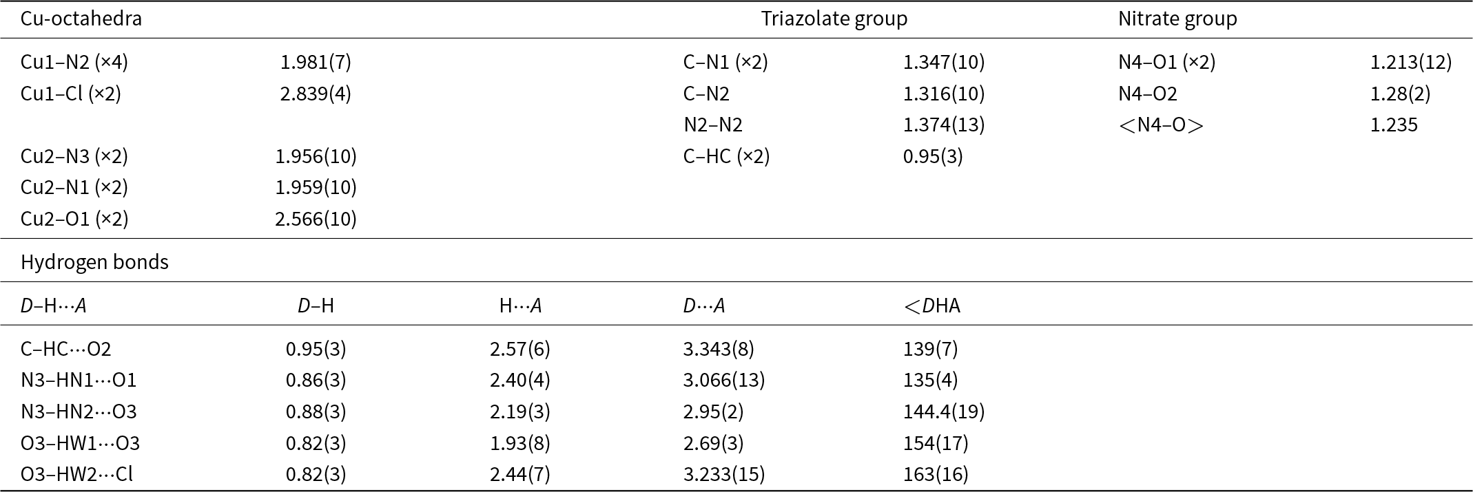
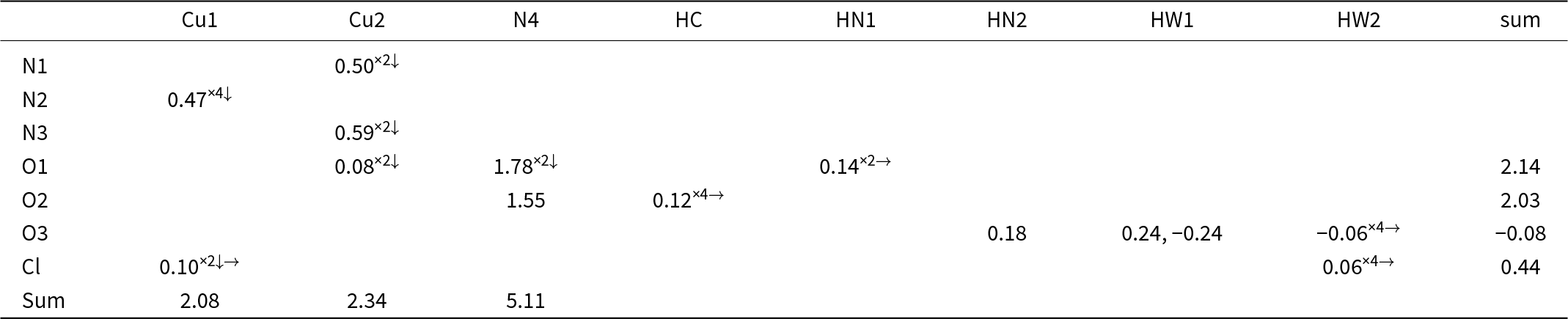
The Rigaku CrystalClear software package was used for processing the structure data, including the application of an empirical absorption correction using the multi-scan method with ABSCOR (Higashi, Reference Higashi2001). The structure was solved using the intrinsic-phasing algorithm of SHELXT (Sheldrick, Reference Sheldrick2015a). SHELXL-2016 (Sheldrick, Reference Sheldrick2015b) was used for the refinement of the structure. The structure solution located all nonhydrogen sites; however, the adjustments had to be made in the assignments of C, N and O sites. The O3 site (H2O) was modelled as split into two half-occupied sites 1.13(4) Å apart. All nonhydrogen sites were refined successfully with anisotropic displacement parameters. Difference-Fourier mapping located all H sites, including those associated with the O3 site. The H sites were refined with the distance restraints C–H 0.95(3), N–H 0.91(3), O–H 0.82(3) and H–H 1.30(3) Å and with U isoH = 1.2U eq(C/N/O). The data collection and refinement details are given inTable 3, atom coordinates and displacement parameters inTable 4, selected bond distances inTable5 and a bond-valence analysis inTable 6. The crystallographic information file has been deposited with the Principal Editor of Mineralogical Magazine and is available as Supplementary material (see below).
Data collection and structure refinement details for pabellóndepicaite

R int = Σ|F o2–F o2(mean)|/Σ[F o2]. GoF = S = {Σ[w(F o2–F c2)2]/(n–p)}1/2. R 1 = Σ||F o|–|F c||/Σ|F o|. wR 2 = {Σ[w(F o2–F c2)2]/Σ[w(F o2)2]}1/2; w = 1/[σ2(F o2)+(aP)2+bP] where a is 0.0633, b is 8.8077 and P is [2F c2+Max(F o2,0)]/3.
Atom coordinates and displacement parameters (Å2) for pabellóndepicaite

* The O3, HW1 and HW2 sites are half-occupied.
Selected bond distances (Å) and angles (°) for pabellóndepicaite

Bond-valence analysis for pabellóndepicaite. Values are in valence units (vu)

The sources for bond-valence parameters are as follows: Cu2+–N3-: Shields etal. (Reference Shields, Raithby, Allen and Motherwell2000); Cu2+–Cl: Brese and O’Keeffe (Reference Brese and O’Keeffe1991); Cu2+–O and N5+–O: Gagné and Hawthorne (Reference Gagné and F.C2015); H···O: Brown (Reference Brown2002); H···Cl: Malcherek and Schlueter (Reference Malcherek and Schlüter2007). Bonds within the triazolate group are not included. Negative values indicate donated bond-valences.
Description of the structure
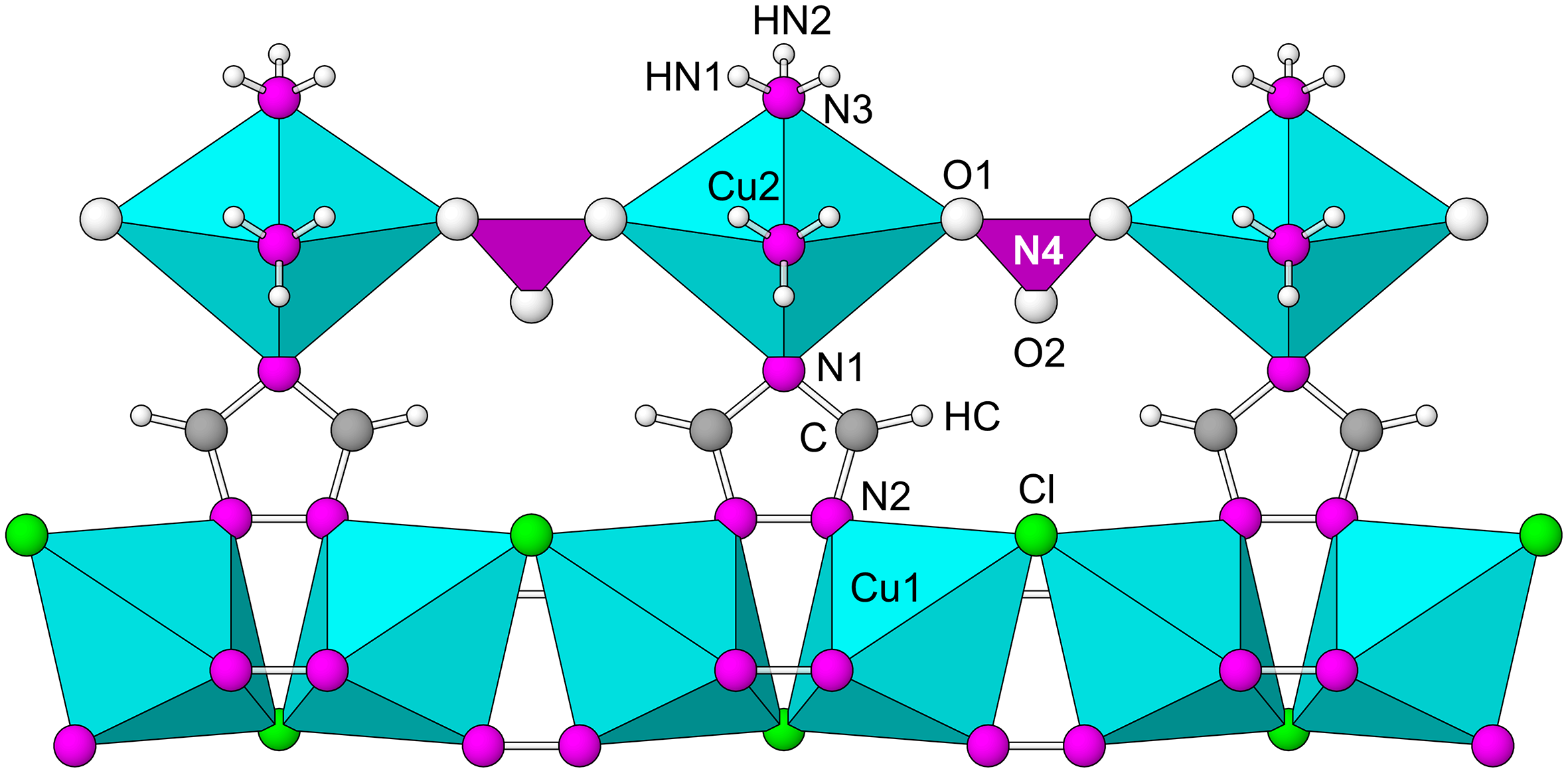
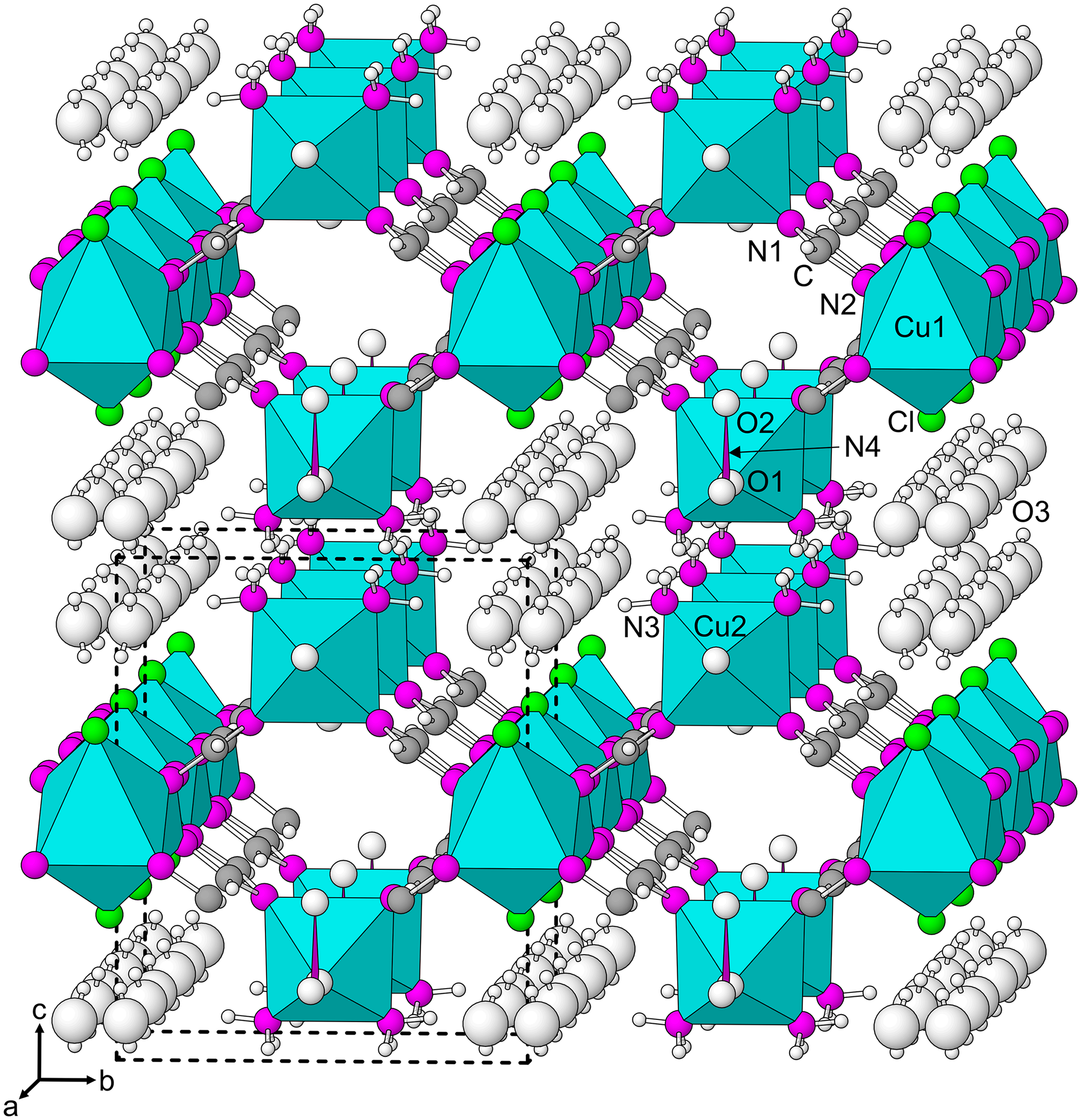
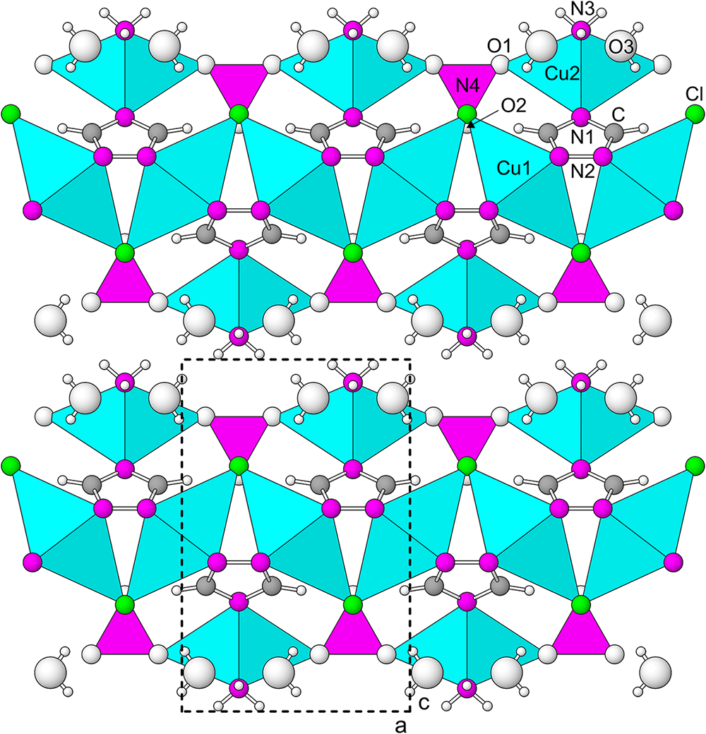
The structure of pabellóndepicaite contains two types of polyhedral chains. CuN4Cl2 octahedra are linked by sharing Cl atoms as well as the N–N edges of triazolate groups to form a zig-zag chain along [100]. This chain is the same as the one in the triazolite structure. A second chain, also along [100], is formed by CuN4O2 octahedra alternating with NO3 triangles. The two types of chains are linked to one another by triazolate groups(Fig. 4). The propagation of this linkage forms layers parallel to {001} with isolated H2O (O3) groups in cavities in the layers(Figs 5 and6). Both the CuN4Cl2 and CuN4O2 octahedra exhibit pronounced 4 + 2 distortion due to the Jahn-Teller effect.
Fragment of the pabellóndepicaite structure showing the two types of polyhedral chains linked together by triazolate rings. The view is down [01 $\overline{1}$] with [100] horizontal.

The structure of pabellóndepicaite viewed slightly canted along [100]. The unit-cell outline is shown by dashed lines. The figures were created using ATOMS, version 6.5 (Shape Software, Kingsport, Tennessee, USA).

The structure of pabellóndepicaite viewed along [010]. The unit-cell outline is shown by dashed lines. The figure was drawn using ATOMS, version 6.5 (Shape Software, Kingsport, Tennessee, USA).

Discussion
Although guano is rich in nitrogen, it does not generally contain azole compounds and specifically compounds containing the 1,2,4-triazole ring. It is likely that the occurrence of 1,2,4-triazolate in minerals, which has only been documented at the Pabellón de Pica guano deposit, is the result of the interaction of N released from the guano with Cu released from the chalcopyrite-bearing gabbro on which the guano has been deposited. Support for this comes from synthetic studies in which Cu2+ has been used to promote (mediate or catalyse) the formation of 1,2,4-triazoles from other N-bearing compounds (e.g. Kuang etal., Reference Kuang, Chen and Ma2014; Romero etal., Reference Romero, Sojo, Arvelo, Calderón, Morales and S.E2020). The affinity of Cu2+ for N is obvious in the structure of pabellóndepicaite, as well as the other 1,2,4-triazolate minerals at Pabellón de Pica, in that all short Cu-ligand bonds in these structures are to N atoms (1,2,4-triazolate and NH3). Furthermore, Haasnoot (Reference Haasnoot2000) notes that Cu2+ shows high affinity for the triazolate ligand.
Based on pseudomorphic relationships, Chukanov etal. (Reference Chukanov, Möhn, Zubkova, Ksenofontov, Pekov, Agakhanov, Britvin and Desor2020b) noted that the other three 1,2,4-triazolate minerals form a transformation series: triazolite NaCu2+2(N3C2H2)2(NH3)2Cl3⋅4H2O → chanabayaite, Cu2+2Cl(N3C2H2)2(NH3,Cl,H2O,□)4 → bojarite Cu2+3(N3C2H2)3(OH)Cl2⋅6H2O, each step involving the loss of one or more components. The presence of bojarite (as an apparent alteration product of chanabayaite and/or triazolite) on the holotype of pabellóndepicaite, which occurs as unaltered crystals, indicates that pabellóndepicaite is not related to this transformation series. Notably, pabellóndepicaite is very rare, having been found on a single specimen, whereas triazolite and chanabayaite are relatively abundant at Pabellón de Pica. Yet triazolite and chanabayaite break down in air with the loss of NH3, whereas pabellóndepicaite is stable. The rarity of pabellóndepicaite may be due the required presence of NO3– during its formation and the NO3– group in the structural unit of pabellóndepicaite presumably contributes to its greater stability.
Supplementary material
The supplementary material for this article can be found at https://doi.org/10.1180/mgm.2024.55.
Acknowledgements
Nikita Chukanov, structures editor Peter Leverett and an anonymous reviewer are thanked for their constructive comments on the manuscript. The EPMA was carried out at the Caltech GPS Division Analytical Facility, which is supported, in part, by NSF Grant EAR-2117942. A portion of this study was funded by the John Jago Trelawney Endowment to the Mineral Sciences Department of the Natural History Museum of Los Angeles County.
Competing interests
The authors declare none.