Introduction
Beck (Reference Beck1967) postulated that early detrimental life events could lead to the development of negative schemas which include themes of loss, failure and abandonment. Dysfunctional attitudes, such as ‘if I fail partly, it is as good as being a complete failure’, are activated during stressful life events and are characteristic of a depressive episode (Haaga et al. Reference Haaga, Dyck and Ernst1991). It has been proposed that depressive symptoms are promoted by dysfunctional attitudes (Sheppard & Teasdale, Reference Sheppard and Teasdale2000) in a reciprocal causal relationship (Burns & Spangler, Reference Burns and Spangler2001). In support, positive associations between depression severity and dysfunctional attitudes have been observed (Beevers et al. Reference Beevers, Keitner, Ryan and Miller2003), which revert to normal during remission (Haaga et al. Reference Haaga, Dyck and Ernst1991), and the magnitude of dysfunctional thinking during a dysphoric mood state is predictive of a subsequent depressive relapse (Segal et al. Reference Segal, Kennedy, Gemar, Hood, Pedersen and Buis2006).
High levels of dysfunctional thinking during a depressive episode have been associated with greater 5-HT2 receptor binding potential in the anterior cingulate, prefrontal regions, thalamus, caudate and putamen (Meyer et al. Reference Meyer, Houle, Sagrati, Carella, Hussey, Ginovart, Goulding, Kennedy and Wilson2004). Administration of the serotonin agonist d-fenfluramine led to a reduction in dysfunctional attitudes, suggesting that serotonin agonism can reduce dysfunctional attitudes by inducing neuronal release of serotonin in depression (Meyer et al. Reference Meyer, McMain, Kennedy, Korman, Brown, DaSilva, Wilson, Blak, Eynan-Harve and Goulding2003). Although a correlation with receptor binding potential and attributions has been observed, subjects were not actively engaged in a dysfunctional attitudes task during the brain scan.
An aim of cognitive–behavioural therapy (CBT) is to address dysfunctional attitudes that contribute to the persistence of depressive symptoms (Dobson & Dozois, Reference Dobson, Dozois and Dobson2001). Following treatment with CBT, increased activity has been noted in the anterior cingulate during a resting state (Goldapple et al. Reference Goldapple, Segal, Garson, Lau, Bieling, Kennedy and Mayberg2004), in response to sad faces (Fu et al. Reference Fu, Williams, Cleare, Scott, Mitterschiffthaler, Walsh, Donaldson, Suckling, Andrew and Steiner2008), and with self-referential processing to positive stimuli though not to negative stimuli (Yoshimura et al. Reference Yoshimura, Okamoto, Onoda, Matsunaga, Okada, Kunisato, Yoshino, Ueda, Suzuki and Yamawaki2014). Additional neural correlates of CBT in depression include normalization of amygdala activity to sad facial expressions (Fu et al. Reference Fu, Williams, Cleare, Scott, Mitterschiffthaler, Walsh, Donaldson, Suckling, Andrew and Steiner2008), increases in ventromedial cortical activity (Ritchey et al. Reference Ritchey, Dolcos, Eddington, Strauman and Cabeza2011), decreases in dorsal frontal cortical activity (Kennedy et al. Reference Kennedy, Konarski, Segal, Lau, Bieling, McIntyre and Mayberg2007), and increases in hippocampal activity during a resting state (Goldapple et al. Reference Goldapple, Segal, Garson, Lau, Bieling, Kennedy and Mayberg2004). The changes in prefrontal, limbic and subcortical activity are generally consistent with models of neurocognitive circuits in depression and the effects of CBT (DeRubeis et al. Reference DeRubeis, Siegle and Hollon2008).
However, the brain regions engaged by dysfunctional thinking in depression and the effects of CBT have not been examined. In the present study, we sought to investigate the neural correlates of dysfunctional attitudes in patients with depression during an acute depressive episode and following treatment with CBT. We expected that patients would show greater endorsement of dysfunctional attitudes during an acute depressive episode, which we expected would improve following treatment with CBT. We hypothesized that major depressive disorder (MDD) patients would show greater activation in the anterior cingulate and regions associated with attention and self-referential processing with extreme attributions relative to healthy controls. We expected to observe increased activity in regions associated with attentional processing of negative stimuli in patients during an acute depressive episode which would resolve following CBT, including increased activity in the amygdala which would normalize following CBT.
Method
Participants
All participants were right handed and fluent in English. Participants were recruited through local newspaper advertisements. The study was approved by the Institute of Psychiatry and South London and Maudsley (SLaM) National Health Services (NHS) Ethics Research Committee, and all participants provided written, informed consent. All procedures contributing to this work comply with the ethical standards of the relevant national and institutional committees on human experimentation and with the Helsinki Declaration of 1975, as revised in 2008.
The patient group consisted of 16 participants [13 women, mean age 40.00 years (s.d. = 9.27)] who met criteria for MDD by the Structured Clinical Interview for DSM-IV (First & Gibbon, Reference First and Gibbon1997) and a clinical interview with a consultant psychiatrist. Inclusion criteria were an acute episode of MDD, unipolar subtype and a score of a minimum of 18 on the 17-item Hamilton Rating Scale for Depression (HAMD; Hamilton, Reference Hamilton1960). The exclusion criteria were a current neurological disorder, history of neurological trauma resulting in a loss of consciousness, history of diabetes or medical disorder, other Axis I disorders including anxiety disorder or history of substance abuse within 2 months of participation in the study. All patients were free of psychotropic medications for a minimum of 4 weeks at the time of recruitment (8 weeks for fluoxetine) and remained medication free throughout the treatment. HAMD score was measured at baseline and following the course of CBT at the end of 16 weeks.
Healthy controls were 16 age-, sex- and intelligence quotient (IQ)-matched healthy participants [13 women, mean age 39.94 years (s.d. = 9.48)] with HAMD scores less than 8 and no history of previous psychiatric illness, neurological disorder or head injury resulting in a loss of consciousness. All healthy controls were free of psychotropic medications. HAMD score was measured at baseline and at the end of 16 weeks.
Dysfunctional Attitude Scale (DAS)
The DAS measures pervasive negative attitudes towards the self, the world and the future. In the present study, the DAS-24 (Power et al. Reference Power, Katz, McGuffin and Duggan1994) was used, which is a shortened version of the DAS (Weissman & Beck, Reference Weissman and Beck1978) consisting of 24 statements. Also, 24 neutral statements were included as a control task for the present study, which we have called the control DAS (cDAS). We have termed the 48-item scale as the ‘modified Dysfunctional Attitude Scale’ (mDAS-48).
During the functional magnetic resonance imaging (fMRI) scan, participants were presented with the mDAS-48 task consisting of statements alternating from the DAS and cDAS. Subjects were asked to respond to each statement using seven-point Likert scales, ranging from totally agree to totally disagree. Extreme responses are a reflection of the endorsement of dysfunctional attitudes (Power et al. Reference Power, Katz, McGuffin and Duggan1994). The fMRI task began with either a DAS or cDAS statement which was presented in a counterbalanced order for consecutive participants, and the same version was used for the same participant. fMRI scans were acquired at baseline (week 0) and upon study completion (week 16). Each MRI scan was up to 1.5 h in duration consisting of fMRI tasks and structural MRI scans, and data from an affective facial processing task have been presented (Fu et al. Reference Fu, Williams, Cleare, Scott, Mitterschiffthaler, Walsh, Donaldson, Suckling, Andrew and Steiner2008).
All behavioural data were recorded during the fMRI scans and analysed using SPSS (version: PASW Statistics 18). Repeated-measures analysis of variance (ANOVA) was used to analyse the main effect of group (patients v. controls), main effect of statement (DAS v. cDAS), main effect of time (week 0 v. week 16) and group × time interactions (i.e. changes in response between baseline and final trials). Percentage change in extreme attributions (total number of extreme DAS scores at week 16 − total number of extreme DAS scores at baseline/total number of extreme DAS scores at baseline × 100) was also calculated for each subject.
CBT treatment
Patients received 16 sessions of CBT with experienced therapists (Fu et al. Reference Fu, Williams, Cleare, Scott, Mitterschiffthaler, Walsh, Donaldson, Suckling, Andrew and Steiner2008). The standard CBT procedures as described by Beck et al. (Reference Beck, Shaw, Rush and Emery1979) were followed, and all therapists met the required level of training and proficiency (Paykel et al. Reference Paykel, Scott, Teasdale, Johnson, Garland, Moore, Jenaway, Cornwall, Hayhurst and Abbott1999). The CBT sessions were audiotaped and reviewed to ensure adherence and competence. HAMD scores were obtained from patients at baseline and after 16 weeks of CBT. We defined treatment response as a minimum reduction of 50% in HAMD score from baseline.
Image acquisition
Gradient echo single-shot echoplanar imaging data were acquired on a neuro-optimized 1.5T IGE LX system (USA) at the Maudsley Hospital, London. A total of 441 T2* weighted images depicting blood oxygenation level-dependent (BOLD) contrast were acquired over 27 min. For each volume, 22 near-axial non-contiguous 3 mm planes parallel to the intercommissural plane; time to echo (TE) = 40 ms; repetition time (TR) = 3.74 s; in-plane resolution = 3.75 mm; interslice gap 0.3 mm; and matrix size 64 × 64 voxels. Four dummy acquisitions were acquired at the beginning of each scan to allow magnetization to reach equilibrium amplitude.
fMRI data analysis
fMRI data analysis was conducted using XBAM software (version 4.1; Institute of Psychiatry, King's College London). Images were first realigned to minimize subject motion and then smoothed using a Gaussian filter (full-width half-maximum = 7.2 mm). Responses to the experimental paradigms were detected by carrying out time-series analysis using two gamma variate functions with peak responses at 4 and 8 s, respectively. The best fit between the weighted sum of these and the time series at each voxel was computed with a goodness of fit at each voxel. The ratio of the sum of squares (SSQ) of deviations from the mean image intensity due to the model over the whole time series to the SSQ of deviations due to the residuals was computed, termed the SSQ ratio. The data were then permuted by a wavelet-based method which permits the calculation of the null distribution of SSQ ratios under the assumption of no experimentally determined response. This distribution was used to calculate the SSQ ratio value and to find the threshold for the activation maps at type I error rate of less than one voxel. The SSQ ratio data for each individual were transformed into standard space of Talairach & Tournoux (Reference Talairach and Tournoux1988).
Group activation maps were computed using the median SSQ ratio at each voxel in the observed and permuted maps. Permutation methods and median statistics were used to obtain the null distribution of SSQ ratios and as well as the critical SSQ ratio to threshold group activation maps at a cluster-level threshold of less than one expected type I error cluster per brain. For the present group analysis, less than one false-positive cluster was expected at p < 0.05 for voxel level and p < 0.01 at cluster level. Only those voxels at which all subjects contributed data were included for analysis (Fu et al. Reference Fu, Williams, Cleare, Scott, Mitterschiffthaler, Walsh, Donaldson, Suckling, Andrew and Steiner2008).
In order to examine the neural correlates of dysfunctional attributions, the fMRI time series corresponding to attributions that corresponded to endorsements of 1, 2, 6 or 7 on the Likert scale were encoded. The fMRI time series associated with regular attributions were encoded by Likert scale responses of 3, 4 or 5. We employed a 2 × 2 ANOVA to examine the main effect of group (patients v. healthy controls across both time points), main effect of time (week 0 v. week 16) and the group × time interaction. The analyses were examined for regular attributions made to DAS relative to control DAS statements and for extreme attributions made to DAS relative to control DAS statements.
Results
Demographics
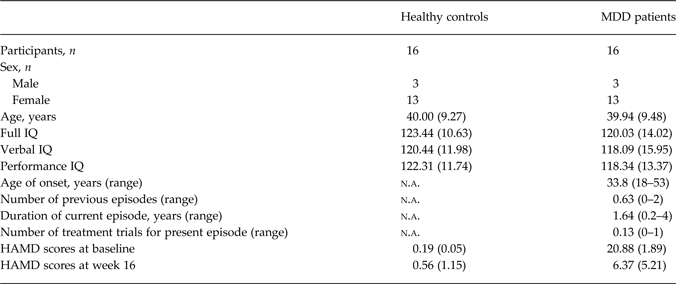
There were no significant group differences in mean age, full IQ, verbal IQ and performance IQ (all p > 0.05) (Table 1). All patients completed a full course of 16 weeks of CBT. There was an expected significant difference in HAMD scores between the groups at week 0 (F 1,30 = 1765.21, p < 0.001) and at week 16 (F 1,30 = 18.96, p < 0.001). Patients showed a significant reduction in mean HAMD scores from baseline to week 16 (F 1,15 = 118.45, p < 0.001).
Demographic and clinical characteristics
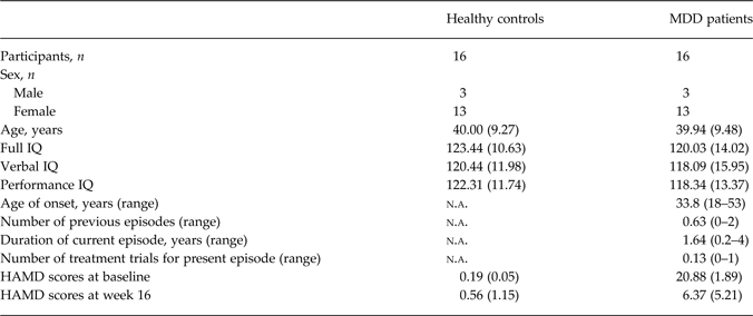
Data are given as mean (standard deviation) unless otherwise specified.
MDD, Major depressive disorder; IQ, intelligence quotient; n.a., not applicable; HAMD, Hamilton Rating Scale for Depression.
Behavioural data
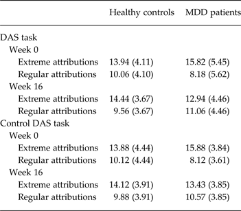
The extreme responses to the DAS statements showed a significant group × time interaction effect (F 1,30 = 7.434, p = 0.011), in which patients showed a significant reduction in mean number of extreme responses following a course of CBT [t = 2.938, degrees of freedom (df) = 15, p = 0.010] while healthy controls did not have a change in extreme scores at the follow-up scan as compared with baseline (t = −0.659, df = 15, p = 0.520) (Table 1). There was also a trend towards a significant effect of time (F 1,30 = 3.681, p = 0.065), as both groups showed a reduction in extreme responses at the follow-up scan. There was no significant main effect of group in extreme responses (F1,30 = 0.016, p = 0.900) (Table 2).
Behavioural performance on the DAS task
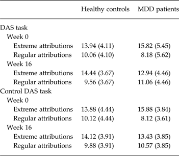
Data are given as mean (standard deviation).
DAS, Dysfunctional Attitudes Scale; MDD, major depressive disorder.
In the control DAS statements, there were no significant main effects of time (F 1,30 = 2.054, p = 0.162), group (F 1,30 = 0.140, p = 0.711) or group × time interaction effects (F 1,30 = 3.343, p = 0.077).
There were no significant correlations between the change in HAMD scores and the change in the number of extreme responses made to the DAS statements in MDD patients (r = 0.465, p > 0.05, one-tailed test). We were also interested in examining the relationship between changes in DAS scores and response to treatment. However, the number of patients who did not respond to treatment (n = 3) was insufficient to compare with those who responded. Hence, we report the mean percentage change in extreme DAS scores in responders (mean = −13.34, s.d. = 33.66) and non-responders (mean = −12.8, s.d. = 15.75).
fMRI results
Neural responses to extreme attributions in DAS
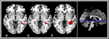
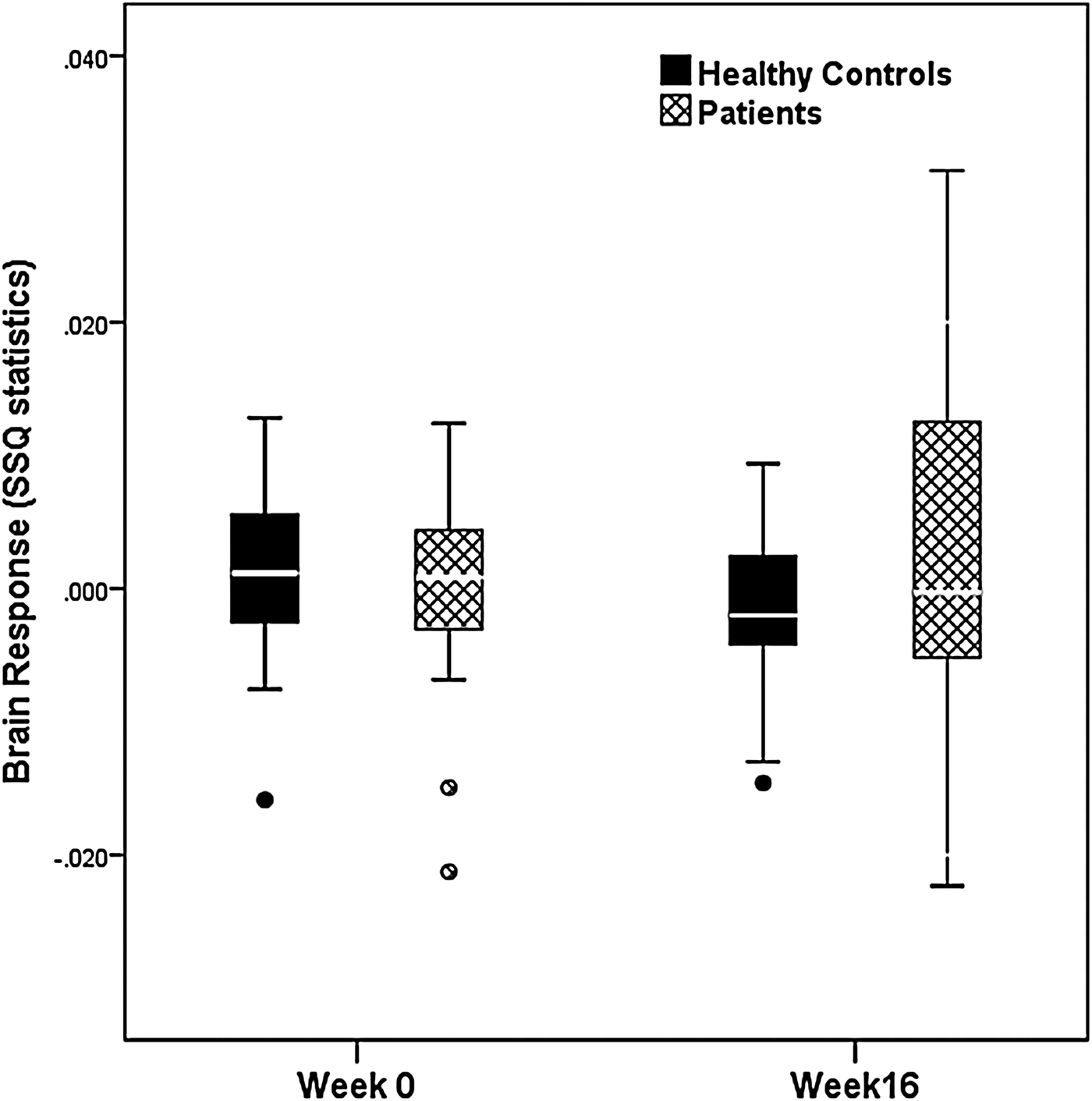
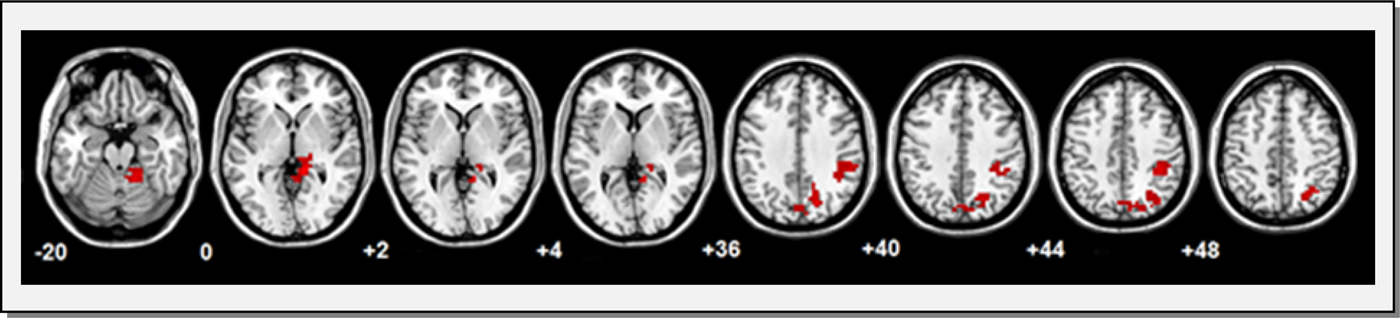
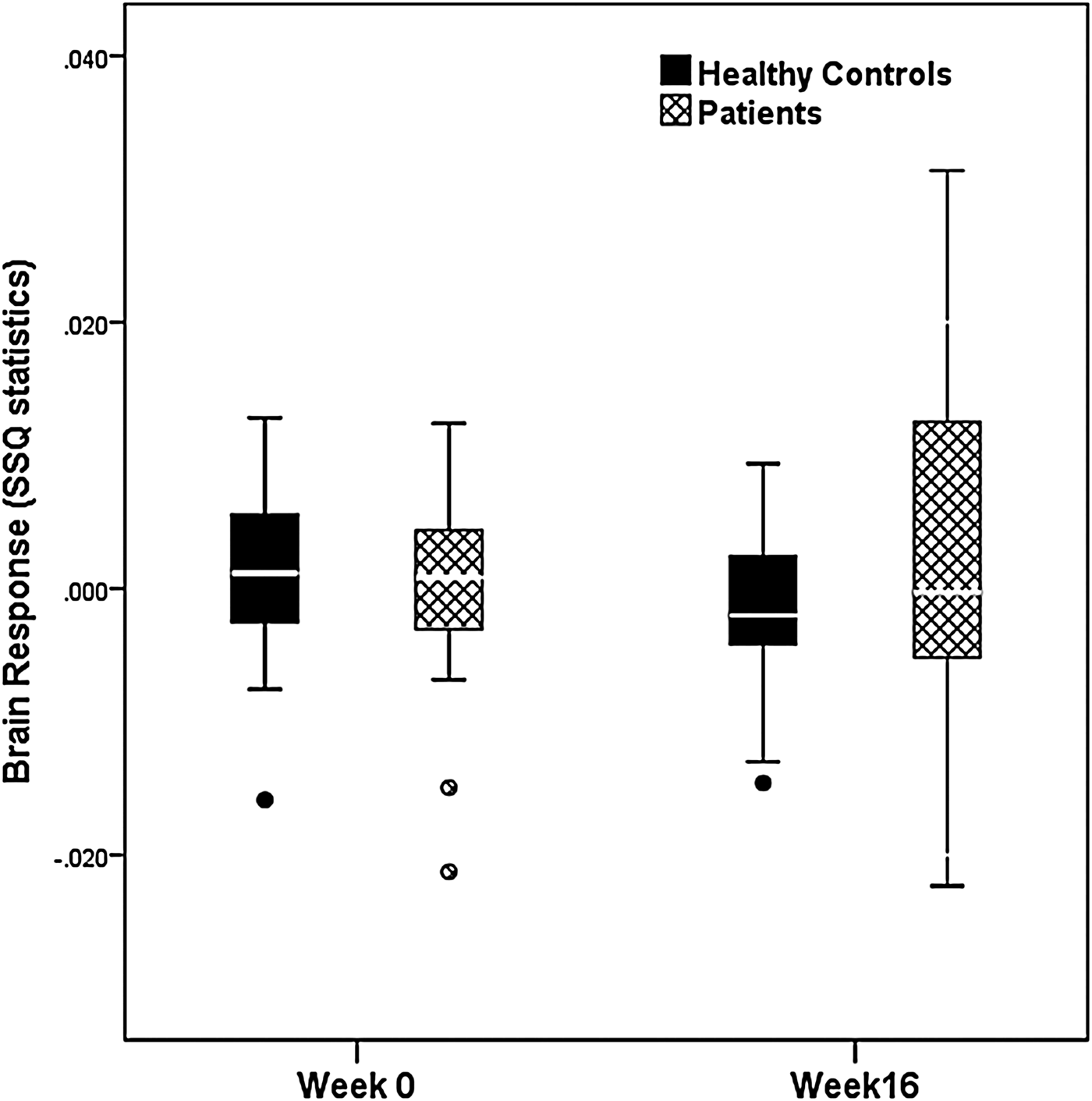
A significant group × time interaction effect for extreme attributions to DAS statements was found in the left parahippocampal gyrus [Brodmann area (BA) 37] (Talairach coordinates: x, y, z = −36, −41, −7, cluster size = 41 voxels, corrected p = 0.0027). This region showed less attenuation in activation in MDD patients as compared with healthy controls at the 16-week scan (Figs 1 and 2).
There was a significant group × time interaction effect in the left parahippocampal region for extreme attributions to Dysfunctional Attitudes Scale statements (corrected p = 0.0027). Both depressed patients and healthy controls showed a decrease in activation in the left parahippocampal gyrus at the follow-up scans but to a lesser extent in patients. Transverse sections of the brain are presented with the Talairach z-coordinates indicated.

The graph presents the group × time interaction effect in the left parahippocampal region. The boxes indicate interquartile range. The horizontal lines in the boxes represent medians. The limit lines indicate ranges excluding outliers, and the circles represent outliers which are defined as points greater than 1.5 times the interquartile range from the limits of the interquartile range. The y-axis sum of squares (SSQ) values represent a normalized statistic of the brain response.
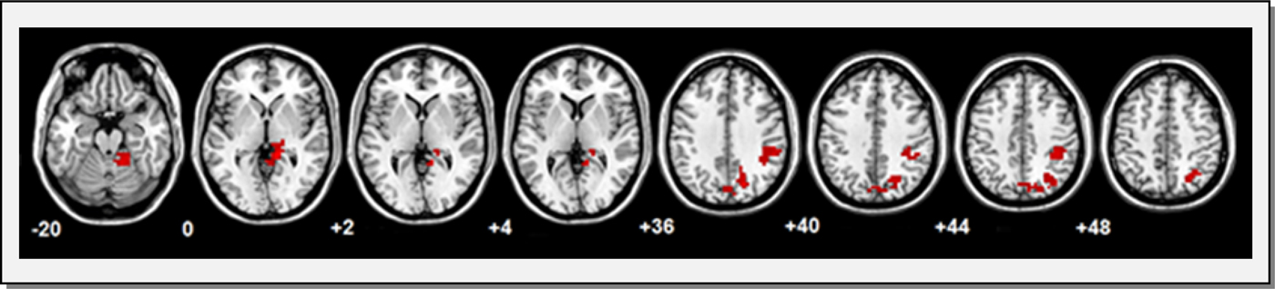
There was a significant main effect of group in which patients showed greater activation in the left hippocampal region (coordinates: x, y, z = −11, −33, −3, cluster size = 27 voxels, corrected p = 0.0016), left inferior parietal lobe (BA 40) (coordinates: x, y, z = −36, −33, 40, cluster size = 55 voxels, corrected p = 0.0013) and left precuneus (BA 7) (coordinates: x, y, z = −14, −67, 33, cluster size = 109 voxels, corrected p = 0.00006) as compared with healthy controls, while in the left cerebellum healthy controls showed greater activation relative to patients (coordinates: x, y, z = −11, −44, −23, cluster size = 45 voxels, corrected p = 0.0016) (Fig. 3).
In the main effect of group, major depressive disorder patients showed significantly greater activation in the left hippocampus (corrected p = 0.0016), left inferior parietal lobe (corrected p = 0.0013) and left precuneus (corrected p = 0.0006), relative to healthy controls. Healthy controls showed a greater activation in left cerebellum (corrected p = 0.0016) compared with depressed patients. Transverse sections of the brain are presented with the Talairach z-coordinates indicated.
In patients, a main effect of time was observed in the right posterior cingulate gyrus (BA 30) (coordinates x, y, z = 11, −44, 23, cluster size = 73 voxels, corrected p = 0.006) which showed decreased activation from week 0 to week 16, while no regions showed greater activation from the initial to the final scan. In healthy controls, no regions showed decreased activation from week 0 to week 16, but there was a significant main effect of time in the left cuneus (BA 18) (coordinates x, y, z = −18, −78, 17, cluster size = 53 voxels, corrected p = 0.005), which showed increased activation from the initial to the final scan.
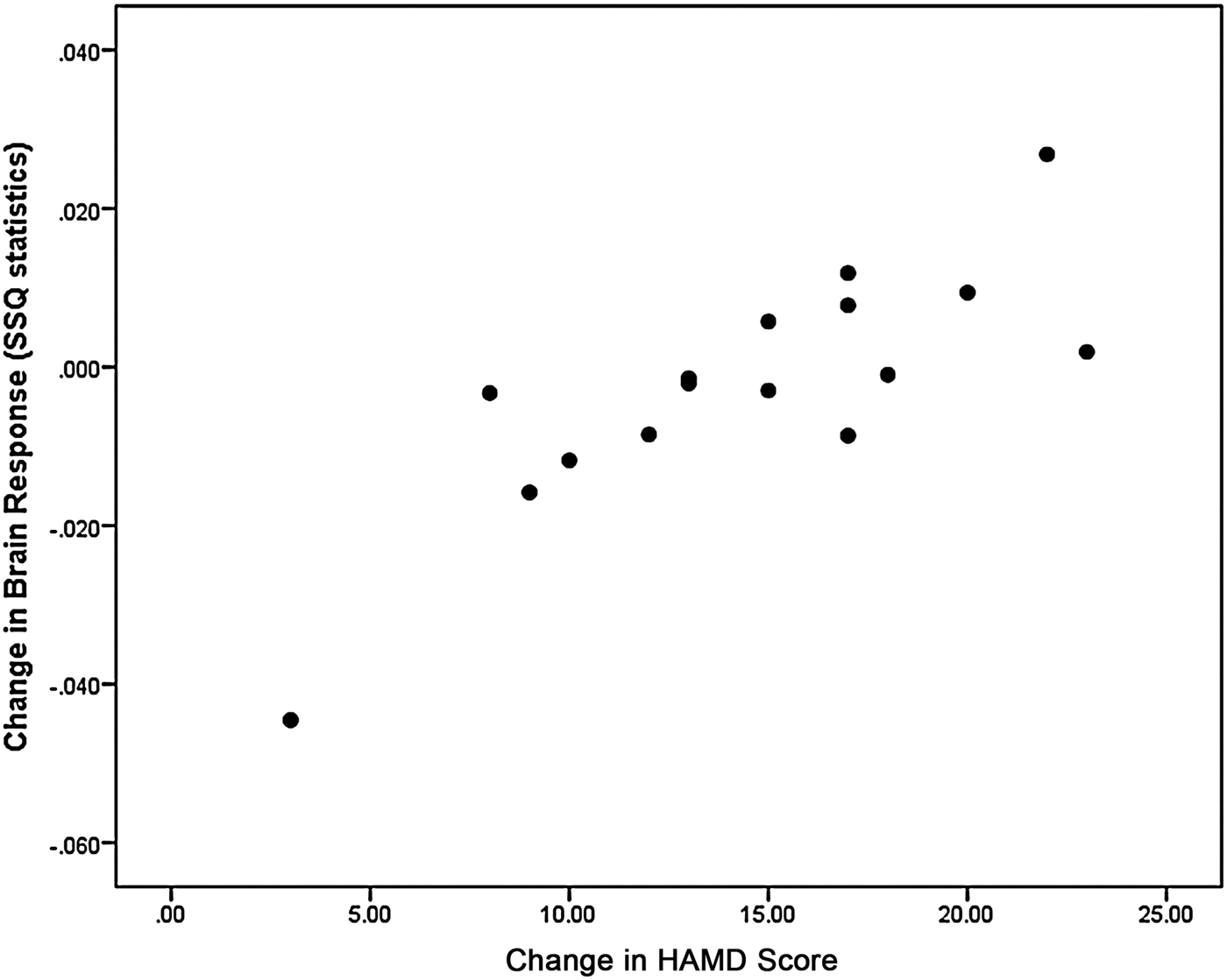
Patients showed a significant positive relationship between changes in HAMD score and overall activity in the left precentral gyrus (BA 6) (coordinates x, y, z = −43, −4, 40; cluster size = 28 voxels, r = 0.739, corrected p = 0.004), in which patients with the greatest improvement in HAMD scores following CBT treatment had the greatest increase in activity in the precentral gyrus (Fig. 4).
A significant correlation was found between the change in the severity of depression as measured by the Hamilton Rating Scale for Depression (HAMD) scores and activity in the left precentral gyrus. Patients who had the greatest change in HAMD score following cognitive–behavioural therapy showed the greatest increase in activity in the left precentral gyrus during processing of dysfunctional attitudes. The y-axis sum of squares (SSQ) values represent a normalized statistic of the brain response.

Neural responses to regular attributions in DAS
There was no significant main effect of group or any group × time interaction effects in the neural responses to regular attributions to the DAS statements.
In patients, no regions showed decreased activation from the initial to final scan, but there was a main effect of time in the left cerebellum (Talairach coordinates x, y, z = −11, −74, −17, cluster size = 26 voxels, corrected p = 0.0025), which showed increased activation from weeks 0 to 16. In healthy controls, main effects of time were observed in the left lingual gyrus (BA 18), left parahippocampal gyrus and bilateral precuneus (BA 7) (corrected p < 0.006), which showed reduced activation from the initial to final scans and in the left inferior frontal gyrus (BA 10) (coordinates x, y, z = −36, 44, 3, cluster size = 36 voxels, corrected p = 0.003) which showed increased activation from the initial to final scans.
Main effects of the DAS task and group are presented in the online Supplementary material.
Discussion
The present study supports a modifying effect of CBT on dysfunctional attitudes (Haaga et al. Reference Haaga, Dyck and Ernst1991; Furlong & Oei, Reference Furlong and Oei2002) as patients endorsed a greater number of extreme responses to DAS statements during an acute depressive episode which normalized following CBT. Dysfunctional attitudes are also seen to reduce following antidepressant treatment (Shankman et al. Reference Shankman, Campbell, Klein, Leon, Arnow, Manber, Keller, Markowitz, Rothbaum and Thase2012), suggesting that they may be a state feature of depression. Parallel decreases in levels of dysfunctional attitudes and the severity of depression following CBT have also been noted (Persons & Burns, Reference Persons and Burns1985), although dysfunctional attitudes have also been observed as a trait feature of depression (Roberts & Gamble, Reference Roberts and Gamble2001). However, we did not observe a correlation between an improvement in depression severity and a reduction in extreme DAS attributions, as all patients showed an improvement in their extreme attributions.
The neural correlates revealed that endorsement of dysfunctional attitudes was associated with left parahippocampal activation in both depressed patients and healthy controls, which decreased at the follow-up scans in both groups but to a lesser extent in patients. The parahippocampal region along with the hippocampus and association areas of the cerebral cortex form the medial temporal lobe system (Eichenbaum & Lipton, Reference Eichenbaum and Lipton2008). There is a bidirectional hierarchy of reciprocal connections in which the cortical association areas connect to the parahippocampal region and in turn to the hippocampus. The output from the hippocampus is then returned to the parahippocampal region and to the cortical regions where the input originated (Eichenbaum & Lipton, Reference Eichenbaum and Lipton2008). The parahippocampal region is associated with contextual associations or episodic memory and shows a familiarity effect during repetition of tasks, with greater activation during novel as compared with familiar tasks (O'Kane et al. Reference O'Kane, Insler and Wagner2005).
Depressed individuals have shown greater activation in the left parahippocampal gyrus relative to controls, during encoding of an associative learning paradigm (Werner et al. Reference Werner, Meindl, Materne, Engel, Huber, Riedel, Reiser and Hennig-Fast2009) and in processing negative pictures (Sheline et al. Reference Sheline, Barch, Price, Rundle, Vaishnavi, Snyder, Mintun, Wang, Coalson and Raichle2009). Reductions in parahippocampal activation have similarly been observed in MDD patient following treatment with antidepressant medication (Kennedy et al. Reference Kennedy, Evans, Krüger, Mayberg, Meyer, McCann, Arifuzzman, Houle and Vaccarino2001; Delaveau et al. Reference Delaveau, Jabourian, Lemogne, Guionnet, Bergouignan and Fossati2011). Behavioural studies of dysfunctional attitudes also show higher endorsement of dysfunctional attitudes by patients relative to controls during negative mood induction (Lau et al. Reference Lau, Haigh, Christensen, Segal and Taube-Schiff2012) and significant improvement in dysfunctional thinking in patients following CBT (Warmerdam et al. Reference Warmerdam, van Straten, Jongsma, Twisk and Cuijpers2010). To date, there has been no fMRI study that has investigated the neural correlates of dysfunctional attitudes in depression, and therefore we cannot make direct comparisons to confirm the role of the parahippocampal gyrus in dysfunctional attitudes. However, left parahippocampal activation seems to be especially associated with negative stimuli (Iidaka et al. Reference Iidaka, Okada, Murata, Omori, Kosaka, Sadato and Yonekura2002; Surguladze et al. Reference Surguladze, Brammer, Keedwell, Giampietro, Young, Travis, Williams and Phillips2005), and activation in this region in both patients and in controls during presentation of DAS statements supports the role of the left parahippocampal gyrus in processing negative information contained in the DAS statements. The reduction in parahippocampal activation at the follow-up scan for both groups is consistent with increased familiarity with repetition of the task, although patients did not demonstrate the same extent in the reduction in activation. This may perhaps reflect patients’ inability to recall the task in the same manner as controls in part due to persistent engagement and contextual associations to the DAS statements.
In the main effect of group across both time points, there was greater activation in a region that encompassed the left hippocampal gyrus, inferior parietal lobe and precuneus in patients relative to healthy controls. The inferior parietal lobe plays a prominent role in attention (Pessoa et al. Reference Pessoa, Kastner and Ungerleider2002), processing of written language (Eckert, Reference Eckert2004), working memory of emotional stimuli (Rämä et al. Reference Rämä, Martinkauppi, Linnankoski, Koivisto, Aronen and Carlson2001), and during episodic memory retrieval (Maddock et al. Reference Maddock, Garrett and Buonocore2001). The increased activation observed in MDD patients relative to controls in the inferior parietal lobe may have reflected their greater attention in the processing of DAS statements along with the retrieval of associated memories. The precuneus is implicated in the visual processing of information including the retrieval of episodic memory which is modulated by attention (Cavanna & Trimble, Reference Cavanna and Trimble2006). In depression, the precuneus has been engaged by visual presentation of negative emotional stimuli (Phillips et al. Reference Phillips, Williams, Heining, Herba, Russell, Andrew, Bullmore, Brammer, Williams and Morgan2004) and by sad relative to happy stimuli (Keedwell et al. Reference Keedwell, Andrew, Williams, Brammer and Phillips2005). The increased activity in the precuneus in MDD patients probably reflects increased attention during visual processing of DAS statements. The circular causality hypothesis (Burns & Spangler, Reference Burns and Spangler2001) proposes that dysfunctional attitudes and negative emotions have a reciprocal causal effect, which may have been induced by the DAS statements.
Furthermore, improvement in the severity of depressive symptoms showed a significant positive correlation with left precentral activity. The precentral gyrus plays an important role in successful response inhibition, while patients in an acute depressive episode tend to show impaired response inhibition (Schmid et al. Reference Schmid, Strand, Årdal, Lund and Hammar2011). Increased activity in the left precental gyrus has been reported in patients following treatment with psychotherapy (Dichter et al. Reference Dichter, Felder, Petty, Bizzell, Ernst and Smoski2009). Larisch et al. (Reference Larisch, Klimke, Vosberg, Löffler, Gaebel and Müller-Gärtner1997) found significant positive correlations between dopamine (D2) binding changes in the left precentral gyrus and an improvement in depression scores following antidepressant treatment, and the left precentral gyrus shows increased functional connectivity with the orbitofrontal cortex at baseline in subsequent responders to antidepressant treatment relative to non-responders (Lisiecka et al. Reference Lisiecka, Meisenzahl, Scheuerecker, Schoepf, Whitty, Chaney, Moeller, Wiesmann and Frodl2011). The positive association between precentral activity and depression scores in the present study could reflect the improvements in inhibitory control in patients as they recovered from an acute depressive episode.
It was notable that the group differences in neural responses to extreme attributions to the DAS statements were not found with the regular attributions to DAS statements, reflecting the specificity of the neural effects to extreme attributions. However, contrary to our hypothesis, we did not find evidence for increased amygdala activity in MDD patients. The probability of amygdala activation is greater during passive processing of emotional stimuli rather than tasks involving any form of attentional effort, and language is associated with a significant reduction in amygdala activity (Costafreda et al. Reference Costafreda, Brammer, David and Fu2008). In the present study, DAS statements were presented as sentences and participants were required to make an active judgement in response, which probably contributed to the low elicitation of amygdala responsivity with the DAS statements. Furthermore, the present study was limited by the lack of a patient group who received a placebo treatment. We are unable to conclude with certainty that the significant difference in brain activation in patients is as a result of treatment with CBT. Future research should also investigate whether a reduction in dysfunctional thinking is evident with antidepressant treatment.
In summary, the present study supports findings that dysfunctional thinking is characteristic of major depression. Extreme attributions to DAS statements are indicative of dysfunctional thinking, and MDD patients showed a significant decrease in extreme attributions following CBT. MDD patients demonstrated persistently greater activity in regions associated with attentional processing and memory retrieval that was induced by the DAS statements. Attenuation of parahippocampal activity was observed at the follow-up scans in both groups, though to a lesser extent in the MDD patients, perhaps reflecting an improvement in dysfunctional thinking with some persistent vulnerability.
Supplementary material
For supplementary material accompanying this paper visit http://dx.doi.org/10.1017/S0033291714002529.
Acknowledgements
The study was funded in part by a NARSAD (Brain & Behavior Research Foundation) Young Investigator Award to C.H.Y.F. We would like to thank the volunteers for their participation in the study, the radiographers for their expert technical assistance with the MRI scans, and Bridget Sensky for her clinical assistance.
Declaration of Interest
C.H.Y.F. has received research grant support from GlaxoSmithKline and Eli Lilly. J.S. has received expenses to attend conferences; she or her supporting institution has received fees for lecturing from AstraZeneca, Janssen-Cilag, Lundbeck and Servier; and her supporting institution has received an unrestricted grant from AstraZeneca. A.S., A.P., V.P.G. and H.S. report no competing interests.