


Introduction
Understanding enzyme conformational dynamics and how these relate to function and inhibitor binding remains a central challenge in structural biology. While X-ray crystallography has been invaluable for resolving protein structures at atomic resolution, it inherently captures only a limited subset of conformational states, typically those that are sufficiently stable, ordered, stabilized by crystal-lattice interactions, and amenable to crystallization (Shi, Reference Shi2014). Conformational heterogeneity and highly flexible protein regions are often poorly represented in crystallographic models. Proteins are not static entities in solution but rather exist as dynamic ensembles that populate different conformational states (Konermann et al., Reference Konermann, Pan and Liu2011). These states may interconvert on a wide range of timescales and differ substantially in activity, ligand affinity, and regulatory potential. Importantly, conformations that are transient, weakly populated, or partially disordered may not be resolvable using conventional structural methods including X-ray crystallography and cryo-EM. This limitation is particularly relevant for intrinsically disordered proteins (IDPs) and proteins containing intrinsically disordered regions (IDRs), many of which have been implicated in human disease and recognized as therapeutically relevant targets (Lazar et al., Reference Lazar, Connor, DeLisle, Burger and Tompa2025).
Deubiquitinases (DUBs) exemplify these challenges. DUBs remove ubiquitin from substrate proteins to regulate protein stability, localization, and signaling, thereby controlling diverse cellular processes which include protein degradation, DNA repair, membrane trafficking, and mitochondrial quality control (Harrigan et al., Reference Harrigan, Jacq, Martin and Jackson2018). While smaller DUBs such as UCHL1 and UCHL3 can often be crystallized in full-length forms (Johnston et al., Reference Johnston, Larsen, Cook, Wilkinson and Hill1997; Das et al., Reference Das, Hoang, Kreinbring, Luchansky, Meray, Ray, Lansbury, Ringe and Petsko2006), most ubiquitin-specific proteases (USPs), the largest and currently most pharmacologically tractable DUB family, resist crystallization as native proteins, with available structures typically representing truncated constructs lacking flexible regions (Gersch et al., Reference Gersch, Gladkova, Schubert, Michel, Maslen and Komander2017). Although these structures define overall catalytic architectures, they may not capture the conformational heterogeneity that governs substrate recognition, regulation, and inhibitor binding in solution. Hydrogen-deuterium exchange mass spectrometry (HDX-MS) provides a powerful complementary approach, enabling peptide-level analysis of solvent accessibility and protein dynamics under near-physiological conditions (Engen, Reference Engen2009). Crucially, HDX-MS can detect protein disorder, slow conformational interconversion, and the coexistence of multiple structural populations that would not be otherwise evident in experimentally determined structures (Masson et al., Reference Masson, Burke, Ahn, Anand, Borchers, Brier, Bou-Assaf, Engen, Englander, Faber, Garlish, Griffin, Gross, Guttman, Hamuro, Heck, Houde, Iacob, Jørgensen, Kaltashov, Klinman, Konermann, Man, Mayne, Pascal, Reichmann, Skehel, Snijder, Strutzenberg, Underbakke, Wagner, Wales, Walters, Weis, Wilson, Wintrode, Zhang, Zheng, Schriemer and Rand2019).
USP30 is a mitochondrial deubiquitinase that opposes PINK1/Parkin-mediated ubiquitination and negatively regulates mitophagy (Cunningham et al., Reference Cunningham, Baughman, Phu, Tea, Yu, Coons, Kirkpatrick, Bingol and Corn2015). Its inhibition promotes ubiquitin accumulation on damaged mitochondria, boosting mitochondrial turnover, and offering promising therapeutic strategies for mitochondrial diseases such as early-onset Parkinsonism and chronic kidney disease (Du et al., Reference Du, Gao, Xue, Zhao, Yang, Chu and Duan2025). Covalent and non-covalent small-molecule USP30 inhibitors are currently in clinical development. We have previously combined HDX-MS with molecular docking and crystallographic data to characterize USP30 conformational dynamics in the presence of both inhibitor types, allowing us to probe regions of the protein that are incompletely resolved or absent in crystal structures (O’Brien et al., Reference O’Brien, Jones, Guenther, Murphy, England, Vendrell, Anderson, Brennan, Davis, Pinto-Fernández, Turnbull and Kessler2023, Reference O’Brien, Jones, Shi, Guenther, Vendrell, Viner, Brennan, Mead, Zarganes-Tzitzikas, Davis, Pinto-Fernández, England, Murphy, Turnbull and Kessler2025). Here we expand on this, identifying conformational subpopulations of USP30 in solution, allowing us to assess how distinct inhibitor classes differentially modulate the USP30 conformational rearrangements involving the blocking loops BL1 and BL2 to accommodate optimal binding. In doing so, we illustrate how HDX-MS can uncover hidden aspects of enzyme regulation and inhibitor binding, providing structural information that is critical for a more complete understanding of inhibitory mechanisms and which can help guide the design of next-generation USP30 and DUB therapeutics.
Materials and methods
HDX-MS
Materials
Recombinant USP30 (residues 64-502Δ179-216 and 288-305, Viva Biotech (Shanghai) Ltd.) was used as described in (O’Brien et al., Reference O’Brien, Jones, Shi, Guenther, Vendrell, Viner, Brennan, Mead, Zarganes-Tzitzikas, Davis, Pinto-Fernández, England, Murphy, Turnbull and Kessler2025). LC-MS grade solvents and reagents were commercially sourced, including D₂O (99 + % D). Non-covalent benzosulfonamide-containing USP30 inhibitor USP30 inh refers to CAS number 2242582-40-5, while covalent cyanopyrrolidine-containing USP30-I-1 refers to that described by Mission Therapeutics (Fang et al., Reference Fang, Sun, Pearce, Eleuteri, Kemp, Luckhurst, Williams, Mills, Almond, Burzynski, Márkus, Lelliott, Karp, Adams, Jackson, Zhao, Ganley, Thompson, Balmus and Simon2023). Compounds were assessed as >95% pure by HPLC-UV.
Sample preparation
For in-solution HDX-MS, holo-USP30 was prepared at a molar ratio of 1:2 (USP30: inhibitor) and diluted to 11 μM for USP30, corresponding to 16 pmol of protein loaded onto the column. Apo-USP30 was prepared identically, but with DMSO in place of the compound. HDX-MS was performed as described in (O’Brien et al., Reference O’Brien, Jones, Shi, Guenther, Vendrell, Viner, Brennan, Mead, Zarganes-Tzitzikas, Davis, Pinto-Fernández, England, Murphy, Turnbull and Kessler2025). Labeling (50 mM HEPES pD 7.2, 400 mM NaCl, 2 mM TCEP in D₂O) and quench (2 M guanidine HCl, 100 mM citric acid, pH 2.3) buffers were prepared and protein mixtures diluted 1:20 in labeling buffer (95% D₂O) and incubated at 20 °C for 30, 60, 600, or 3600 sec (in triplicate), then quenched 1:1 to pH 2.5. No maxD control was performed and so interpretation is based on relative HDX changes, rather than absolute deuterium incorporation.
LC-MS/MS
Analysis was performed using an Ultimate 3000 UHPLC coupled to an Orbitrap Exploris™ 480 mass spectrometer (both Thermo Fisher Scientific). Quenched samples were digested online at 8 °C with pepsin/protease XIII, desalted, and separated on a C18 column over a 10-40% ACN gradient. All steps were performed near 0 °C using a Chronnect HDX workstation (Trajan).
Data analysis
Prior to HDX-MS, a peptide database was generated from non-deuterated USP30 using data-dependent HCD-MS2. Peptides were identified with BioPharmaFinder (v5.1), and HDX-MS data were processed in HDExaminer (Trajan) using a curated USP30 peptide list, selecting a single high-quality charge state per peptide across all time points. Statistical analysis was performed in HDExaminer, based on an unpaired Welch’s t-test (n = 3 per condition). Significant D-uptake differences between protein states were defined as peptides exceeding a global confidence threshold derived from pooled standard deviations under an unpaired model at a 95% confidence interval with p < 0.05 (Masson et al., Reference Masson, Burke, Ahn, Anand, Borchers, Brier, Bou-Assaf, Engen, Englander, Faber, Garlish, Griffin, Gross, Guttman, Hamuro, Heck, Houde, Iacob, Jørgensen, Kaltashov, Klinman, Konermann, Man, Mayne, Pascal, Reichmann, Skehel, Snijder, Strutzenberg, Underbakke, Wagner, Wales, Walters, Weis, Wilson, Wintrode, Zhang, Zheng, Schriemer and Rand2019).
Molecular docking
The crystal structure of the human USP30 catalytic domain (residues 64 to 502) in complex with ubiquitin-propargylamide at 2.34 Å resolution (PDB code: 5OHK) was used as the target receptor for docking the selective non-covalent USP30 inhibitor, USP30 inh , using AutoDock Vina implemented in AMDock 1.5.2. The crystal structure of the human USP30 catalytic domain in complex with a Fab fragment antibody and covalent inhibitor 552 at 2.94 Å resolution (PDB code: 8D1T), where the Fab fragment antibody and covalent inhibitor have been removed, was used as the target receptor for docking the covalent inhibitor, USP30-I-1, using the covalent docking method in ICM-Pro (MolSoft LLC).
Results
HDX-MS was employed to locate the binding site of covalent and non-covalent USP30 inhibitors, providing novel structural insights into the dynamics of complex formation in solution. In HDX-MS, backbone amide hydrogens exchange with deuterium upon dilution in deuterated solvent, with exchange rates reflecting regional solvent accessibility and structural flexibility (Engen, Reference Engen2009). Regions that become shielded from exchange following ligand binding are indicative of direct or proximal interaction sites. Following USP30 digestion, a total of 161 peptides were reliably identified across all replicates and shortlisted for data analysis, covering 98.3% of the USP30 sequence (Supplementary Tables S1 and S2, and Supplementary Figure S1) (O’Brien et al., Reference O’Brien, Jones, Shi, Guenther, Vendrell, Viner, Brennan, Mead, Zarganes-Tzitzikas, Davis, Pinto-Fernández, England, Murphy, Turnbull and Kessler2025).
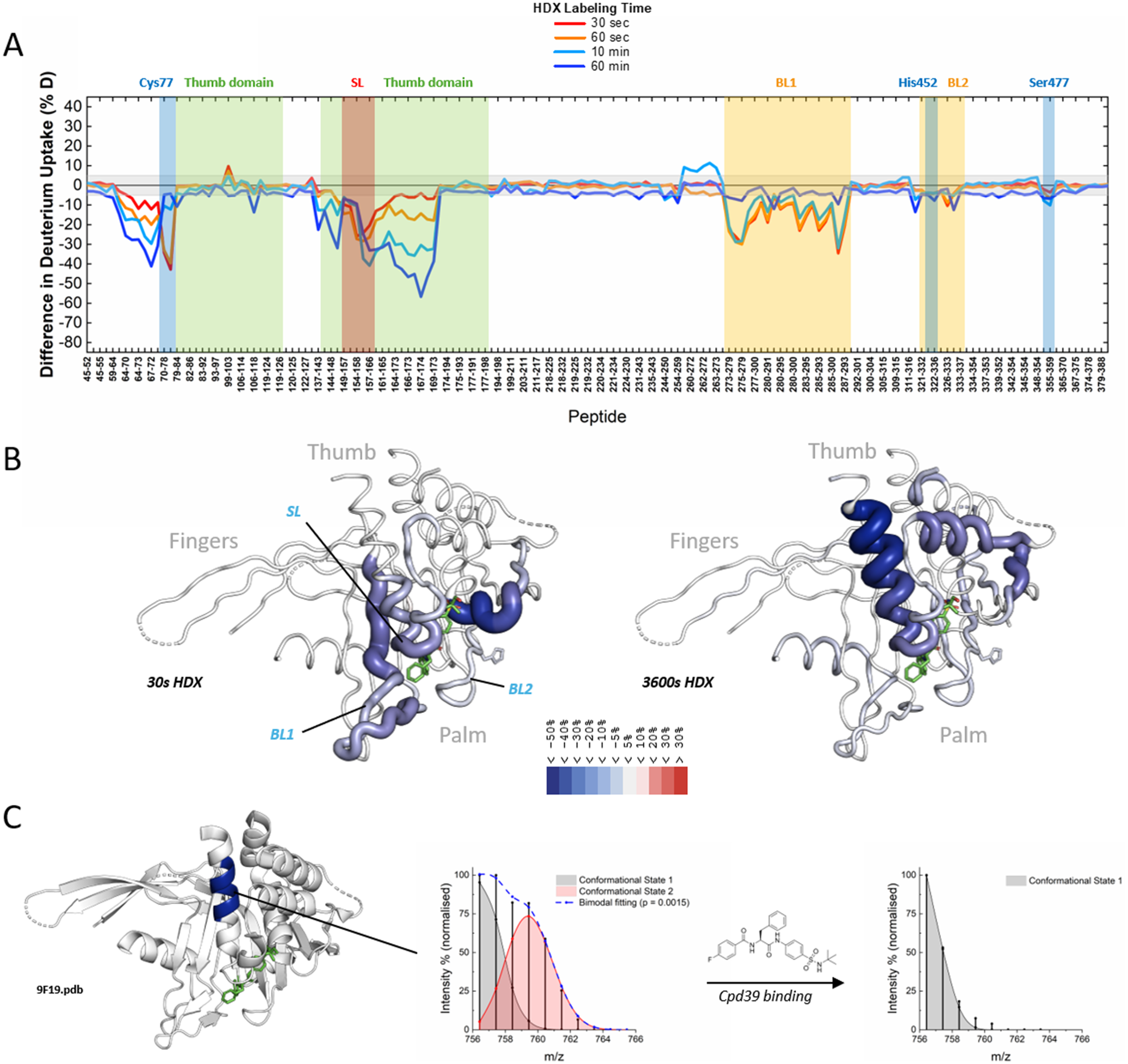
After USP30 incubation with the USP30 non-covalent inhibitor, USP30 inh , HDX-MS data confirmed that binding of the benzosulfonamide inhibitor induces changes that are consistent with localized structural stabilization and/or altered solvent accessibility within USP30. Comparison of apo- and holo-USP30 showed significant solvent protection clustered at the interface of the thumb and palm subdomains (Figure 1a, b and Supplementary Figure S2), a region known to guide the ubiquitin C-terminus into the active site (O’Brien et al., Reference O’Brien, Jones, Guenther, Murphy, England, Vendrell, Anderson, Brennan, Davis, Pinto-Fernández, Turnbull and Kessler2023). The most pronounced reductions in deuterium uptake mapped to peptides spanning the catalytic Cys77 (G69-F78), regions within USP domain 2 (D145-M149 and E155-E174), and blocking loop 1 (BL1; residues I323-L328), whereas more modest, yet significant, protection was observed for residues encompassing the blocking loop 2 (BL2; H449-Y456). In contrast, much of the protein, including the catalytic His452 and the region surrounding Ser477, exhibited no measurable change in deuterium uptake, indicating that USP30 inh binding perturbs a discrete functional pocket rather than altering the overall solvent accessibility or backbone dynamics of the protein. Interestingly, the most pronounced protection (up to 50%) was observed for peptides spanning residues His167-Glu174, a region distal from both the catalytic site and the inhibitor-binding pocket (Figure 1b) (O’Brien et al., Reference O’Brien, Jones, Guenther, Murphy, England, Vendrell, Anderson, Brennan, Davis, Pinto-Fernández, Turnbull and Kessler2023). Notably, apo-USP30 displays a bimodal deuterium uptake distribution in this region (Figure 1c), consistent with an underlying equilibrium between two distinct conformational states (Weis et al., Reference Weis, Wales, Engen, Hotchko and Ten Eyck2006). Binding of USP30 inh is associated with a shift toward the conformational subpopulation with lower exchange, accompanied by reduced solvent accessibility (Figure 1c).
Characterization of USP30inh binding to USP30. (a) Residual plot showing USP30 inh -induced differences in deuterium uptake compared to apo-USP30. All time points are individually colored, with USP30 peptides displayed on the x-axis. Peptides are sorted from the USP30 N- to C-terminus. Deuterium incorporation is mapped to the y-axis. Positive and negative values indicate differential deuterium uptake, compared to the apo-state. (b) 3D putty visualization showing the 3D (9F19.pdb) differential heatmap of deuterium uptake between holo- and apo-USP30 after 30 sec and 1 h exchange. Thickness correlates with the magnitude of deuterium uptake. USP30 inh analogue, compound NK036, is shown in green. Only peptides passing the significance criteria (Welch’s t-test, p < 0.05, and a global confidence threshold of ±0.28 Da) were used for 3D mapping. (c) A bimodal distribution of the MS spectrum was observed after 600 sec of exchange for peptide 167-173 (blue) in apo-USP30 (left) compared to a single distribution for its holo-USP30 counterpart (right). Upon inhibitor binding, only the more solvent-protected protein state is detected.

Figure 1. Long description
Panel A is a line graph titled Difference in Deuterium Uptake (percent D). The y-axis ranges from negative 80 to positive 40. The x-axis lists U S P 30 peptides from N-terminus to C-terminus. Four lines represent H D X labeling times: 30 seconds (red), 60 seconds (orange), 10 minutes (cyan), and 60 minutes (dark blue). Significant negative dips, indicating protection upon inhibitor binding, occur at the Thumb domain, S L, B L 1, and B L 2 regions. Specific residues Cys 77, His 452, and Ser 477 are marked with vertical blue bars.
Panel B shows two 3 D putty visualizations of U S P 30 labeled 30 s H D X and 3600 s H D X. The protein structure is shown in gray ribbons with regions of deuterium uptake colored in shades of blue. The thickness of the ribbon increases with the magnitude of protection. Labels indicate the Fingers, Thumb, and Palm domains. The inhibitor N K 0 3 6 is shown as green sticks in the binding pocket. A color scale at the bottom indicates protection levels from 5 percent to less than negative 50 percent.
Panel C displays a 3 D structure on the left with a specific peptide highlighted in dark blue. To the right are two mass spectra showing Intensity percent (normalized) versus m forward slash z. The first spectrum for apo-U S P 30 shows a bimodal distribution with two overlapping peaks labeled Conformational State 1 (gray) and Conformational State 2 (red), with a dashed blue line for bimodal fitting. An arrow labeled Cpd 39 binding points to the second spectrum, which shows a single gray peak for Conformational State 1, indicating the protein has shifted to a single solvent-protected state.
Binding of a covalent inhibitor, USP30-I-1, was then assessed. USP30-I-1 is a potent, selective cyanopyrrolidine-containing covalent small-molecule inhibitor, designed to enhance mitophagy by irreversibly suppressing USP30 deubiquitinating activity (O’Brien et al., Reference O’Brien, Jones, Shi, Guenther, Vendrell, Viner, Brennan, Mead, Zarganes-Tzitzikas, Davis, Pinto-Fernández, England, Murphy, Turnbull and Kessler2025). In cellular activity-based protein profiling experiments, USP30-I-1 demonstrated strong, concentration-dependent engagement of endogenous USP30 with minimal off-target activity against more than 40 other deubiquitinases, with a cellular IC₅₀ of approximately 94 nM (O’Brien et al., Reference O’Brien, Jones, Shi, Guenther, Vendrell, Viner, Brennan, Mead, Zarganes-Tzitzikas, Davis, Pinto-Fernández, England, Murphy, Turnbull and Kessler2025). A crystal structure of the protein-inhibitor complex has not yet been resolved; therefore, HDX-MS was employed to define the compound binding site and the conformational dynamics associated with covalent inhibition of USP30.
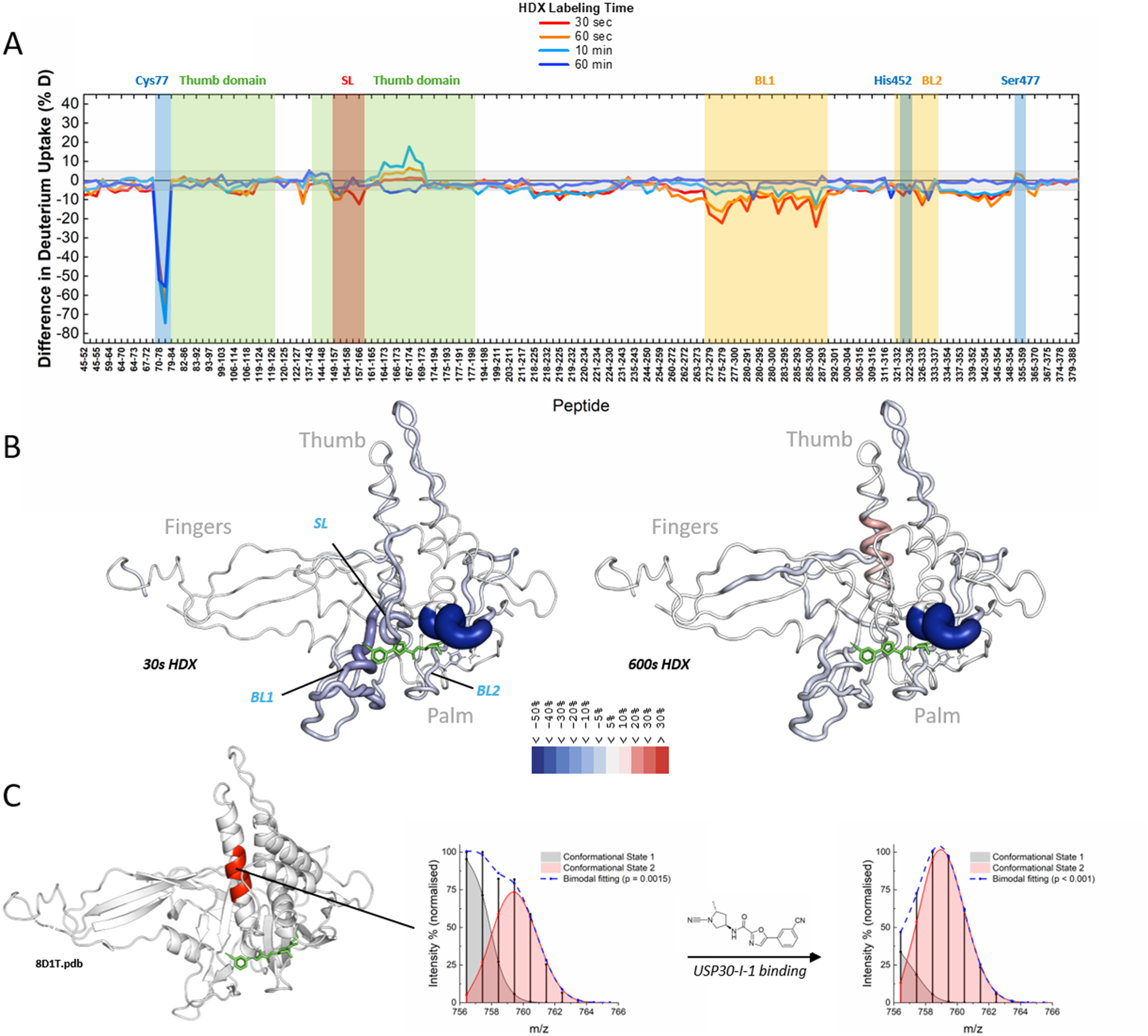
Comparative analysis of apo- and holo-USP30 revealed that inhibitor binding induces highly localized solvent protection centered around the catalytic region of the enzyme, rather than global structural rearrangements (Figure 2a, b and Supplementary Figure S2). As expected, the most pronounced reduction in deuterium uptake matches peptides encompassing the catalytic cysteine Cys77 (residues 70-78, unmodified peptide), with mean perturbations approaching 60% across labeling time points. Additional, though less extensive, solvent protection was observed in regions corresponding to the switching loop (SL; residues 150-157), BL 1 and 2 (residues 322-340 and 446-456, respectively), and adjacent elements of the palm subdomain, including residues flanking His452 and the β-strand preceding Ser477 (O’Brien et al., Reference O’Brien, Jones, Shi, Guenther, Vendrell, Viner, Brennan, Mead, Zarganes-Tzitzikas, Davis, Pinto-Fernández, England, Murphy, Turnbull and Kessler2025). In contrast, the majority of the USP30 sequence exhibited no significant difference in deuterium exchange, indicating that covalent modification by USP30-I-1 does not globally alter protein dynamics or solvent accessibility, but instead stabilizes a discrete catalytic-site-centered conformation. Interestingly, in contrast to the non-covalent inhibitor, incubation with the covalent inhibitor does not result in protection of residues 166-174; instead, this region shows increased solvent exposure, together with a clear perturbation in the corresponding MS isotopic distribution (Figure 2c).
Characterization of USP30-I-1 binding to USP30. (a) Residual plot showing USP30-I-1-induced differences in deuterium uptake compared with apo-USP30. All time points are individually colored and peptides sorted as above. Deuterium incorporation is mapped to the y-axis. Positive and negative values indicate differential deuterium uptake, respectively, compared with the apo-state. (b) 3D Putty Visualization showing the 3D (8D1T.pdb) differential heatmap of deuterium uptake between holo- and apo-USP30 after 30 sec and 10 min exchange. Thickness correlates with the magnitude of deuterium uptake. The docked inhibitor is shown in green. Only peptides passing the significance criteria (Welch’s t-test, p < 0.05, and a global confidence threshold of ±0.34 Da) were used for 3D mapping. (c), A bimodal distribution of the MS spectral plot was observed after 600 sec of exchange for peptide 167-173 in apo-USP30 (left) compared to an almost single distribution for its holo-USP30 counterpart (right). Upon inhibitor binding, the equilibrium between these two states is shifted towards the most solvent-exposed substrate.

Figure 2. Long description
Panel A is a line graph showing the difference in deuterium uptake percentage on the y-axis against peptide sequences on the x-axis. Four colored lines represent time points: red for 30 seconds, orange for 60 seconds, light blue for 10 minutes, and dark blue for 60 minutes. Significant negative peaks are highlighted in blue and yellow vertical bands, corresponding to Cys 77, B L 1, and B L 2 regions. A positive peak is highlighted in a red band labeled S L. Green bands indicate the Thumb domain.
Panel B shows two 3D putty visualizations of the protein structure labeled 30 s H D X and 600 s H D X. The protein is shown in a gray ribbon format with thickness and color indicating deuterium uptake. A green molecule representing the docked inhibitor is at the center. Labels point to the Fingers, Thumb, and Palm regions, as well as S L, B L 1, and B L 2 loops. A color scale at the bottom ranges from dark blue (less than -50 percent) to dark red (greater than 30 percent).
Panel C contains a 3D ribbon model on the left with a red helical segment. An arrow points from this segment to two mass spectra plots. The x-axis is m over z and the y-axis is Intensity percentage (normalized). The left plot (apo-state) shows a bimodal distribution with two peaks shaded gray (Conformational State 1) and pink (Conformational State 2). An arrow labeled U S P 30-I-1 binding points to the right plot (holo-state), which shows a shift to a single dominant pink peak.
Molecular docking studies were performed to validate the binding mode of the covalent cyanopyrrolidine-containing inhibitor to USP30. As illustrated in Figure 2b, USP30-I-1 is predicted to bind within the thumb-palm cleft of USP30, which physiologically guides the ubiquitin C-terminal tail into the active site. USP30-I-1 is predicted to form a covalent thioimidate adduct with the catalytic Cys77. In the docked pose, the imine nitrogen of the covalent adduct accepts a hydrogen bond from the side chain of Asn72, while donating a hydrogen bond to the backbone carbonyl of Gly74. In addition, the amide moiety of the inhibitor forms a hydrogen bond with the backbone carbonyl of Gln160. The inhibitor is further accommodated by residues located in BL1, BL2, and the SL, which all flank the predicted binding site.
Discussion
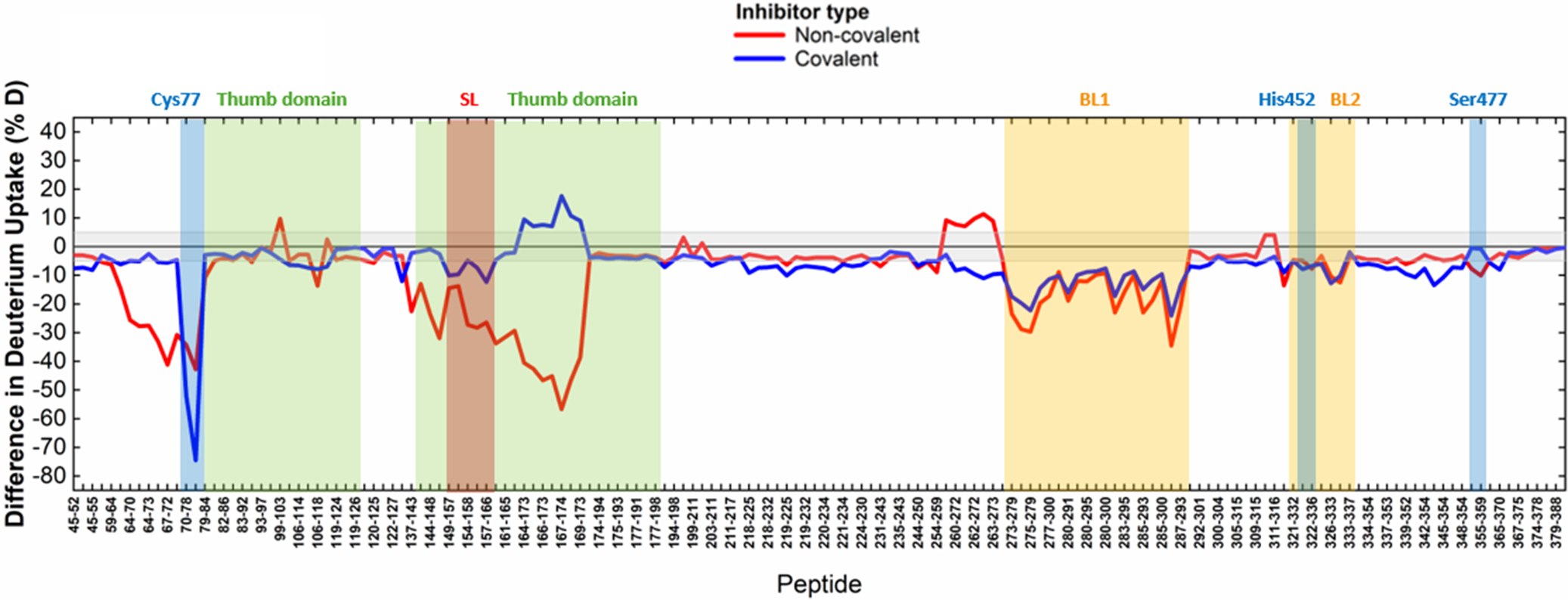
Herein, we integrated HDX-MS and molecular docking and compared them to available X-ray crystallographic data to investigate how chemically distinct small-molecule inhibitors engage USP30. USP30 is a mitochondrial deubiquitinating enzyme that counteracts PINK1-Parkin-mediated ubiquitination on the outer mitochondrial membrane, thereby limiting mitophagy. Inhibition of USP30 enhances mitochondrial quality control by promoting clearance of damaged mitochondria, potentially boosting their turnover and promoting neuroprotection in conditions such as Parkinson’s Disease (Bingol et al., Reference Bingol, Tea, Phu, Reichelt, Bakalarski, Song, Foreman, Kirkpatrick and Sheng2014). By probing USP30 inhibition in solution, HDX-MS enables the characterization of dynamic features and low-population conformational states that might be otherwise incompletely resolved or absent in crystal structures and provides insight into how different types of small-molecule inhibitors differentially modulate the enzyme. Both covalent and non-covalent inhibitor classes suppress USP30 activity by targeting the thumb-palm cleft and blocking ubiquitin access to the catalytic site, yet this is achieved through distinct modes of interaction for each (Figure 3). Binding of the non-covalent benzosulfonamide-containing inhibitor USP30 inh induces protection across an extended region of the thumb-palm interface, suggesting a broad range of interactions within the binding pocket. This HDX-MS profile closely mirrors the recently published crystallographic structure of the USP30 catalytic domain in complex with compound NK036, a solubility-enhanced derivative of USP30 inh (Kazi et al., Reference Kazi, Klink, Gallant, Kipka and Gersch2025). In contrast, the covalent cyanopyrrolidine-containing inhibitor USP30-I-1 produces a more localized stabilization centered on the catalytic Cys77 and adjacent loops (Figure 3), reflecting an inhibition mechanism that is primarily driven by irreversible active-site engagement. In this context, the HDX-MS data are in good agreement with the proposed docking pose of USP30-I-1 and closely align with the available X-ray structures of a covalently inhibited USP30 catalytic domain (PDB codes 8D1T and 8D0A, unpublished).
Comparative HDX-MS analysis of covalent and non-covalent USP30 inhibitors. Residual plots showing the strongest differences in deuterium uptake measured across all labeling time points for each USP30 peptide following binding of the non-covalent (USP30 inh , in red) or covalent (USP30-I-1, in blue) inhibitor, relative to apo-USP30. For each peptide, the maximum absolute difference observed at any time point is reported, providing a summary view of the regions most affected by inhibitor binding. Peptides are sorted from the USP30 N- to C-terminus. Deuterium incorporation is mapped to the y-axis. This representation highlights the distinct patterns and spatial extent of conformational perturbations induced by covalent versus non-covalent inhibition.

Figure 3. Long description
A line graph with the y-axis labeled Difference in Deuterium Uptake percent D ranging from negative 80 to 40. The x-axis is labeled Peptide and lists specific amino acid sequences from 45-52 to 379-388. A legend at the top identifies a red line as Non-covalent and a blue line as Covalent.
Moving from left to right along the x-axis, the following regions are highlighted with vertical colored bands and labels.
* Cys 77 is a blue band where the blue line shows a sharp negative peak reaching negative 75 percent.
* Thumb domain is a green band spanning peptides 79-84 to 119-126 where both lines fluctuate near zero.
* A second green Thumb domain region spans 144-148 to 177-191. Within this, a red band labeled S L contains a deep negative trough for the red line reaching negative 55 percent, while the blue line shows a positive peak reaching nearly 20 percent.
* B L 1 is a yellow band spanning 273-279 to 287-293 where both lines show significant negative uptake differences between negative 10 and negative 35 percent.
* His 452 is a narrow blue-grey band near peptide 321-332.
* B L 2 is a yellow band spanning 326-333 to 333-337.
* Ser 477 is a blue band near peptide 355-359.
Overall, the red line shows more frequent and deeper negative deviations across the sequence compared to the blue line, except at the Cys 77 site where the covalent inhibitor shows the most extreme negative difference.
From recent structural studies, a common emerging theme in DUB biology is the conformational plasticity that characterizes the SL and its central role as a determinant of inhibitor engagement (Mevissen and Komander, Reference Mevissen and Komander2017). It is known that the USP30 SL can adopt both ‘in’ or ‘out’ conformations, for example, when in complex with covalent inhibitors (PDB codes 8D1T and 8D0A), or in the presence of a ubiquitin-propargylamide moiety (PDB code 5OHK), respectively. In addition, the X-ray structure of a human USP30 chimera bound to NK036 highlights how this loop can undergo an unanticipated loop-to-helix transition involving residues 154-159 (Kazi et al., Reference Kazi, Klink, Gallant, Kipka and Gersch2025). This transition leads to the formation of a cryptic pocket which is engaged by the compound and is absent from any ubiquitin- or covalent inhibitor-bound structures. However, in the absence of a published apo-USP30 structure, it remains unresolved whether this conformation is fully induced by ligand binding or reflects stabilization of a pre-existing USP30 sub-population.
Here, HDX-MS provides important complementary evidence for the presence of an underlying conformational equilibrium in solution involving residues that surround the SL. Specifically, we observe a bimodal deuterium uptake distribution for residues 167-173 within the thumb domain (Figure 1c), a feature that is indicative of the presence of two discrete conformational states coexisting in solution, which may correspond to active/inactive forms or allosteric intermediates. Although this region is not directly contacted by the inhibitor and is located far from the active site, it is in close spatial proximity to the SL (residues 154-159). Upon binding of USP30 inh , this bimodal distribution is suppressed in favor of a more rigid, solvent-protected structural architecture (Figure 1c). Alongside this shift, increased protection is observed for the α-helix spanning residues 141-150, suggesting a local structural reorganization that supports the rearrangement of the newly formed cryptic pocket (Figure 2b). On the other hand, the covalent inhibitor USP30-I-1, while forcing the SL to adopt the ‘in’ conformation and inducing pronounced protection around the active site, does not stabilize the same thumb-domain conformational state as the non-covalent compound. Instead, its binding shifts the equilibrium towards a conformational state that is more solvent exposed (Figure 2c and 3). Taken together, these observations suggest that the thumb-domain architecture of USP30 may be transiently heterogenous prior to ligand binding.
Speculation box
Overall, our findings support a model in which USP30 exists as an ensemble of interconverting conformations, with the SL and surrounding thumb-domain regions potentially acting as a dynamic hub. By resolving bimodal exchange regimes and long-range conformational coupling, HDX-MS uncovers hidden regulatory states that, together with the findings of a cryptic pocket formation, might help rationalize the diversity of inhibitor-bound USP30 structures, highlighting the importance of conformational equilibria in guiding rational inhibitor design.
Open peer review
To view the open peer review materials for this article, please visit http://doi.org/10.1017/qrd.2026.10023.
Supplementary material
The supplementary material for this article can be found at http://doi.org/10.1017/qrd.2026.10023.
Data availability statement
HDX-MS data are accessible through the PRIDE Archive under identifier PXD054057.
Acknowledgements
We thank Dr Yuqi Shi from Thermo Fisher Scientific for their assistance with HDX-MS data acquisition.
Author contribution
Conceived and designed the study: A.P.-F., E.J.M., A.P.T., B.M.K. and D.P.O.B.; conducted data gathering: A.P., A.P.T. and D.P.O.B.; performed data analyses: A.P., A.P.T. and D.P.O.B.; wrote the first draft of the article: A.P. and D.P.O.B. All authors reviewed and revised the final article.
Financial support
This work was supported by Alzheimer’s Research UK (grant no. ARUK-2021DDI-OX). A.P.-F. and B.M.K. labs were supported by the Chinese Academy of Medical Sciences (CAMS) Innovation Fund for Medical Science (CIFMS), China (grant number: 2024-I2M-2-001-1).
Competing interests
The authors declare none.


 Open access


Comments
To the Editors,
Please find enclosed our report entitled "Uncovering Mechanisms of Drug Action Through Protein Structural Dynamics: Insights from Deubiquitinase Inhibitors” which we would like to submit for critical evaluation and potential publication in QRB Discovery. We were invited to submit this report following participation and oral presentation at the inaugural Uncovering Hidden Biology Conference which was held in Worcester College, University of Oxford in autumn last year. Our submission will form part of a “Proceedings of” collection which will be guested edited by Professor Dame Carol Robinson, Sophie Lawrence and Jack Bennett.
Ubiquitin specific protein 30 (USP30) is a mitochondrial associated deubiquitylating enzyme which has been implicated in a wide range of pathophysiological disorders through its participation in PINK1/Parkin-mediated mitophagy, BAX/BAK-dependent apoptosis, oncogenesis, and pexophagy. Conditions linked to USP30 biology include chronic kidney and cardiovascular diseases, and hepatocellular carcinoma and peroxisome biogenesis disorders. Most notably, the protein has been suggested as an effective therapeutic strategy against neurodegenerative disorders such as Parkinson’s Disease. USP30 inhibition may counteract the deleterious effects of impaired turnover of damaged mitochondria which is inherent to both familial and sporadic forms of the disease. Small molecule inhibitors targeting USP30 are currently in development, but little is known about their precise mode of action.
In our report, we integrate Hydrogen-Deuterium eXchange Mass Spectrometry (HDX-MS) with molecular docking and available crystal structures to characterize the solution-state conformational dynamics of USP30. HDX-MS reveals structural dynamics that are not resolved crystallographically, uncovering hidden mechanisms of drug action. Our findings provide mechanistic insights into USP30 inhibitor binding and underscore the utility of HDX-MS as a tool for guiding DUB-targeted drug discovery.
We thank you in advance for considering our work for publication in QRB Discovery.
On behalf of all authors,
Darragh O’Brien, PhD