Introduction
Haplorchis taichui is a small heterophyid intestinal fluke of fish-eating birds and mammals, and is an occasional parasite of humans (Faust & Nishigori, Reference Faust and Nishigori1926; Yamaguti, Reference Yamaguti1958). Humans acquire infections from eating raw or undercooked freshwater fish containing metacercariae of H. taichui. Cyprinid fish are an important intermediate host of this fluke. Haplorchis taichui infects various fish that are part of the human diet in northeast and northern Thailand (Kliks & Tantachamrun, Reference Kliks and Tantachamrun1974; Srisawangwong et al., Reference Srisawangwong, Sithithaworn and Tesana1997; Sukontason et al., Reference Sukontason, Piangjai, Muangyimpong, Sukontason, Methanitikorn and Chaithong1999; Kumchoo et al., Reference Kumchoo, Wongsawad, Chai, Vanittanakom and Rojanapaibul2005).
Treatment of H. taichui is still a great problem and there is a high prevalence of H. taichui infection in the northern part of Thailand. The pathogenicity of worm infections has been reported from humans in Lampang Province, northern Thailand. Three heavily infected cases presented mucosal ulceration, mucosal and submucosal haemorrhages, fusion and blunting of villi, crypt hyperplasia, chronic inflammation, and fibrosis of the submucosa (Sukontason et al., Reference Sukontason, Sukontason, Muanguimpong and Piangjai2005). The drugs of choice for treatment of this parasite, such as praziquantel, have been reported by many investigators (Scholz et al., Reference Scholz, Ditrich, Tuma and Giboda1991; Pungpak et al., Reference Pungpak, Radomyos, Radomyos, Schelp, Jongsaksuntigul and Bunnag1998; Radomyos et al., Reference Radomyos, Wongsaroj, Wilairatana, Radomyos, Praevanich, Meesomboon and Jongsuksuntikul1998). Niclosamide is effective in the treatment of many kinds of trematode and cestode infections. This drug inhibits oxidative phosphorylation in the mitochondria of cestodes and has low side-effects because it is not absorbed from the gastrointestinal tract (Katz, Reference Katz1986). Only a few cases of nausea, vomiting, diarrhoea, abdominal pain and dizziness were reported from some patients infected with Fasciolopsis buski and treated with niclosamide (Suntharasamai et al., Reference Suntharasamai, Bunnag, Tejavanij and Harinasuta1974; Garcia, Reference Garcia2001; Anon., 2003). Niclosamide at a dosage of 6 g over 3 alternate days has been recommended for Heterophyes heterophyes infection (Khalil & Rifaat, Reference Khalil and Rifaat1964) and 100–125 mg kg− 1 for Metagonimus yokogawai infection (Rim, Reference Rim1975). Also, niclosamide has been used to treat Diplozoon paradoxum and Gyrodactylus aculeati parasitizing the gills and skin of fish (Schmahl & Taraschewski, Reference Schmahl and Taraschewski1987). This drug inhibits the uptake of glucose by the helminth, stops the production of its energy and can cause a paralytic effect on the worm. Niclosamide works by killing worms which are then passed in the stool. Occasionally worms cannot be collected after treatment because they are destroyed in the intestine of the host (Anon., 2003).
Little is known on the effect of drugs against H. taichui. Apart from spine damage and swollen basal spines, H. taichui appear to be resistant to praziquantel (Scholz et al., Reference Scholz, Ditrich, Tuma and Giboda1991). However, a 100% cure was found with niclosamide at a dose of 40 mg kg− 1 body weight of mice (Sukontason et al., Reference Sukontason, Sukontason, Muanguimpong and Piangjai2000). Albendazole has also been used in H. taichui infections, with a 100% cure at 40 mg kg− 1 (Waikagul et al., Reference Waikagul, Nuamtanong and Visiadsuk2003). Scanning electron microscopy has proved to be a useful tool for evaluating surface changes to flukes in general resulting from anthelmintic action. This is the first morphological study of the effects of niclosamide on H. taichui. The sequence of changes in the tegumental surface of adult H. taichui after in vitro exposure to various concentrations of niclosamide is described using scanning electron microscopy.
Materials and methods
Encysted metacercariae of Haplorchis taichui were obtained from naturally infected cyprinid fish (Heinecorhynchus siamensis) collected from Chiang Mai, northern Thailand. Encysted metacercariae were isolated by 1% (w/v) pepsin solution (pepsin 1:2500 Sigma® 1 g: NaCl solution 99 ml: hydrochloric acid 1 ml), pH 0.29–0.70, for 2 h at 37°C. The digested material was passed through graded sieves, rinsed with 0.85% (w/v) NaCl, and metacercariae were collected and examined under a stereo microscope. Three day-old chicks (Gallus gallus domesticus) were experimentally fed with approximately 1000 metacercariae. At the end of day 7 post-infection, adult worms were collected from the small intestine by the Baermann technique. Worms were washed in Tyrode's solution and used immediately for the experiment. A stock solution of niclosamide was prepared by dissolving 500 mg of niclosamide (Hexane®, Thailand) in 5 ml of absolute ethanol and diluting this stock solution with distilled water to give a concentration of 50 μg ml− 1. Niclosamide was prepared in various concentrations of 0.01, 0.1, 1.0 and 10.0 μg ml− 1. For each experiment, 5 ml of solution and 100 active worms were introduced to each Petri dish and incubated at 37°C, including the control group with Tyrode's solution without niclosamide. After 30 min, 1, 6, 12 and 24 h-treated worms were washed three times in 0.1 m phosphate buffer, pH 7.2. The movement of worms was observed and recorded under a stereo microscope. For scanning electron microscopy, treated worms were fixed in 2.5% (w/v) glutaraldehyde at 4°C for 24 h and post-fixed with 1% osmium tetroxide for 2 h. They were dehydrated in a graded alcohol series, dried in a critical-point dryer, gold-coated, and observed under a Dual Stage DS-130C and a JEOL-JSM2001LV scanning electron microscope.
Results
Activity study
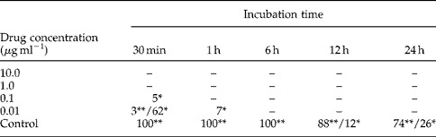
Niclosamide was effective against H. taichui after in vitro treatment. One hundred active worms were treated in each experiment. Exposure of worms to 0.01 μg ml− 1 of niclosamide after 30 min showed 3 active, 62 slightly active and 35 inactive worms and, 1 h later, 7 worms were slightly active and 93 worms were inactive. In 0.1 μg ml− 1 of niclosamide solution after 30 min, only 5 of 100 worms were slightly active and all worms were inactive after 1 h. On the other hand, worms exposed to 1.0 and 10.0 μg ml− 1 of niclosamide were all inactive after 30 min. Control groups in Tyrode's solution showed active and slightly active worms over a 24 h (table 1).
Activity of Haplorchis taichui after incubation with concentrations ranging from 0.01 to 10 μg ml−1 of niclosamide.
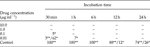
100 active worms were used for each experiment.
** Active movement (1–5 s per time);
* slightly active movement (6–10 s per time); –inactive (>10 s no movement).
Scanning electron microscopy
Niclosamide caused progressive alterations to the tegumental surface of H. taichui and the degree of damage was correlated with the concentration of drug and exposure times.
Control groups: Tyrode's solution
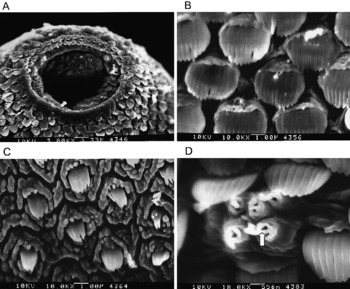
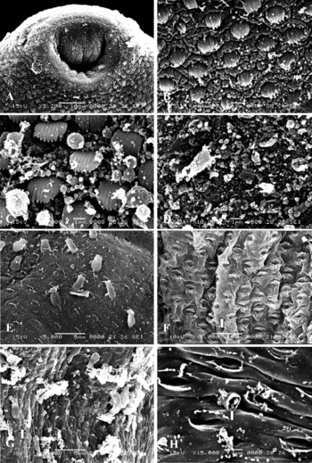
The surface morphology of the tegument appeared normal, covered with scale-like multi-pointed spines and numerous sensory papillae. The tegument in the anterior region of worms is densely covered with large spines, which decrease in size and number posteriorly (fig. 1A–D).
Tegumental surface of adult Haplorchis taichui, 24 h after incubation in Tyrode's solution. A, Surface around the oral sucker ( × 3000); B, anterior region with large spines ( × 10,000); C, posterior region with smaller size and number of spines ( × 10,000); D, the sensory papillae (arrowed) ( × 18,000).

Treatment with 0.01 μg ml−1 niclosamide
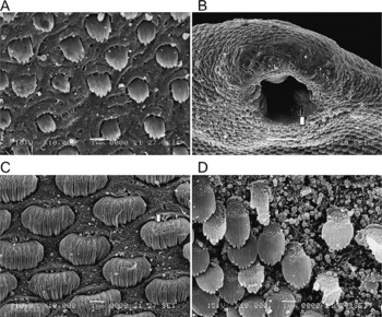
In worms observed after 30 min and 1 h, most of the tegument and sensory papillae on both surfaces retained a normal topography. After 6 and 12 h the surface became swollen with the development of small blebs and in some areas the spines, which appeared sunken, were supported by swellings on the tegument (fig. 2A). In some specimens after 12 h, the tegument surrounding the oral sucker showed areas of erosion (fig. 2B). After 24 h, the margin and base of each spine had begun to erode (fig. 2C) and numerous blebs had ruptured to produce further erosion of the tegument and spines (fig. 2D).
Tegumental surface of adult Haplorchis taichui after incubation in 0.01 μg ml− 1 niclosamide solution. A, Surface is swollen and covered with small blebs after 6 h ( × 10,000); B, damage on the surface around the oral sucker with the occurrence of lesions (arrowed) after 12 h ( × 2000); C, spine damage and lesions (arrowed) at the base of spines after 24 h ( × 10,000); D, numerous small blebs resulting in some lesions and erosion of spines after 24 h ( × 10,000).

Treatment with 0.1 μg ml−1 niclosamide
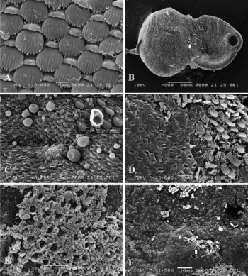
After 30 min, although the sensory papillae remained unaffected, the surface showed signs of erosion with swelling of the tegument around the spines (fig. 3A). After 1 h, blebs of various sizes appeared above the ventrogenital sac which was more severely affected than other parts. Some blebs had collapsed and shrivelled, and some spine sinking had occurred (fig. 3B–C). After 6 and 12 h, the blebs had increased in number with some blebs being in the process of disrupting (fig. 3C inset). By 12 h, spines around the ventrogenital sac and in the mid-body to posterior region became eroded (fig. 3D) and after 24 h, extensive blebbing had occurred and surface was completely swollen (fig. 3E). Some worms had lost the apical plasma membrane on both the ventral and dorsal surfaces in the middle to posterior regions, exposing empty spine sockets. The tegument on both surfaces showed similar changes (fig. 3E–F).
Tegumental surface of adult Haplorchis taichui after treatment with 0.1 μg ml− 1 niclosamide solution. A, Anterior region with swelling of tegument between the spines after 30 min ( × 10,000); B, spherical blebs more prominent above the ventrogenital sac (arrowed) after 1 h ( × 500); C, larger blebs and sunken spines appear after 1 h, (inset, × 10,000) ruptured blebs occur after 6 h ( × 3000); D, loss of spines on the mid-body surface and the appearance of spine sockets after 12 h ( × 3000); E, numerous blebs on the surface after 24 h ( × 5000); F, loss of spines and the occurrence of spine sockets around the ventrogenital sac (arrowed) after 24 h ( × 1500).

Treatment with 1.0 μg ml−1 niclosamide
After 30 min, spherical blebs appeared on the tegument, heavily between the oral sucker and ventrogenital sac (fig. 4A) and after 1 h, extensive blebbing occurred on the body surface (fig. 4B). Most of the tegumental surface was very swollen and the spines appeared sunken. Six hours after exposure, the surface blebs had become more widespread and in some cases severe, especially on the surface between the spines (fig. 4C–D). Some blebs had ruptured to form tegumental lesions, resulting in the surface beginning to slough off (fig. 4D). After 12 h, the number of blebs and lesions had increased resulting in damage and loss of the tegumental surface and empty spine sockets appearing in the middle to posterior region of the body (fig. 4E–F). After 24 h, severe damage to the tegument occurred, with the loss of the apical plasma membrane from both ventral and dorsal surfaces, leaving empty spine sockets (fig. 4F). More severe damage occurred in the posterior region of the body with numerous small perforations occurring in areas where the tegument had been removed (fig. 4F–G) and most sensory papillae had ruptured (fig. 4H).
Tegumental surface of adult Haplorchis taichui after incubation with 1.0 μg ml− 1 niclosamide solution. A, Anterior region with numerous small blebs at 30 min ( × 2200); B, posterior region with numerous blebs after 1 h ( × 10,000); C, various size of blebs in anterior region after 6 h ( × 10,000); D, posterior region eroded with some loss of spines, and the surface covered with blebs after 6 h ( × 10,000); E, spines lost with the appearance of spine sockets after 12 h ( × 5,000); F, loss of spines presence of spine sockets and small perforations (arrowed) on the basal lamina after 12 h ( × 5000); G, numerous small perforations (arrowed) on the basal lamina after 24 h ( × 5000); H, sensory papillae (arrowed) ruptured after 24 h ( × 15,000).

Treatment with 10.0 μg ml−1 niclosamide
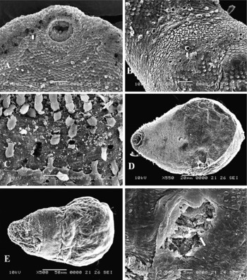
After 30 min, most worms exhibited swelling of the tegument around the spines and after 1 h, some blebs which had burst causing lesions were scattered over the inter-spine areas (fig. 5A–B). After 6 h, more blebs had ruptured and the surface was eroded on both the ventral and dorsal surfaces (fig. 5C). In some specimens, spines had been lost and the tegument was covered with numerous small blebs resulting in an eroded surface with lesions, which, after 12 h- had increased on both dorsal and ventral surfaces (fig. 5D–E). Numerous small perforations were found throughout the tegument, especially in the mid-body to posterior region. Similar changes occurred after 24 h and the apical plasma membrane completely disappeared from both surfaces, revealing empty spine sockets and loss of spines (fig. 5F). The tegumental surface changes were similar on both surfaces, with the anterior region of worms less damaged than the posterior region.
Tegumental surface of adult Haplorchis taichui after incubation with 10.0 μg ml− 1 niclosamide solution. A, Some blebs ruptured and lesions (arrowed) in the anterior region after 1 h ( × 1500); B, numerous blebs ruptured causing lesions (arrowed) after 1 h ( × 1500); C, numerous blebs scattered on the tegument and loss of the plasma membrane in some regions after 6 h ( × 5000); D, loss of spines on the ventral surface after 12 h ( × 550); E, loss of spines on the dorsal surface after 12 h ( × 500); F, complete loss the apical plasma membrane and some lesions (arrowed) after 24 h ( × 3000).

Discussion
The present study has shown that niclosamide is highly effective in killing adults of Haplorchis taichui in vitro at low concentrations. All worms become inactive after 1 h in 0.01 μg ml− 1 and after 30 min in 0.1 μg ml− 1 of niclosamide. This finding is similar to the effects of praziquantel on some trematodes such as Fibricola seoulensis (Lee, Reference Lee1985) and Paragonimus westermani (Lee et al., Reference Lee, Park, Hong, Chai and Hong1987). It is suggested that niclosamide can kill intestinal flukes with high efficiency and can cure heterophyidiasis. In addition, niclosamide has been reported to cure some fluke infections such as Metagonimus yokogawai (Rim, Reference Rim1975) Fasciolopsis buski (Suntharasamai et al., Reference Suntharasamai, Bunnag, Tejavanij and Harinasuta1974) and Haplorchis taichui (Sukontason et al., Reference Sukontason, Sukontason, Muanguimpong and Piangjai2000).
The present study has shown that niclosamide progressively causes severe disruption to the tegumental surface of H. taichui with longer exposure times. After 30 min to 6 h of exposure to 0.01 μg ml− 1 and 30 min to 1 h in 0.1, 1 and 10 μg ml− 1 niclosamide, morphological changes were observed in the anterior region, especially around the oral sucker and ventrogenital sac of the fluke. Various-sized blebs developed in the area between the oral sucker and ventrogenital sac. After 6 h in 0.1, 1.0 and 10 μg ml− 1 niclosamide, more severe disruption was evident in the posterior region. Movement and feeding of worms are disrupted since the drug penetrates the pharynx and the early part of the intestine whereas changes in the posterior region of the gut are more severe on exposure over a longer period of the incubation. The more rapid anterior blebbing is probably due to niclosamide being absorbed with great efficacy and over longer incubation times, worms become less active and finally immobile. Damage in the posterior region of worms is due to drug accumulation in the posterior part of the gut. The results support the concept that oral ingestion of niclosamide is the main route of entry, which agrees with the work of Meaney et al. (Reference Meaney, Fairweather, Brennan and Forbes2004, Reference Meaney, Haughey, Brennan and Fairweather2005) on some fasciolicides. In addition, the rapid tegumental disruption observed in vitro is probably due to worms being in more direct contact with the drug. There is a more immediate effect of this drug via oral uptake and worms quickly become paralysed and rapidly killed (Fairweather et al., Reference Fairweather, Holmes and Threadgold1984; McKinstry et al., Reference McKinstry, Fairweather, Brennan and Forbes2003). However, some drugs such as benzimidazoles can penetrate the tegument of the parasite with high efficiency. Mottier et al. (Reference Mottier, Alvarez, Ceballos and Lanusse2006) reported that passive diffusion though the external helminth surface is a major mechanism of benzimidazole penetration into the trematode, Fasciola hepatica, the cestode, Moniezia benedeni and the nematode, Ascaris suum.
Regional differences in tegumental disruption were observed in treated H. taichui. The dorsal surface showed a similar disruption to the ventral surface and the posterior region was more severely disrupted than the anterior part. The findings are similar to the effects of praziquantel (Apinhasmit & Sobhon, Reference Apinhasmit and Sobhon1996), albendazole sulphoxide (Buchanan et al., Reference Buchanan, Fairweather, Brennan, Trudgett and Hoey2003), and 5-chloro-2-methylthio-6-(1-naphthyloxy)-1-H-benzimidazole (Rivera et al., Reference Rivera, Ibarra, Zepeda, Fortoul, Hernandez, Castillo and Canto2004, Reference Rivera, Ibarra, Zepeda, Fortoul, Canto, Hernandez and Castillo2005). The regional differences in damage may be due to tegumental structure, route of drug uptake and drug metabolism (McKinstry et al., Reference McKinstry, Fairweather, Brennan and Forbes2003). The drug is more rapidly absorbed when worms are contacted in the earlier incubation times, resulting in the occurrence of various-sized blebs between the oral sucker and ventrogenital sac. The anterior region is less damaged than posteriorly probably because this area is more densely covered with larger spines compared with smaller spines in the posterior region.
Surface blebbing and swelling of the tegument of H. taichui were observed after short exposure times. McKinstry et al. (Reference McKinstry, Fairweather, Brennan and Forbes2003) suggested that blebbing is an initial stress reaction by worms to replace the surface membrane. Similar observations were made for other anthelmintic drugs (Lee et al., Reference Lee, Park, Hong, Chai and Hong1987; Stitt & Fairweather, Reference Stitt and Fairweather1993; Apinhasmit & Sobhon, Reference Apinhasmit and Sobhon1996; Meaney et al., Reference Meaney, Fairweather, Brennan, Ramasamy and Subramanian2002, Reference Meaney, Fairweather, Brennan, McDowell and Forbes2003; Buchanan et al., Reference Buchanan, Fairweather, Brennan, Trudgett and Hoey2003). Blebbing and swelling are early changes leading to loss of the tegument. It is suggested that the anthelmintic drug can penetrate into deeper-lying tissues, following breaching of the apical plasma membrane, thereby producing more severe worm disruption. Similar swelling of the tegument and blebbing, then rupturing and lesions, have been reported for praziquantel (Lee, Reference Lee1985; Lee et al., Reference Lee, Park, Hong, Chai and Hong1987; Apinhasmit & Sobhon, Reference Apinhasmit and Sobhon1996). The swelling of the tegument is probably due to the disruption of ion pumps on the apical plasma membrane. This action decreases the production of ATP in the mitochondria and affects the ion pump, leading to an influx of water and swelling of the tegument (Skuce et al., Reference Skuce, Anderson and Fairweather1987). When tegumental surface disruption becomes more severe, transport is disrupted throughout the syncytium so the plasma membrane cannot be replaced quickly enough to prevent separation of the tegument, leading to complete loss of the syncytium. The drug exposes the basal lamina and creates small perforations, then penetrates the internal tissues, causing greater worm damage with some lesions (Meaney et al., Reference Meaney, Fairweather, Brennan, Ramasamy and Subramanian2001). In the present study, blebs were formed on the tegumental surface of treated worms. The removal of the apical plasma membrane and spine sockets occurred on both surfaces. Surface destruction was more pronounced posteriorly than anteriorly with both surfaces. Numerous small perforations occurred posteriorly but fewer anteriorly. These findings suggest that worms exposed to niclosamide showed severe disruption, although further work using transmission electron microscopy are need to describe internal structural changes and confirm the route of drug transport.
Acknowledgements
This investigation was supported by BGJ Program no. BGJ 47K0010 and RGJ PhD Program no. PHD/0081/2544 from the Thailand Research Fund. The authors would like to thank Asoc. Prof. Dr Kom Sukontason, Mr Tanu Marayong, Mr Thanapon Yooyen, and colleagues in the Parasitology Research Laboratory Department of Biology, Faculty of Sciences, Chiang Mai University for their assistance. The Electron Microscopy Research and Service Center, Faculty of Science, Chiang Mai University, Chiang Mai, Thailand and the Electron Microscopy Research and Service Center, Seoul National University, Seoul, Korea, are gratefully thanked for electron microscopy facilities. Special thanks are given to Mrs Bussabong Kantalue, Mr Anantachai Kamsaiyai and Mr Sung-Yil Choi for their technical assistance in electron microscopic operation and photography.