Introduction
Atrial standstill represents a rare cardiac arrhythmia characterised by the complete absence of atrial electrical and mechanical activity, manifesting electrocardiographically as the absence of P waves accompanied by compensatory junctional or ventricular escape rhythms. Reference Tan, Giglia and Cecchin1 The epidemiological burden of atrial standstill remains poorly characterised, with an estimated prevalence of less than 0.1% in the general population; however, precise incidence data are limited due to the scarcity of comprehensive population-based studies. The clinical presentation of atrial standstill in paediatric populations typically encompasses bradycardia, exercise intolerance, syncope, and congestive heart failure symptoms. Electrocardiographic findings are pathognomonic, demonstrating a complete absence of P waves with resultant junctional or ventricular escape mechanisms. Echocardiographic assessment characteristically reveals significant atrial chamber dilatation with complete absence of atrial contractile function, reflecting the underlying electrical silence. Therapeutic management of atrial standstill necessitates a dual approach: identification and treatment of reversible underlying aetiologies where feasible, coupled with comprehensive management of associated cardiovascular complications. Given the complete absence of atrial electrical activity precluding effective atrial capture, cardiac device therapy should be optimised for ventricular-based pacing modalities, specifically ventricular demand pacing mode, to ensure adequate heart rate support and haemodynamic stability.
In paediatric populations, atrial standstill demonstrates a predominantly genetic aetiology, with pathogenic variants in the SCN5A gene—encoding the cardiac voltage-gated sodium channel Nav1.5—representing the most frequently implicated molecular mechanism. Additional channelopathy genes, including SCN10A and other ion channel-encoding loci, have been identified as causative factors, underscoring the genetic heterogeneity of this condition. The SCN5A gene product, sodium channel protein type 5 subunit alpha (Nav1.5), serves as a critical determinant of cardiac electrophysiology by mediating voltage-dependent sodium ion permeability across excitable membranes. This transmembrane protein undergoes conformational transitions between opened and closed states in response to membrane voltage fluctuations, thereby forming a sodium-selective pore that facilitates Na+ ion flux according to prevailing electrochemical gradients. Reference Gellens, George and Chen2–Reference Chakrabarti, Wu and Yang4 The resultant rapid sodium influx during the depolarisation phase of the cardiac action potential constitutes the fundamental trigger for myocardial contraction, establishing Nav1.5 as an indispensable component of cardiac excitation-contraction coupling. Pathogenic SCN5A variants exhibit remarkable phenotypic heterogeneity, manifesting across a broad spectrum of cardiac arrhythmias and structural heart diseases. These include congenital atrioventricular heart block, Reference Smits, Eckardt and Probst5–Reference Groenewegen, Firouzi and Bezzina11 long QT syndrome 3, Reference Millat, Chevalier and Restier-Miron12–Reference Priori, Napolitano and Gasparini14 Brugada syndrome 1, Reference Kapplinger, Tester and Salisbury13,Reference Priori, Napolitano and Gasparini14 sick sinus syndrome, Reference Kapplinger, Tester and Alders7,Reference Benson, Wang and Dyment15 familial paroxysmal ventricular fibrillation 1, Reference Millat, Chevalier and Restier-Miron12,Reference Otagiri, Kijima and Osawa16 atrial standstill, Reference Meregalli, Tan and Probst6,Reference Groenewegen, Firouzi and Bezzina11,Reference McNair, Ku and Taylor17 dilated cardiomyopathy, Reference Chakrabarti, Wu and Yang4,Reference McNair, Ku and Taylor17 and atrial fibrillation, Reference Ellinor, Nam, Shea, Milan, Ruskin and MacRae18,Reference Darbar, Kannankeril and Donahue19 reflecting the pleiotropic effects of sodium channel dysfunction. Therapeutic intervention necessitates permanent cardiac pacemaker implantation; however, paediatric implementation presents distinctive clinical challenges encompassing device miniaturisation constraints, anatomical considerations for optimal lead positioning, and the heightened risk of long-term device-related complications. The clinical phenotype may present as an isolated arrhythmic disorder or progress to encompass broader conduction system deterioration and associated cardiomyopathy. Reference Dong, Xiao and Cao20 Consequently, early diagnostic recognition coupled with comprehensive genetic counselling assumes paramount importance, particularly given the propensity for familial clustering in hereditary variants. Long-term clinical outcomes are contingent upon the specific underlying genetic aetiology and the timeliness of therapeutic intervention.
This report presents a rare case of atrial standstill secondary to compound heterozygous SCN5A variants, which was incidentally discovered during attempted upgrade from single-chamber to dual-chamber pacemaker therapy. The clinical scenario underscores the diagnostic complexities inherent in identifying atrial standstill, particularly when masked by existing ventricular pacing dependency. Our investigation delineates the critical diagnostic modalities essential for the comprehensive evaluation of paediatric patients presenting with atrioventricular heart block and sick sinus syndrome, particularly those with congenital onset. These include systematic intracardiac electrophysiological assessments to characterise atrial electrical activity and comprehensive genetic screening to identify underlying channelopathy variants. Such multimodal diagnostic approaches are fundamental for establishing accurate diagnoses in very young patients where clinical presentations may be subtle or atypical. Furthermore, this case highlights the pivotal role of advanced echocardiographic strain analysis in monitoring myocardial mechanics and predicting the deleterious haemodynamic consequences of chronic right ventricular pacing in ventricular demand mode. Given the inability to achieve dual-chamber pacing due to absent atrial capture, consideration should be given to physiological pacing alternatives, specifically left bundle branch area pacing, which may provide superior haemodynamic outcomes while maintaining ventricular synchrony in patients with atrial standstill. The clinical implications extend beyond immediate pacing considerations, emphasising the necessity for long-term cardiac function monitoring and the potential benefits of conduction system pacing techniques in preserving myocardial performance in this vulnerable patient population.
Case presentation
Initial clinical presentation and laboratory examination
The study was approved by the ethics committee of West China Second University Hospital of Sichuan University (approval number 2021-069). In addition, we obtained written informed consent from the patients’ parents prior to performing WES and for inclusion of the clinical and imaging details of the patients in the publication.
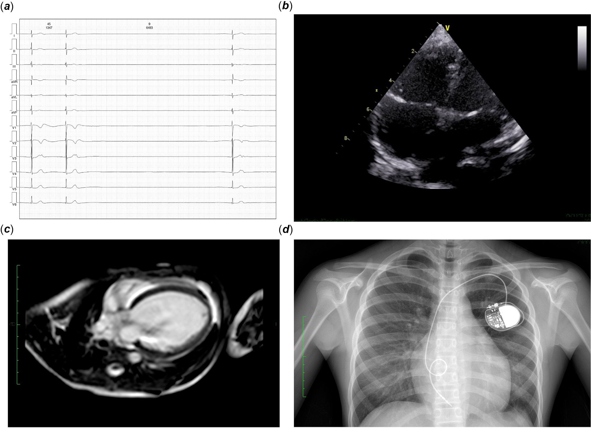
The proband was initially diagnosed with bradycardia during a routine kindergarten physical examination at 5 years of age. The patient remained asymptomatic, denying dyspnoea, chest discomfort, palpitations, syncopal episodes, seizure activity, or exercise intolerance. Physical examination revealed heart rate variability ranging from 32–61 beats per minute, with nocturnal resting bradycardia of 30–40 bpm. Vital signs demonstrated a respiratory rate of 23 breaths per minute and a blood pressure of 110/66 mmHg. The patient appeared chronically ill but maintained comfort with preserved mental status. Physical examination was notable for the absence of generalised oedema, lymphadenopathy, respiratory distress, or oropharyngeal abnormalities. Cardiovascular examination revealed irregular rhythm with accentuated first heart sound (S1) and absence of murmurs. Water-hammer pulse and positive capillary pulsation sign were present. Abdominal and neurological examinations yielded normal findings. Initial electrocardiogram (ECG) demonstrated severe bradycardia (32 bpm) with sinus arrest, junctional escape rhythm, intermittent sinus capture, left axis deviation, nonspecific ST-T wave abnormalities, and QT interval prolongation. Twenty-four-hour Holter monitoring confirmed severe bradycardia with prolonged sinus arrest episodes (maximum duration 6.5 s) (Figure 1( a )). Transthoracic echocardiography revealed mild left ventricular enlargement (Figure 1( b )). Cardiac magnetic resonance imaging demonstrated left ventricular dilatation with mildly reduced systolic function (LVEF 47.31%), without evidence of delayed myocardial enhancement in the interventricular septum or left ventricular perfusion defects (Figure 1( c )). Laboratory investigations, including stool analysis, hepatorenal function, serum electrolytes, rheumatologic markers, metabolic parameters (pyruvate, β-hydroxybutyrate), comprehensive thyroid function panel, and cardiac biomarkers, were within normal limits. Parental cardiac screening revealed normal cardiac structure and function. Family history was negative for sudden cardiac death or significant cardiovascular disease.
The clinical manifestation of the proband at the initially pacemaker implantation. ( a ). ECG demonstrated long duration sinus arrest and severe bradycardia. ( b ). Echocardiography demonstrated mild left ventricular enlargement. ( c ). Cardiac MRI presented no delayed myocardial enhancement or perfusion defects. ( d ). The chest X-ray after pacemaker implantation.

Figure 1. Long description
The image contains four distinct medical images. The first image is an electrocardiogram (ECG) showing long duration sinus arrest and severe bradycardia. The second image is an echocardiogram demonstrating mild left ventricular enlargement. The third image is a cardiac MRI scan presenting no delayed myocardial enhancement or perfusion defects. The fourth image is a chest X-ray taken after pacemaker implantation, showing the device in place. These images collectively illustrate the clinical manifestation of a patient at the time of initial pacemaker implantation.
Subsequently, the patient underwent single-chamber pacemaker implantation (Figure 1( d )) with the establishment of a structured follow-up protocol. At 11 years of age, the patient was scheduled for a dual-chamber pacemaker system upgrade due to battery depletion and concerns regarding potential adverse effects associated with chronic right ventricular apical pacing in the ventricular demand mode. This upgrade was indicated to optimise haemodynamic function and mitigate long-term complications of single-chamber ventricular pacing, including pacemaker syndrome and deleterious ventricular remodelling.
Intraoperative electrophysiological assessment and clinical decision-making
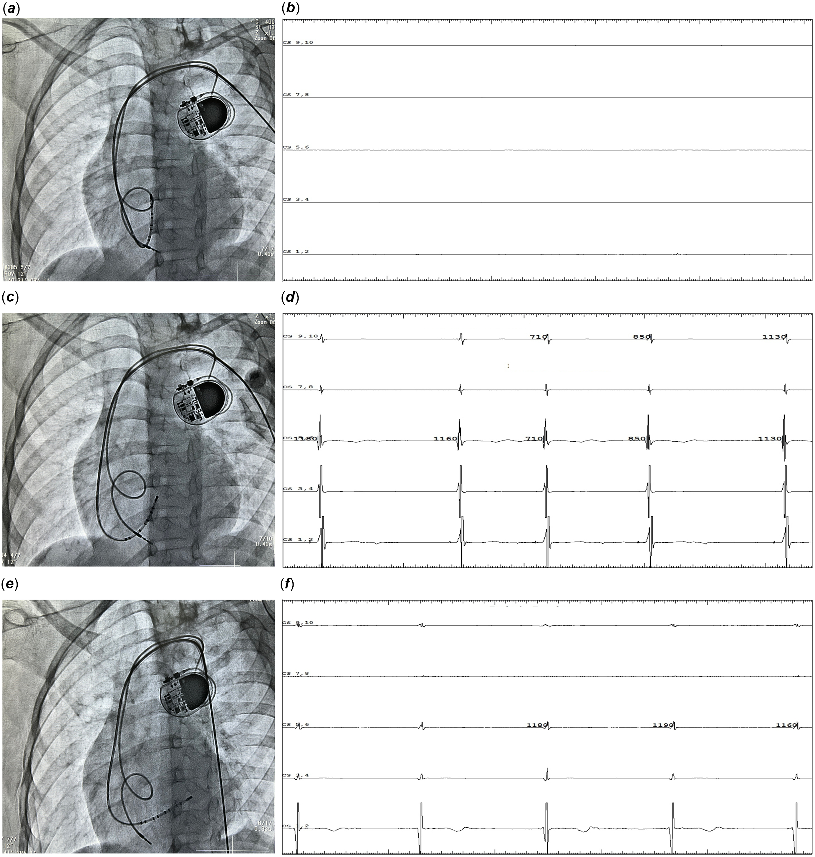
During the planned dual-chamber pacemaker upgrade procedure, initial attempts were made to establish atrial pacing capability through right atrial lead placement. Despite systematic exploration of multiple anatomical sites within the right atrium, adequate atrial capture could not be achieved, raising suspicion for extensive atrial conduction abnormalities involving both atria. To comprehensively evaluate atrial electrophysiological function, coronary sinus electrode mapping was performed. Initial positioning of the coronary sinus electrode within the right atrium (Figure 2( a )) demonstrated a complete absence of electrical activity along the anterior right atrial wall (Figure 2( b )). Subsequently, the electrode was advanced across the mitral valve (Figure 2( c )), revealing only ventricular electrograms (V waves) that corresponded temporally with surface ECG QRS complexes, confirming ventricular electrical activity while demonstrating absent atrial signals. Further assessment involved coronary sinus electrode placement within the coronary sinus ostium (Figure 2( e )), which similarly demonstrated absence of left atrial electrical potentials, with only left ventricular electrograms (V waves) detectable (Figure 2( f )). Comprehensive pacing attempts from both the coronary sinus and right atrial positions failed to achieve atrial capture, even at maximum output parameters (20 mA), confirming the diagnosis of atrial standstill. These intraoperative findings demonstrated disease progression from the initial presentation of severe sinus bradycardia with prolonged sinus arrest in early childhood to complete bi-atrial standstill by adolescence. This aggressive pathological evolution precluded dual-chamber pacemaker implantation due to the absence of viable atrial tissue for pacing. Consequently, the therapeutic strategy was modified to single-chamber ventricular pacemaker replacement, utilising the existing ventricular pacing lead while maintaining the original pacing configuration.
Intraoperative electrophysiological assessment of the proband. ( a )–( b ). CS electrode mapping demonstrated complete absence of electrical activity along the anterior right atrial wall. ( c )–( d ). Electrode was advanced across the mitral valve, revealing only ventricular electrograms that corresponded temporally with surface ECG QRS complexes, confirming ventricular electrical activity while demonstrating absent atrial signals. ( e )–( f ). CS electrode placement within the coronary sinus ostium, which similarly demonstrated absence of left atrial electrical potentials, with only left ventricular electrograms detectable.

Figure 2. Long description
The image shows an intraoperative electrophysiological assessment of the proband. The first set of images (a and b) displays CS electrode mapping, demonstrating the complete absence of electrical activity along the anterior right atrial wall. The second set (c and d) shows the electrode advanced across the mitral valve, revealing only ventricular electrograms that correspond temporally with surface ECG QRS complexes, confirming ventricular electrical activity while demonstrating absent atrial signals. The third set (e and f) illustrates the CS electrode placement within the coronary sinus ostium, which similarly demonstrates the absence of left atrial electrical potentials, with only left ventricular electrograms detectable.
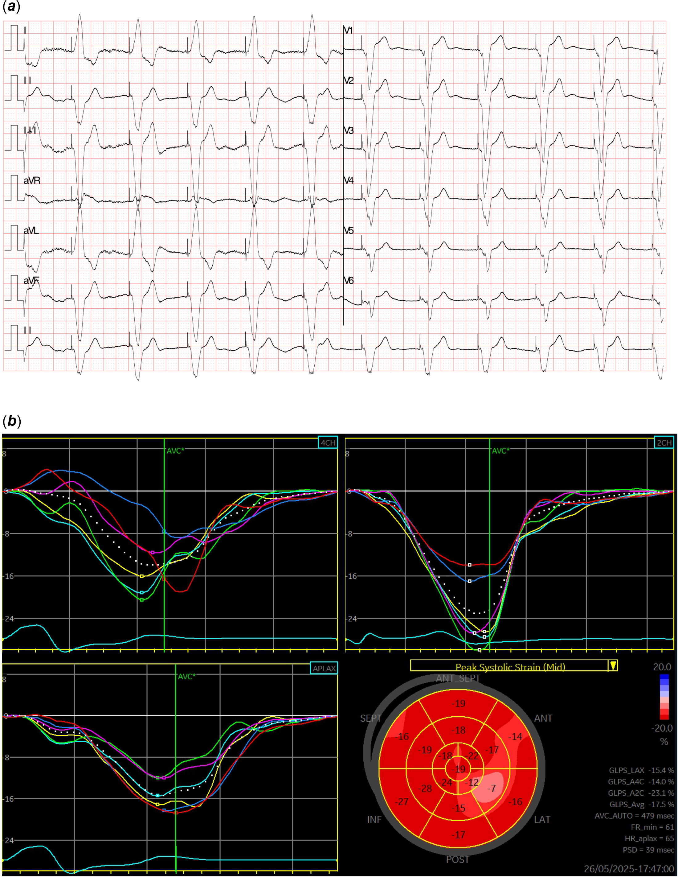
To evaluate the impact of chronic right ventricular apical pacing on myocardial function, a comprehensive assessment of cardiac mechanics was performed. The ventricular demand pacing strategy with the electrode positioned at the right ventricular apex generated an electrocardiographic pattern resembling complete left bundle branch block morphology (Figure 3( a )). Left bundle branch area pacing was not feasible given the patient’s young age at initial implantation (5 years old), necessitating conventional right ventricular apical pacing with potential for electromechanical dyssynchrony and subsequent cardiac dysfunction. Myocardial strain analysis was performed to quantify regional and global left ventricular function. The assessment demonstrated the absence of significant paradoxical motion between the interventricular septum and left ventricular free wall, indicating preserved mechanical synchrony despite the non-physiological activation pattern. However, speckle-tracking echocardiography revealed mildly reduced global longitudinal strain (GLS = −7%) with particular impairment noted in the lateral myocardial segments (Figure 3( b )). This finding suggests subclinical myocardial dysfunction potentially attributable to chronic right ventricular pacing-induced electromechanical dyssynchrony, though overt mechanical dissociation was not observed.
Myocardial function assessment on VVI strategy. ( a ). ECG pattern resembling CLBBB morphology. ( b ). The speckle-tracking echocardiography revealed mildly reduced global longitudinal strain (GLS = −7%) with particular impairment noted in the lateral myocardial segments.

Figure 3. Long description
The image contains two main sections. The first section (a) displays an electrocardiogram (ECG) pattern resembling complete left bundle branch block morphology. It shows multiple leads, including I, II, III, aVR, aVL, aVF, V1, V2, V3, V4, V5, and V6, each with distinct waveforms. The second section (b) presents three speckle-tracking echocardiography graphs and a polar map. These graphs illustrate the strain curves for different views: two-chamber, four-chamber, and apical long-axis. The polar map visually represents the global longitudinal strain (GLS) with values distributed across different myocardial segments. The GLS is mildly reduced at -7 percent, with notable impairment in the lateral myocardial segments. The graphs and map together provide a detailed assessment of myocardial function.
Molecular results
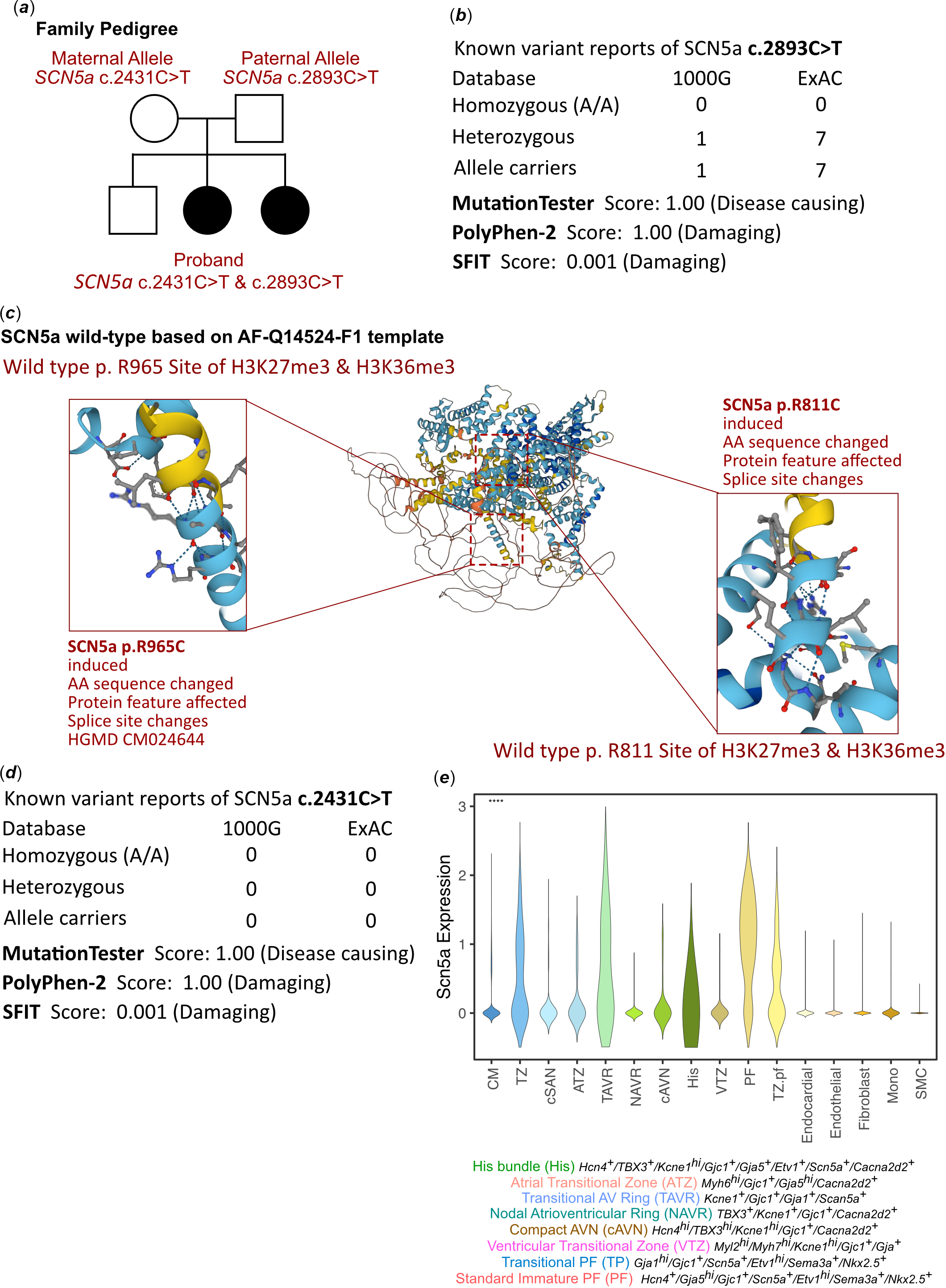
As an inherited cardiomyopathy had been suspected, a genetic test was involved to explore any associated variants. Whole-exon sequencing had been performed for this proband and her parents. According to the analysis result of WES, a compound heterozygous variant had been identified as c.2431C>T (p.R811C) and c.2893C>T (p.R965C) of the SCN5A gene. (Figure 4( a )) The allele c.2431C>T was maternally inherited and c.2893C>T was paternally inherited. And the variant of SCN5A c.2431C>T had never been reported once in ExAC, 1000G, and ClinVar database, which was also considered as a novel genotype. (Figure 2( b )–( d )) As there was no available full-length protein crystal structure for SCAN5A, which had been analysed by X-ray or cryo-EM, the AlphaFold3 protein structure software (https://alphafoldserver.com/) was used to predict protein crystal structure. The protein structure of SCN5A has been built and named AF-P21333-F1. Reference Varadi, Anyango and Deshpande21,Reference Jumper, Evans and Pritzel22 (Figure 4( c )) Then, we performed modelling analysis using AlphaFold3 for the mutant site in wild type with the AF-Q14524-F1. Then, the molecular function of the variant site had been annotated (Figure 4( c )). This would highlight how the mutation alters the charge distribution of the protein. Changes in charge may affect protein interactions with other molecules, stability, and overall function. In accordance with the ACMG/AMP 2015 guidelines for the interpretation of sequence variants, the novel c.2431C>T (p.R811C) variant was classified as likely pathogenic. This classification is supported by the following evidence: (1) it is a de novo or compound heterozygous variant in a gene with a known recessive disease mechanism (PM2, PM3); (2) multiple lines of computational evidence, including AlphaFold3 structural modelling and charge distribution analysis, support a deleterious effect on the gene product (PP3); and (3) The patient’s phenotype of atrial standstill is highly consistent with a monogenic disorder caused by variants in the SCN5A gene, which is a known disease-causing gene for atrial standstill and cardiac arrhythmia disorders (PP4). And the c.2893C>T (p.R965C) variant, inherited from the father, is a known missense variant reported in ClinVar with conflicting interpretations of pathogenicity, but its presence in trans with a novel likely pathogenic variant supports its contribution to the compound heterozygous state. Using scRNA-seq data (GSE132658), Reference Goodyer, Beyersdorf and Paik23 SCN5A were identified as highly expressed in the His bundle and Purkinje fibre across the transitional zone (Figure 4( e )). So, the loss of function of SCN5A c.2431C>T and c.2893C>T resulted in protein damage, which mainly affected the transduction manner of the His bundle and Purkinje fibre. The variants had been deposited in ClinVar (RCV006547764.1 and RCV006547587.1).
The variants of SCN5A molecular features. ( a ). The proband exhibited a compound heterozygous variant of SCN5A (c.2431C>T and c.2893C>T). ( b ). The variant of SCNA5A c.2893C>T had been reported in ExAC, 1000G and ClinVar. And it has predicted protein damaging by MutationTaster. ( c ). The protein structure of SCN5A has been built and named AF-P21333-F1. ( d ). The variant of SCNA5A c.2431C>T had not been reported in ExAC, 1000G and ClinVar. And it has predicted protein damaging by MutationTaster. ( e ). Murine scRNA-seq data (GSE132658) revealed cardiac Scan5a expression is confined to the conductive bundles and fibres.

Figure 4. Long description
The illustration presents various aspects of SCN5A gene variants. Panel (a) shows a family pedigree indicating a proband with compound heterozygous variants SCN5A c.2431C>T and c.2893C>T. Panel (b) lists the known variant reports of SCN5A c.2893C>T from databases 1000G and ExAC, along with scores from MutationTester, PolyPhen-2, and SFIT indicating its damaging potential. Panel (c) displays the protein structure of SCN5A based on the AF-Q14524-F1 template, highlighting the wild-type and induced variants p.R965C and p.R811C with changes in protein features and splice sites. Panel (d) provides data on the SCN5A c.2431C>T variant, which has not been reported in major databases and is also predicted to be damaging. Panel (e) shows a violin plot of Scn5a expression in various cardiac regions based on murine scRNA-seq data, indicating confined expression in conductive bundles and fibers.
Final diagnosis and treatment
Based on comprehensive clinical evaluation, advanced cardiac imaging, and genetic analysis, the patient was diagnosed with hereditary atrial standstill characterised by aggressive disease progression, manifesting initially as severe bradycardia in early childhood with subsequent evolution to complete bi-atrial electrical silence. This rare inherited cardiomyopathy demonstrates a particularly rapid clinical course in this paediatric case. A structured long-term surveillance protocol has been established, incorporating serial monitoring of GLS as a sensitive marker of subclinical myocardial dysfunction and disease progression. Regular assessment of cardiac mechanics will facilitate early detection of pacing-induced cardiomyopathy and guide therapeutic optimisation. For future device replacement procedures, left bundle branch area pacing is under consideration as a more physiological pacing modality to minimise electromechanical dyssynchrony and preserve long-term cardiac function. This approach aims to mitigate the potential adverse effects of chronic right ventricular apical pacing while maintaining adequate cardiac support in this complex paediatric case with progressive atrial standstill. While left bundle branch area pacing represents a promising physiological pacing strategy to mitigate the deleterious effects of chronic right ventricular pacing, its application in an 11-year-old child with lifelong device dependency presents several unique technical challenges. These include (1) the smaller cardiac dimensions and thinner interventricular septum in a child, which increase the risk of septal perforation and coronary artery injury; (2) the lack of dedicated, paediatric-sized delivery sheaths and pacing leads optimised for left bundle branch area pacing; (3) the long-term uncertainty regarding the impact of a fixed pacing lead in the left bundle branch region on ventricular function and valvular competence over decades. So, a long-term surveillance protocol should be established, incorporating serial echocardiography with strain imaging to assess ventricular synchrony and function, as well as periodic device interrogation to monitor lead parameters.
Discussion
This case exemplifies the clinical spectrum and therapeutic challenges associated with atrial standstill, a rare inherited cardiac conduction disorder characterised by complete cessation of atrial electrical and mechanical activity. The pathophysiology involves progressive loss of atrial automaticity and conductivity, resulting in profound bradycardia with compensatory emergence of junctional or ventricular escape rhythms to maintain cardiac output. Both patients harboured pathogenic variants in the SCN5A gene, which encodes the α-subunit of the voltage-gated cardiac sodium channel (Nav1.5). Reference Marini, Iseppi and Quintarelli24,Reference Surget, Hermida, Maltret, Champ-Rigot, Gandjbakhch and Extramiana25 SCN5A mutations are associated with a broad spectrum of cardiac channelopathies, with atrial standstill representing one of the more severe phenotypic manifestations. The loss-of-function mutations likely result in impaired sodium channel activity, leading to progressive atrial electrical silence and conduction system dysfunction. Reference Kohli and Nayak26–Reference Anand, Shah, Sabnis and Mahajan28 The case demonstrated a characteristic biphasic clinical course, initially presenting with persistent atrial tachycardia during early childhood, potentially representing a compensatory arrhythmic response to underlying atrial conduction abnormalities. Reference Tan, Giglia and Cecchin1,Reference Howard, Chiang and Ceresnak29 Subsequently, progressive deterioration of atrial electrical function culminated in severe bradycardia, necessitating single-chamber ventricular pacemaker implantation. The requirement for device replacement and detection of prolonged sinus pauses during surveillance monitoring underscores the dynamic and inexorably progressive nature of this condition.
The primary therapeutic intervention for atrial standstill involves permanent ventricular pacemaker implantation to restore adequate chronotropic support and maintain haemodynamic stability. In the absence of identifiable reversible aetiologies, cardiac pacing represents the sole definitive treatment modality for atrial standstill. Reference John and Kumar30 The complete loss of atrial electrical activity renders cardiac rhythm entirely dependent on unreliable ventricular escape mechanisms, which are frequently inadequate during episodes of severe bradycardia or asystolic events, potentially precipitating life-threatening haemodynamic compromise. These cases emphasise the critical importance of prompt recognition of pathognomonic electrocardiographic features characteristic of atrial standstill, including complete absence of P-wave activity, profound bradycardia, and dependence on junctional or ventricular escape rhythms. Early diagnosis coupled with expeditious pacemaker implantation is essential to ensure adequate haemodynamic support and prevent catastrophic complications, particularly in the vulnerable paediatric population, where delayed recognition may result in irreversible cardiac dysfunction or sudden cardiac death. The identification of pathogenic SCN5A variants in both patients underscores the significant genetic contribution to atrial standstill pathogenesis. Comprehensive genetic testing should be considered the standard of care in atrial standstill cases, as molecular diagnosis provides crucial insights into disease mechanisms, enables cascade family screening, and may inform future therapeutic strategies. The genetic characterisation facilitates risk stratification, guides surveillance protocols, and potentially opens avenues for targeted molecular therapies as our understanding of channelopathy pathophysiology advances. Reference Kong, Zhou and Wang31 Furthermore, genetic counselling becomes imperative for affected families to assess recurrence risks and implement appropriate preventive measures. However, right ventricular pacing is known to worsen heart failure, especially in patients with preexisting left ventricular dysfunction. Reference Sweeney, Bank and Nsah32 In patients presenting with atrial standstill, the long-term cardiovascular consequences of mandatory ventricular pacemaker dependency remain poorly defined and inherently unpredictable. The absence of physiological atrioventricular synchrony, coupled with non-physiological ventricular activation patterns, may predispose to progressive cardiac dysfunction over extended follow-up periods, particularly given the young age at implantation and anticipated lifelong device dependence. Critical evaluation of the initial management strategy reveals that comprehensive intracardiac electrophysiological assessment during the primary pacemaker implantation procedure could have facilitated earlier recognition of underlying atrial standstill. Had systematic evaluation of atrial electrical activity been performed through multi-site atrial mapping and stimulation protocols at the time of initial device placement, the complete absence of atrial function could have been definitively established. This enhanced diagnostic approach would have precluded the necessity for subsequent surgical intervention, thereby reducing cumulative procedural morbidity, minimising repeated anaesthetic exposure, and enabling more comprehensive long-term management planning from the initial presentation. These findings emphasise the critical importance of thorough electrophysiological characterisation during primary device implantation, particularly in paediatric patients with complex inherited conduction disorders where disease evolution may be unpredictable.
This report presents the comprehensive diagnosis and management of a patient with hereditary atrial standstill secondary to pathogenic SCN5A gene variants, illustrating the complex clinical trajectory associated with this rare inherited cardiac conduction disorder. This case demonstrated characteristic evolution, initially manifesting with sinus bradycardia followed by progressive high-grade atrioventricular conduction block. Intraoperative electrophysiological assessment during attempted dual-chamber pacemaker implantation revealed a complete absence of atrial electrical activity across multiple anatomical sites, precluding atrial lead placement and necessitating single-chamber ventricular pacing as the definitive therapeutic strategy. Molecular genetic analysis identifying pathogenic SCN5A variants provided crucial diagnostic confirmation and enabled cascade family screening to identify at-risk relatives prior to symptom onset. Early genetic identification facilitates proactive surveillance strategies, timely therapeutic intervention, and personalised management approaches, representing a paradigm shift toward precision medicine in inherited cardiac conduction disorders. Ongoing research into SCN5A-associated channelopathies continues to enhance understanding of genotype-phenotype correlations and disease progression patterns. Current investigations are expanding therapeutic options through targeted molecular therapies, optimisation of physiological pacing strategies, and refined risk stratification algorithms, collectively contributing to improved long-term outcomes in patients with rare inherited cardiac conduction disorders.
Data availability statement
All the data had been presented in the manuscript and supplementary information. Other data sets used in this study are available from the corresponding author upon reasonable request. The variants had been deposited in ClinVar (Accession Numbers: RCV006547764.1 and RCV006547587.1).
Acknowledgements
None.
Author contributions
Qiu Y and Liu F contributed equally to this work. Qiu Y, Liu F, Zhou K, and Li Y collected the data. Qiu, Liu F, and Lin S performed the analysis. Hua Y and Li Y conceptualised and designed the study, coordinated and supervised data collection, and critically reviewed the manuscript for important intellectual content. Hua Y and Li Y were responsible for the revision of the manuscript for important intellectual content; all authors issued final approval for the version to be submitted.
Financial support
This work was supported by grants from Technology Project of Sichuan Province of China (2024YFFK0078 and 2024YFFK0367) and the National Natural Science Foundation of China (82470249).
Competing interests
The authors declare that they have no conflict of interest.
Ethical standards
The study was approved by the Ethics Committee of West China Second University Hospital, Sichuan University (approval number: 2025-172). Written informed consent was obtained from the parents or legal guardians of all participating children prior to sample collection.
Clinical trial number
Not applicable.
Consent for publication
Not applicable.