Acute rheumatic fever is a non-suppurative, multi-systemic, autoimmune disease developing secondary to group A streptococcal pharyngitis.Reference Baykal, Sağlam and Turan 1 Although the importance of echocardiography is indisputable in demonstrating and monitoring heart involvement, its extensive usage is limited by the fact that it is not easily accessible, is expensive and requires experienced staff to carry it out.
Natriuretic peptides were discovered in the 1980s and include natriuretic and vasodilator peptides. The measurement of N-terminal pro-brain natriuretic peptide, which belongs to the natriuretic peptide family and is released particularly from the ventricle myocytes because of haemodynamic changes such as volume and pressure loading of the heart, is an easily applicable and cheap biochemical reagent method that produces results in a short time and does not require interpretation. Plasma brain natriuretic peptide levels are increased in left heart failure as a result of ventricular hypertrophy and loading. Studies on adults have shown that brain natriuretic peptide levels are increased in rheumatic valvular diseases. This has been associated with atrial dilation.Reference Sutton, Stewart and Gerber 2 – Reference Iltumur, Karabulut and Yokus 5
Unlike the studies in the literature, this study was carried out before the treatment of carditis and in the long-term follow-up. This study aims to investigate the importance of N-terminal pro-brain natriuretic peptide in the follow-up and prognosis of patients with a diagnosis of acute rheumatic carditis.
Materials and methods
This prospective study was carried out at Erciyes University Medical Faculty between November, 2009 and October, 2010 with the approval of the Ethics Committee. A total of 20 patients diagnosed with acute rheumatic carditis at the Department of Pediatric Cardiology in the university’s Children’s Hospital were included in the study. The control group comprised 20 healthy children with similar age/gender distribution presenting at the Cardiology Outpatient Clinic with chest pain and found to have normal examination, including electrocardiographic, biochemical and echocardiographic findings.
The diagnosis of acute rheumatic fever was made according to the modified Jones’ criteria updated in 1992: the presence of two major or one major and two minor findings.Reference Baykal, Sağlam and Turan 1 Patients diagnosed with acute rheumatic fever underwent colour Doppler echocardiography by the same paediatric cardiologist, and those with a definite diagnosis of carditis but without hypertension, renal and hepatic dysfunction were included in the study. Antistreptolysin O, C-reactive protein, erythrocyte sedimentation rate, and N-terminal pro-brain natriuretic peptide levels were tested at diagnosis. Teleradiography and electrocardiography were carried out. Serum C-reactive protein, erythrocyte sedimentation rate, and N-terminal pro-brain natriuretic peptide tests and echocardiography were repeated in the first week, first month and third month.
Left ventricle functions, ejection fraction, fractional shortening, diameters of the left atrium, aortic root and the diastolic and end-systolic diameters of the left ventricle were measured during echocardiography at diagnosis and in the first week, first month and third month. Mitral regurgitation, aortic regurgitation and tricuspid regurgitation were detected based on measuring the diameter of the vena contracta with colour Doppler. If this value was<0.3 cm, it was considered as first degree, if between 0.3 and 0.7 cm as second degree, and if >0.7 cm as third degree. Patients with moderate and severe carditis were given steroids at 2 mg/kg/day – maximum 60 mg/day – for 2–4 weeks as anti-inflammatory therapy. When the dose was tapered, aspirin 80–100 mg/kg/day was added to the treatment. Patients with mild carditis – six patients – were given only aspirin – 80–100 mg/kg/day. All patients had normal systolic functions at diagnosis, and as this continued during therapy none of our patients received anticongestive therapy.
The control group comprised 20 healthy children with similar age/gender presenting at the Pediatric Cardiology Outpatient Clinic for chest pain, and found to have normal electrocardiographic, biochemical, teleradiographic and echocardiographic findings. The control group was tested once for N-terminal pro-brain natriuretic peptide levels and once for echocardiography.
Statistical analysis
SPSS for Windows 15.0 program was used for statistical analysis. Distribution of variables was determined using the Shapiro–Wilks test. The χ2 test was used for comparing inter-group proportional distributions. For comparisons of inter-group means, the independent t-test was used for variables showing normal distribution and Mann–Whitney U test for variables showing abnormal distribution. For comparisons of intra-group means, the paired t-test was used for variables showing normal distribution and Wilcoxon test for those showing abnormal distribution. Friedman analysis was performed for the evaluation of the change of variables in the baseline, first week, first month and third month of the abnormally distributed variables in dependent groups. In this study, correlation analysis was done. The Pearson and Spearman correlation coefficients were calculated. Linear regression analysis was performed by using enter model. In all statistical analyses, values of p<0.05 were considered to be significant.
Results
The study group consisted of 20 patients aged between 5 and 16 years (mean age 11.6±2.7 years), with nine girls (45%) and 11 boys (55%). The control group was aged between 6 and 16 years (mean age 11.6±2.7 years). In the control group, there were nine girls and 11 boys. There were no differences between the study and control groups regarding age (p=0.15) and gender (p=0.12) distribution. At diagnosis, six patients had isolated carditis; nine had carditis and arthritis; four had carditis and chorea; and one had arthritis, carditis, and chorea. There was no significant difference between the presentation forms of the patients and gender (p=0.10). None of the patients had erythema marginatum or subcutaneous nodules. Of the minor findings, all patients had elevated acute phase reactants, seven patients had fever, 16 had arthralgia and 15 had prolonged PR in the electrocardiography.
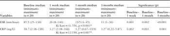
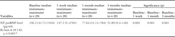
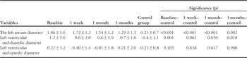
Erythrocyte sedimentation rate and C-reactive protein levels measured at diagnosis were significantly higher than those measured in the first week, first month and third month (p=0.001) (Table 1). The N-terminal pro-brain natriuretic peptide levels of children with acute rheumatic carditis were significantly higher than those of the control group at diagnosis, first week, and first month (p<0.05) (Table 2). When the N-terminal pro-brain natriuretic peptide levels of the patient group were compared, the N-terminal pro-brain natriuretic peptide levels at diagnosis were significantly higher than those in the first week, first month and third month (p=0.001) (Table 3). When echocardiographic investigations at baseline, in the first week, first month and third month were compared with the control group, left atrium diameter and left ventricular end-diastolic diameter values were found to be statistically significant (Table 4).
Erythrocyte sedimentation rate and C-reactive protein values during the working of the patient group.

CRP=C-reactive protein; ESR=erythrocyte sedimentation rate
* Friedman test
Comparison of serum N-terminal pro-brain natriuretic peptide levels in patients and control subjects.

NT-ProBNP=N-terminal pro-brain natriuretic peptide
N-terminal pro-brain natriuretic peptide values during the working of the patient group.

* Friedman test
Comparison of echocardiograpic values during the working of the patients and control subjects (z-scores).

A strong relationship was found between left atrium diameter and brain natriuretic peptide level at the time of diagnosis (r=0.57, p=0.008). However, in the first month the same relationship was found to be intermediate (r=0.40, p=0.007). On the other hand, there was a significant positive correlation between N-terminal brain natriuretic peptide levels and erythrocyte sedimentation rate values at diagnosis and in the first month (p<0.001). At diagnosis, all patients had mitral failure: it was first degree in 30%, second degree in 30% and third degree in 40% of the patients. Whereas the first degree mitral failure rate rose to 80% in the third month, second degree mitral failure regressed to 5% and third degree mitral failure to 15%.
Discussion
Although acute phase reactants are used in the follow-up of acute rheumatic fever, new parameters are needed for the follow-up of carditis. It still remains a clinical syndrome for which no specific diagnostic test exists. When the distribution of the major and minor criteria of acute rheumatic fever was investigated in the patient group, all patients were noted to have carditis, 50% had arthritis, and 25% had Sydenham’s chorea. C-reactive protein, which is one of the acute phase reactants, is useful in showing the activity of the disease at diagnosis, response to therapy, and exacerbation of the disease after cessation of therapy. It returns to normal levels faster than the sedimentation rate. Elevated erythrocyte sedimentation rate and C-reactive protein levels in our patients at diagnosis and regression with therapy are compatible with classical knowledge.Reference Tani 6 – Reference Gerber, Stewart and French 10
In acute rheumatic carditis, endocardial involvement shows itself with valvular involvement. Whereas Potocki et alReference Potocki, Mair and Weber 11 did not show a correlation between N-terminal pro-brain natriuretic peptide secretion and mitral failure severity and left ventricular remodelling in patients with mitral failure, Gerber et alReference Gerber, Stewart and French 10 showed that as the severity of aortic leakage is increased brain natriuretic peptide levels are increased, reflecting the volume and function of the left ventricle.Reference Gerber, Stewart and French 10 In a study by Davutoglu et alReference Davutoglu, Celik and Aksoy 4 carried out on 92 adult patients with chronic rheumatic valvular disease, N-terminal pro-brain natriuretic peptide levels increased with the severity of mitral failure and tricuspid failure. In our study, all patients had mitral failure at diagnosis. There was no significant correlation between N-terminal pro-brain natriuretic peptide and mitral failure. The results were compatible with the results of the study by Potocki et al.Reference Potocki, Mair and Weber 11 This may be due to the low number of cases and the fact that cases received treatment at an early stage.
Although rheumatic carditis is also an important cause of heart failure in developing countries, there are few studies investigating brain natriuretic peptide levels in these patients.Reference Cimen, Oran and Cimen 12 In the study by Cimen et al,Reference Cimen, Oran and Cimen 12 elevation of N-terminal pro-brain natriuretic peptide levels in patients before treatment was significant compared with the control group, whereas post-treatment levels were not significantly different from the control group.
After the discovery of B-type natriuretics in the diagnosis of heart failure and in showing the prognosis of cardiovascular diseases, the correlation between brain natriuretic peptide levels and echocardiography measurements has become a major subject of research interest. In the assessment of cardiac function, clinical status and outcomes, measurement of peptide levels can provide a supplemental contribution to echocardiography measurements. Nir et alReference Nir, Bar-Oz and Perles 13 showed that brain natriuretic peptide and N-terminal pro-brain natriuretic peptide levels are elevated in increased ventricular pressure and volume loading. Tavlı et alReference Tavlı, Sarıtas and Saylan 14 showed that brain natriuretic peptide is a successful biochemical marker of ventricular dysfunction, especially the ones leading to volume and pressure elevation in the left ventricle. In a study by Fried et al,Reference Fried, Bar-Oz and Perles 15 N-terminal pro-brain natriuretic peptide levels were shown to be positively correlated with left ventricular diameter, volume and muscular mass in 10 patients diagnosed with left ventricular functional disorder and seven patients with chronic dilated cardiomyopathy, and they were inversely correlated with ejection fraction. Owing to the fact that all of the patients included in our study had normal left ventricle functions, there was no significant correlation between ejection fraction, fraction shortening values, and N-terminal pro-brain natriuretic peptide levels. Sutton et alReference Sutton, Stewart and Gerber 2 compared the brain natriuretic peptide and N-terminal pro-brain natriuretic peptide levels in symptomatic and asymptomatic mitral failure patients with normal ejection fraction. In our study, a significant correlation was found between left atrium diameters at the time of diagnosis and in the first month and the N-terminal pro-brain natriuretic peptide levels of the same period.
In our study including 20 children with active carditis, elevated serum levels of N-terminal pro-brain natriuretic peptide at diagnosis returned to normal levels with anti-inflammatory therapy. N-terminal pro-brain natriuretic peptide was found to be valuable in the follow-up of anti-inflammatory therapy, just like C-reactive protein and erythrocyte sedimentation rate. In particular, there was a significantly positive correlation between N-terminal pro-brain natriuretic peptide and erythrocyte sedimentation rate. This shows that in addition to being an important indicator of heart failure, N-terminal pro-brain natriuretic peptide can also be used as an easy and cheap inflammatory marker in the follow-up of carditis. Studies with a longer follow-up and with more cases and including children with acute rheumatic carditis with valvular involvement at different levels are needed.
Acknowledgment
All authors contributed extensively and equally to this work.
Financial Support
This research received no specific grant from any funding agency, commercial or not-for-profit sectors.
Conflicts of Interest
None.
Ethical Standards
The ethical aspects were respected and the research was approved by the Committee of Ethics and Research of Erciyes University.