

Introduction
Delayed radiation-induced vasculopathy is an increasingly recognised cause of stroke in cancer survivors (Kang, Kwon, & Kim, Reference Kang, Kwon and Kim2002). In most reported cases, this involves either a single haemorrhagic stroke – often secondary to rupture of a radiation-induced cavernous haemangioma – or a single ischaemic stroke (either large-vessel or lacunar) related to premature atherosclerosis (Keezer & Del Maestro, Reference Keezer and Del Maestro2009; Plummer, Henderson, O’Sullivan, & Read, Reference Plummer, Henderson, O’Sullivan and Read2011). In this article, we report a unique case presenting with a progressive arteriopathy resulting in numerous haemorrhagic and small-vessel ischaemic strokes over a 4-month period.
Case
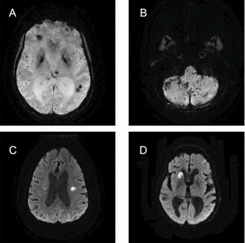
A 40-year-old woman presented with sudden-onset dysphagia, dysarthria and expressive aphasia. Head computed tomography (CT) scan showed an acute left temporo-parietal haemorrhage. Brain magnetic resonance imaging (MRI) showed areas of chronic infarction in the left basal ganglia, midbrain and pontomesencephalic junction. White matter hyperintensities on T2-weighted images were in keeping with microangiopathic disease. The gradient-recalled echo (GRE) sequences revealed hundreds of supra- and infra-tentorial micro-haemorrhages (Figure 1A, B) consistent with cavernous malformations. She improved neurologically, but chronic sequelae included dysarthria and dysphagia.
(A) and (B) Gradient-recalled echo sequences showing multiple microbleeds in the cerebellum, brainstem, cerebral hemispheres and deep grey structures. (C) Diffusion-weighted imaging (DWI) from the second hospital presentation showing an acute infarct in the left corona radiata. (D) DWI from the third hospital presentation showing an acute infarct in the right caudate nucleus.

Three months later, the patient returned with subacute worsening of dysarthria and dysphagia. Brain MRI revealed acute infarction (restricted diffusion) in the left corona radiata (Figure 1C) and right subinsular region. One month later she returned with dysarthria, decreased level of consciousness and ptosis, and MRI demonstrated acute infarcts in the right caudate head (Figure 1D), centrum semiovale, parasagittal frontal lobe and middle cerebellar peduncle, as well as the central midbrain. An MR angiogram with gadolinium, a trans-oesophageal echocardiogram (with injection of agitated saline), Holter monitor and hypercoagulable work-up (including antiphospholipid antibody testing, homocysteine, protein C and S deficiencies, antithrombin III deficiency, Factor V Leiden mutation and prothrombin 220210 mutation) were negative. Fasting triglycerides were 7.01 mmol/l. She started aspirin 81 mg and atorvastatin 10 mg daily.
Her past medical history included childhood leukaemia (aged 12), treated with whole-body and cranio-cervical radiation. Unfortunately, detailed records of her radiotherapy course were not available. Subsequently she had a bone marrow transplant, contracting hepatitis C from blood products. In 2007 she underwent liver transplantation for cirrhosis. She had hypothyroidism and was diagnosed with mild hypertension 6 months prior to presentation. Medications included ciclosporin, azathioprine, prednisone, cotrimoxazole, omeprazole, levothyroxine, amlodipine, ramipril and alendronate. She was a non-smoker. There was no family history of stroke.
Discussion
This patient presented with recurrent haemorrhagic and ischaemic strokes over a 9-month period. Her medical history was most significant for remote whole-body and cranio-cervical radiation. Serial MRI imaging demonstrated multiple cavernous malformations throughout the brain, with chronic and acute haemorrhage. She had multiple, small, acute, ischaemic infarcts (all under 1 cm in diameter) involving the subcortical white matter, basal ganglia and brainstem. Multiple investigations failed to demonstrate large-artery atherosclerosis, a cardioembolic source or a thrombophilic state.
The location and size of this patient's ischaemic lesions are most consistent with small-vessel disease. In the relative absence of traditional vascular risk factors, delayed radiation-induced small-vessel arteriopathy is the most likely aetiology. Delayed radiation vasculopathy is a recognised cause of ischaemic and haemorrhagic stroke, with several implicated pathologies (Shobha, Smith, Demchuk, & Weir, Reference Shobha, Smith, Demchuk and Weir2009).
Many authors have reported accelerated atherosclerosis as a result of cranio-cervical radiation, causing progressive occlusion of large arteries supplying the brain. The mechanism is thought to be via endothelial cell injury within the extracranial carotid artery (Plummer et al., Reference Plummer, Henderson, O’Sullivan and Read2011; Shobha et al., Reference Shobha, Smith, Demchuk and Weir2009). More rarely, there can be atherosclerotic stenosis of the intracranial internal carotid artery (Keezer & Del Maestro, Reference Keezer and Del Maestro2009; Plummer et al., Reference Plummer, Henderson, O’Sullivan and Read2011), or its main branches. A recent review of 12 consecutive cases of acute stroke in patients who had previously received radiotherapy for head and neck cancers determined that large artery atherosclerosis, either artery-to-artery embolism or atherothrombosis of the parent vessel, was the stroke mechanism in 11 of the 12 cases (Kang et al., Reference Kang, Kwon and Kim2002).
Cavernous haemangiomas (or cavernomas) are vascular sinusoids lined by a single layer of endothelium and lack intervening brain tissue. Within the past 20 years there has been increased recognition that cavernomas can be acquired lesions secondary to cranial radiation (Plummer et al., Reference Plummer, Henderson, O’Sullivan and Read2011). There is an increased rate of haemorrhage in cavernomas secondary to cranial radiation, compared to sporadic or familial cavernomas (Keezer & Del Maestro, Reference Keezer and Del Maestro2009). The most common symptoms of cavernoma haemorrhage include seizures, headache and motor dysfunction; however, the majority of bleeds are asymptomatic, with only radiological evidence (Keezer & Del Maestro, Reference Keezer and Del Maestro2009).
Finally, evidence of radiation-induced damage to small vessels, such as arterioles and capillaries, has been observed pathologically (Shobha et al., Reference Shobha, Smith, Demchuk and Weir2009). Predictably, this may manifest as small-vessel infarction, seen on imaging as lacunar strokes. This phenomenon is relatively poorly characterised, and only a few reports have linked lacunar stroke and radiation-induced vasculopathy (Salih, Higgins, Warburton, & Baron, Reference Salih, Higgins, Warburton and Baron2007; Shobha et al., Reference Shobha, Smith, Demchuk and Weir2009). Interestingly, in the case series mentioned above (Kang et al., Reference Kang, Kwon and Kim2002), the one case of acute stroke not attributed to large-vessel atherosclerosis was characterised by left striatocapsular infarction. This may have represented a lacunar infarction secondary to radiation-induced small-vessel disease. One suggested mechanism for post-radiation lacunar infarction is radiation-induced parent vessel (e.g., middle cerebral artery) atheroma causing occlusion of the origin of a perforating branch (Kang et al., Reference Kang, Kwon and Kim2002).
We could find evidence of only a single case-report describing a patient who had received brain radiotherapy presenting with recurrent small-vessel infarctions, with these occurring over the course of 14 years, starting 10 years post-radiation (Shobha et al., Reference Shobha, Smith, Demchuk and Weir2009). In that report, the patient also sustained a thalamic haemorrhage.
The present case is unique for a number of reasons. Our patient had evidence of innumerable cavernous haemangiomas and small-vessel ischaemic infarctions, without other traditional vascular risk factors, with both pathologies felt to be related to her prior cranial irradiation. To our knowledge, no other reported case has presented with such a fulminant clinical course, over a period of months, characterised by both haemorrhagic and multiple ischaemic strokes. It is of interest to note that our patient presented with prominent and worsening dysarthria, which appears to be a prominent feature of other published accounts of lacunar infarctions due to radiation vasculopathy (Kang et al., Reference Kang, Kwon and Kim2002; Shobha et al., Reference Shobha, Smith, Demchuk and Weir2009).
In summary, cranial irradiation is an important risk factor for both large- and small-vessel ischaemic strokes. Delayed radiation-induced vasculopathy can present in a fulminant manner with recurrent strokes over a short period of time.
Disclosure statement
The authors have no conflicts of interest or financial disclosure.