Introduction
Cyantraniliprole, a widely used anthranilic diamide insecticide, disrupts calcium homeostasis by targeting ryanodine receptor (RyR) in the sarcoplasmic reticulum (SR). Reference Samurkas, Yao and Hadiatullah1 While mammals possess multiple RyR isoforms, insects have a single type that shares 47% homology to the mammalian RyRs. Reference Takeshima, Nishi and Iwabe2 Despite its higher affinity for insect RyR, cyantraniliprole’s potential for off-target effects in mammals requires further investigation.
The SR acts as the primary calcium store within cells, with its release tightly regulated by RyRs, inositol trisphosphate (IP3), SR calcium pumps, and calcium-binding proteins. Reference Arruda and Hotamisligil3 Disruption of RyR function by diamides leads to intracellular calcium depletion, potentially causing thermoregulatory disturbances, muscle hyperactivity, and cardiac arrhythmias. Reference Selby, Lahm and Stevenson4 The European Food Safety Authority (EFSA) reports hepatic alterations in mammals exposed to cyantraniliprole in both short-term and long-term studies. 5 However, oral absorption is estimated to be 70%, with rapid excretion within 48 hours and no bioaccumulation. The acceptable daily intake of cyantraniliprole for humans is 0.01 mg/kg body weight. Reference da Silva Scarton, Tsuzuki and Guerra6
Previous research highlights the potential mammalian toxicity of cyantraniliprole, with studies showing oxidative stress and altered liver weight in Wistar rats exposed to 10 and 150 mg/kg body weight for 28 days postnatally (day 60). Reference da Silva Scarton, Tsuzuki and Guerra6 The liver, a key organ for xenobiotic metabolism, is susceptible to imbalances between antioxidant defenses and reactive oxygen species (ROS) production. Reference Marí, Colell and Morales7 The kidneys play a vital role in maintaining body homeostasis through blood filtration and calcium regulation. Reference Faria, Ahmed, Gerritsen and Mihaila8–Reference McMahon9 ROS can disrupt calcium homeostasis by increasing cytosolic calcium influx, leading to altered cellular signaling pathways and potentially activating or repressing protein and gene expression. Reference Ermak and Davies10 Conversely, elevated intracellular calcium can further enhance ROS formation. This vicious cycle underlies cell death and carcinogenesis models, where aberrant calcium signaling exacerbates ROS production and overwhelms antioxidant defenses. Reference Hempel and Trebak11
Additionally, evidence demonstrates that maternal exposure to agrochemicals during lactation or pregnancy may contribute to the development of physiological changes in organisms. Reference Firmin, Bahi-Jaber and Abdennebi-Najar12 Pesticide exposure has been shown to induce physiological alterations in exposed individuals and their offspring. Given the lack of data on the transgenerational effects of cyantraniliprole on liver and kidney histophysiology, this study aims to evaluate the impact of maternal gestational and lactational exposure on these organs of pubescent and adult male offspring.
Methods
Drug preparation
Cyantraniliprole, a chemical compound also known as 3-Bromo-1-(3-chloro-2-pyridinyl)-N-[4-cyano-2-methyl-6[(methylamino)carbonyl]phenyl]-1H-pyrazole-5-carboxamide (BENEVIA® 10% w/v – Du Pont Brasil S.A., Barueri, SP, Brazil), was sourced from the Agronomy Department of the Agricultural Sciences Center at the State University of Londrina (UEL). Tap water was used to dilute the toxic agent to a final pesticide concentration of 10 mg/kg. Given the lack of prior studies on the systemic toxicity of cyantraniliprole on liver development, the dosage was determined based on the pesticide’s data sheet registered with the Ministry of Agriculture, Livestock, and Supply (registration number 13,915), which classifies a dose of 10 mg/kg as low. Additionally, the European Food Safety Authority (EFSA) establishes a no observed adverse effect level (NOAEL) of 1000 mg/kg body weight for both maternal and fetal organisms in rats. Thus, the 10 mg/kg/day dose used in this study corresponds to only 1% of the NOAEL defined by EFSA, ensuring a 100-fold safety margin – a common practice in regulatory toxicology to account for inter- and intraspecies variability and to maintain a conservative approach in risk assessment. 13 Moreover, considering that animals are generally more resistant, the 10 mg/kg/day dose represents a level approximately 1,000 times higher than the Acceptable Daily Intake (ADI) established for humans, as reported by ANVISA, when extrapolated to Wistar rats. 14 Furthermore, studies conducted by our research laboratory have already demonstrated that oral exposure to 10 mg/kg of cyantraniliprole for 28 days was capable of inducing hepatic alterations in adult Wistar rats. This study adheres to guideline 443 of the Organization for Economic Co-operation and Development (OECD) concerning the extended one-generation reproductive toxicity study. 15
Animals
Animal care and handling complied with the National Institute of Health’s Guide for the Care and Use of Laboratory Animals, and all experimental procedures received approval from the Ethics Committee of Animal Use of UEL (OF. CIRC. CEUA n° 20/2020, process no. 211037.2019.93). Adult Wistar rats, males (n = 15; 90 days old) and females (n = 33; 80 days old), were obtained from the Central Animal Facility of UEL. The animals were housed in the animal facility of the Laboratory of Toxicology and Metabolic Disorders of Reproduction of the General Biology Department of UEL. They were maintained under a 12-hour light/dark cycle at 22°C ± 3°, with water and standard chow provided ad libitum.
Experimental protocol
For mating, one male was introduced into a cage containing two females (1:2 ratio) in the late afternoon after determining the estrous phase. The next morning, vaginal smears were performed to check for the presence of sperm. Day zero of gestation (GD 0) was determined for females with sperm detected in the vaginal smears. These females (n = 10 per group) were placed in individual cages and divided into two groups: control dams (n = 10) and dams administered cyantraniliprole (n = 10). Cyantraniliprole administration began on the GD 5, in order not to interfere on embryo implantation, Reference Milesi, Alarcón and Ramos16 with a dose of 10 mg/kg/day, administered by gavage in a final volume of 0.5 ml daily, throughout gestation until the end of lactation on postnatal day (PND) 21 of the offspring. Control dams received water in the same volume and manner. All animals were monitored daily for general well-being and signs of toxicity (tearing, salivation, nasal discharge, diarrhea, excessive urination, tremors, agitation, and piloerection).
After birth, the litter was reduced to 8 pups, 4 males and 4 females, whenever possible. After weaning, 1 male rat from each of the 10 females’ offspring was randomly selected to be included in one of the following groups: pubertal offspring of control dams (n = 9), pubertal offspring of dams treated with cyantraniliprole (n = 10), adult offspring of control dams (n = 9), and adult offspring of dams treated with cyantraniliprole (n = 10). Only one male pup per litter was included in the study to minimize the “litter effect,” with each litter considered an independent experimental unit. Offspring were maintained until PND 55 and PND 90, representing pubertal and adult stages, respectively. During the postnatal period, one pup from each control group died, and these losses were considered in the final sample size. Postnatal days (PND) 55 and 90 were selected to represent the pubertal and adult stages, respectively, as these time points coincide with critical periods of endocrine modulation and sexual maturation in rats. This developmental framework allows for the investigation of age-dependent susceptibilities to environmental exposures. Reference Perobelli17 Blood glucose levels were measured 30 minutes before euthanasia, without fasting, using a drop of blood from the animals’ caudal vein in a glucometer (Accu-Chek Active strips, Roche, Taquara, RJ, Brazil). After weighing, the animals were anesthetized with intraperitoneal injection of ketamine (75 mg/kg b.w., Sedomin® 10%, Avellaneda, Argentina) and xylazine (10 mg/kg b.w., Anasedan®, Paulínia, Brazil). Blood was collected via puncture of the inferior vena cava, followed by euthanasia through cardiac puncture. Blood plasma was obtained through centrifugation at 4000 r.p.m. for 20 minutes at 4°C and preserved at 20°C until analysis. Liver and kidneys were dissected and weighed. The left lateral lobe of the liver was sectioned into several pieces for histopathological analysis, evaluation of oxidative stress biomarkers, and inflammatory profile. The right kidney was designated for histopathological analyses, while the left kidney was used for oxidative stress analysis.
Histopathological evaluation
Non-serial histological sections, 5 µm thick, were prepared using a semi-automatic microtome. In hematoxylin and eosin (HE) stained liver sections, 10 images each of the periportal and centrilobular regions were obtained per animal at 40x magnification. Semiquantitative histopathological analysis, conducted by two blinded experienced researchers, assessed centrilobular necrosis, lobular inflammation, sinusoidal dilation, hydropic degeneration, congestion, and eosinophilic hepatocyte cytoplasm, grading findings as absent, mild, moderate, or severe. Reference Tektemur, Güzel and Gül18 In HE-stained kidney sections, 20 images each of the cortical and medullary regions at 40x magnification were captured per animal, evaluating Bowman’s capsule dilation, tubular dilation, cellular degeneration–necrosis, and cellular infiltrate, with findings graded similarly. Reference da Silva JV, Erthal and Vercellone19
The Periodic acid-Schiff (PAS) technique was applied to liver sections to highlight neutral glycoconjugates, Reference Du, Ding and Zou20 while Azan trichrome and Picrosirius Red stains were used to assess total and type I collagen, respectively. Reference Horai, Utsumi and Ono21–Reference Singh, Singh and Singh22 In kidneys, PAS and Azan trichrome techniques were used for similar analyses. Reference da Silva JV, Erthal and Vercellone19 For PAS, 10 images per animal/organ were obtained at 40x magnification, while 10x magnification was used for other techniques. ImagePro Plus 4.5 software (Media Cybernetics, Silver Spring, MD, USA) analyzed the images, with results expressed as a percentage of pixel area per liver/renal region.
Biochemical dosages
Plasma samples were analyzed for aspartate aminotransferase (ALT) and alanine aminotransferase (AST) to assess liver injury, using commercial kits (Labtest® Diagnóstico S.A., Lagoa Santa, MG, Brazil) according to manufacturer instructions. Renal function was evaluated by measuring plasma creatinine and urea levels with commercial kits (VIDA Biotecnologia, Brazil). The tests were conducted using a Multiskan GO Microplate Spectrophotometer (Thermo Scientific) from the Physiological Sciences Department of UEL. ALT and AST results were reported in U/L of plasma, while creatinine and urea were expressed in mg/dL.
Oxidative stress analysis
Protein quantification
Protein concentration in samples was determined using the Bradford method. Samples were homogenized in phosphate buffered saline (PBS) (pH 7.2) and diluted at 1:25. In a 96-well plate, 10 μL of diluted samples and 250 μL of Bradford reagent were pipetted in duplicate, with absorbance measured at 595 nm to adjust sample dilution to a final protein concentration of 1 mg/mL. Reference Bradford23
Lipid peroxidation
Lipid peroxidation was assessed using the thiobarbituric acid reactive substances (TBARS) method according to Federici et al. (2007). 50 μL of normalized samples were pipetted in duplicate into a microplate, followed by 5 μL each of FeCl3, ascorbic acid, trichloroacetic acid (TCA), and thiobarbituric acid (TBA). The reaction was conducted at 90°C for 15 minutes in a water bath, cooled to stop the reaction, and absorbance was read at 535 and 572 nm. Results were expressed as nmol of TBARS per mg of protein. Reference Federici, Shaw and Handy24
Catalase activity
Catalase (CAT) activity was determined by hydrogen peroxide degradation into oxygen and water. Samples and 297 μL of reaction medium (Tris-HCl buffer and H2O2) were placed in a UV microplate in duplicate and read at 240 nm at 15-second intervals for 1 minute. Data were presented as nmol.min−1.mL−1.mg protein−1. Reference Aebi25
Glutathione-S-transferase activity
Glutathione S-transferase (GST) activity was determined by monitoring thioether formation from reduced glutathione (GSH) interaction with 1-chloro-2,4-dinitrobenzene (CDNB) at 340 nm for 5 minutes at 40-second intervals, as described by Keen et al. (1976). Values were expressed as nmol.min−1.mL−1.mg protein−1. Reference keen, Habig and Jakoby26
Reduced glutathione quantification
GSH concentration was measured using the method by Rahman et al. (2006), with modifications. The oxidation of GSH by 5,5′-dithiobis 2-nitrobenzoic acid (DTNB) generates a yellow color measured at 412 nm, with results expressed as μmol/mg of protein. Reference Rahman, Kode and Biswas27
Inflammatory profile
Myeloperoxidase (MPO) activity
Liver samples (approximately 100 mg) were removed and stored in a cryotube with 200 μL of potassium phosphate (5mM; pH 6,0) buffer with hexadecyl trimethylammonium bromide (0,5%) at −80°. The samples were homogenized in ice condition using a Tissue-Tearor (Biospec®). Subsequently, the homogenates were centrifuged, the supernatant (15 μL) was transferred to microtubes with 200 μL 50 mM K2PO4 buffer, pH 6.0, containing 0.167 mg/mL of o-dianisidine dihydrochloride and 0.015% hydrogen peroxide and analyzed for MPO activity. Each sample was assayed in triplicate and read at 450 nm. The results were expressed as the number of neutrophils per mg of protein using a standard curve of neutrophils (196–400.000 neutrophils). Reference Ferraz, Manchope and Andrade28
N-acetyl-β-D-glucosaminidase (NAG) Activity
For this technique, 10 μL of the supernatant obtained from the MPO activity procedure was separated and added to a 96-well plate, followed by the addition of 40 μL of 50 mM phosphate buffer, pH 6.0. The reaction was initiated by adding 2.24 mM of 4-nitrophenyl NAG. The plate was incubated at 37°C for 10 minutes, and the reaction was stopped by adding 100 μL of 0.2 M glycine buffer, pH 10.6. NAG activity was determined at 400 nm using a spectrophotometer (Multiskan GO Microplate, Thermo Fisher Scientific, Vantaa, Finland). NAG activity results were expressed as the number of macrophages per mg of protein using a macrophage standard curve (196–400,000 macrophages). Reference Ferraz, Manchope and Andrade28
Statistical analysis
Data normality and homogeneity were verified using Kolmogorov–Smirnov and Levene tests, respectively. Student’s t-test or Kruskal–Wallis tests were applied for parametric and non-parametric data, respectively, with Tukey’s or Dunn’s post hoc tests. A significance level of 5% was adopted for all statistical analyses. Statistical analyses were performed using GraphPad Prism version 9.0 (GraphPad Software, San Diego, CA, USA).
Results
Capillary blood glucose, animal, and tissue weight
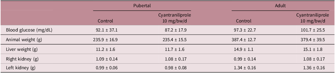
No significant differences were observed in body weight, capillary blood glucose levels, or liver and kidney weights in the offspring of dams that were administered cyantraniliprole during the pubertal and adult stages (Table 1). There were no differences in food and water consumption (data not shown).
Effects of cyantraniliprole administration on capillary glucose concentration, body weight, and tissue weight in Wistar rats at pubertal and adult ages

Results presented as mean ± standard deviation of the mean. T-test (P < 0.05).
Histopathological and morphometric analysis of rat liver and kidney
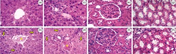
Pubertal animals exposed to cyantraniliprole at a dose of 10 mg/kg/day during pregnancy and lactation exhibited severe vacuolization of hepatocytes in both the centrilobular region and the portal space, as well as moderate congestion in the portal space (Fig. 1 – B and D). In adult animals, moderate vacuolization of hepatocytes and vascular congestion were observed in both regions (Fig. 2 – B and D). Morphometric analyses did not reveal significant differences between the groups.
Photomicrographs of the centrilobular region (A and B), periportal region (C and D), cortex (E and F), and medulla (G and H) of pubertal offspring from control dams that received potable water/vehicle (A, C, E, and G) and from dams that received Cyantraniliprole 10 mg/kg/d (B, D, F, and H). The arrow indicates the vacuolization of hepatocytes (B) and the increase in Bowman’s space (F). The asterisk indicates the congestion of the portal vein (D). Staining: hematoxylin and eosin, magnification 40x. Scale bar: 20 µm.

Photomicrographs of the centrilobular region (A and B), periportal region (C and D), cortex (E and F), and medulla (G and H) of adult offspring from control dams that received potable water/vehicle (A, C, E, and G) and from dams that received Cyantraniliprole 10 mg/kg/d (B, D, F, and H). The arrow indicates the vacuolization of hepatocytes (B and D). The asterisk indicates the congestion of centrilobular vein and portal space (B and D). Staining: hematoxylin and eosin, magnification 400x. Scale bar: 20 µm.

In the kidney analysis (Fig. 1 – F), the administration of cyantraniliprole 10 mg/kg/day resulted in a 20% increase in Bowman’s space in the pubertal group (p < 0.05). No other alterations were observed in the pubertal and adult groups.
Histochemical analyses of rat liver
In pubertal animals treated with cyantraniliprole, no differences were observed in the Azan and PAS analyses. Type I collagen fibers in the portal spaces were reduced by approximately 14.6% in the treated group compared to the control (p < 0.05). (Fig. 3 – J, K, and L).
Azan Heidenhain trichrome staining for total collagen in the centrilobular and periportal regions of pubertal offspring from control dams that received potable water/vehicle (A and D) and from dams that received Cyantraniliprole 10 mg/kg/d (B and E). Picrosirius red staining for type I collagen (red) in the centrilobular and periportal regions of pubertal offspring from control dams that received potable water/vehicle (G and J) and from dams that received Cyantraniliprole 10 mg/kg/d (H and K). PAS reaction for neutral glycoconjugates in the centrilobular and periportal regions of pubertal offspring from control dams that received potable water/vehicle (M and P) and from dams that received Cyantraniliprole 10 mg/kg/d (N and Q). Photomicrographs with magnification 400x. Scale bar: 20 µm. Quantitative histochemical analysis of the respective regions for total collagen percentage (C and F), type I collagen (I and L), and neutral glycoconjugates (O and R). Values expressed as mean ± standard deviation of the mean. t-test (P < 0.05).

In adult animals, a 25.92% increase in glycoconjugate staining was observed in PAS analysis (p < 0.05).(Fig. 4 – M, N, and O).
Azan Heidenhain trichrome staining for total collagen in the centrilobular and periportal regions of pubertal offspring from control dams that received potable water/vehicle (A and D) and from dams that received Cyantraniliprole 10 mg/kg/d (B and E). Picrosirius red staining for type I collagen (red) in the centrilobular and periportal regions of pubertal offspring from control dams that received potable water/vehicle (G and J) and from dams that received Cyantraniliprole 10 mg/kg/d (H and K). PAS reaction for neutral glycoconjugates in the centrilobular and periportal regions of pubertal offspring from control dams that received potable water/vehicle (M and P) and from dams that received Cyantraniliprole 10 mg/kg/d (N and Q). Photomicrographs with magnification 400x. Scale bar: 20 µm. Quantitative histochemical analysis of the respective regions for total collagen percentage (C and F), type I collagen (I and L), and neutral glycoconjugates (O and R). Values are expressed as mean ± standard deviation of the mean. T-test (P < 0.05).

Histochemical analyses of rat kidney
Azan staining revealed a 16.95% and 28.54% reduction in total collagen in the cortical and medullary regions, respectively, in pubertal animals (p < 0.05).(Fig. 5 – A, B, C, D, E, and F). However, there were no differences in glycoconjugate staining between the groups.
Azan Heidenhain trichrome staining for total collagen in the cortical and medullary regions of pubertal offspring from control dams that received potable water/vehicle (A and D) and from dams that received Cyantraniliprole 10 mg/kg/d (B and E). PAS reaction for neutral glycoconjugates in the cortical and medullary regions of pubertal offspring from control dams that received potable water/vehicle (G and J) and from dams that received Cyantraniliprole 10 mg/kg/d (H and K). Photomicrographs with magnification 400x. Scale bar: 20 µm. Quantitative histochemical analysis of the respective regions for total collagen percentage (C and F) and neutral glycoconjugates (I and L). Values are expressed as mean ± standard deviation of the mean. T-test (P < 0.05).

In adult animals (Fig. 6 -D, E, and F), a 29.79% decrease in total collagen was observed in the medullary region (p < 0.05). No differences were detected in PAS analyses.
Azan Heidenhain trichrome staining for total collagen in the cortical and medullary regions of adult offspring from control dams that received potable water/vehicle (A and D) and from dams that received Cyantraniliprole 10 mg/kg/d (B and E). PAS reaction for neutral glycoconjugates in the cortical and medullary regions of pubertal offspring from control dams that received potable water/vehicle (G and J) and from dams that received Cyantraniliprole 10 mg/kg/d (H and K). Photomicrographs with magnification 400x. Scale bar: 20 µm. Quantitative histochemical analysis of the respective regions for total collagen percentage (C and F) and neutral glycoconjugates (I and L). Values are expressed as mean ± standard deviation of the mean. T-test (P < 0.05). Data (L) are presented as median and interquartile range (IQR), statistical comparisons were performed using the Mann–Whitney U test (P < 0.05).

Evaluation of hepatic and kidney parameters in rats
In the pubertal group, plasma ALT levels increased by 59.68%, while creatinine levels decreased by 53.83%. No significant changes were observed in AST and urea levels (p < 0.05). In the adult group, no differences were observed between control animals and those administered cyantraniliprole 10 mg/kg/day (Fig. 7 – A and C).
Plasma concentrations of the liver enzymes alanine aminotransferase (ALT) and aspartate aminotransferase (AST) in pubertal (A and B) and adult (E and F) offspring from control dams and from dams that received Cyantraniliprole 10 mg/kg/d. Concentrations of creatinine and urea in pubertal (C and D) and adult (G and H) offspring from control dams and from dams that received Cyantraniliprole 10 mg/kg/d. Values are expressed as mean ± standard deviation of the mean. T-test (P < 0.05). Data (C) are presented as median and interquartile range (IQR), statistical comparisons were performed using the Mann–Whitney U test (P < 0.05).

Biomarkers of oxidative stress
No differences were observed in the concentrations of TBARS. In the liver of pubertal animals, catalase activity increased by 66.36%, accompanied by a 28.93% increase in GSH levels (p < 0.05) (Fig. 8 – B and D). In the kidneys of pubertal animals, no differences were observed in the analyses performed. In the liver of adult animals, TBARS concentrations increased by 20.27% (p < 0.05) (Fig. 9 – A). Conversely, no differences were observed in the kidney analyses of the same group. In the livers of adult animals, GST activity increased by 16.13% (p < 0.05) (Fig. 9 – C). Oppositely, no differences were observed in the kidney analyses and no differences were observed in the GSH levels in the analyses performed.
Biomarkers of oxidative stress in the liver (A, B, C, and D) and kidneys (E, F, G, and H) of pubertal offspring from control dams that received potable water/vehicle and from dams that received Cyantraniliprole 10 mg/kg/d. Values are expressed as mean ± standard deviation of the mean. T-test (P < 0.05). Data (B, C, D, and H) are presented as median and interquartile range (IQR), statistical comparisons were performed using the Mann–Whitney U test (P < 0.05).

Biomarkers of oxidative stress in the liver (A, B, C, and D) and kidneys (E, F, G, and H) of adult offspring from control dams that received potable water/vehicle and from dams that received cyantraniliprole 10 mg/kg/d. Values are expressed as mean ± standard deviation of the mean. T-test (P < 0.05). Data (C) are presented as median and interquartile range (IQR), statistical comparisons were performed using the Mann–Whitney U test (P < 0.05).

Inflammatory profile analysis
In the liver, MPO activity was similar among the experimental groups, whereas the NAG assay revealed a 113.07% increase exclusively in the pubertal group (p < 0.05) (Fig. 10 – B).
Activity of the enzymes myeloperoxidase (MPO) and N-acetyl-β-D-glucosaminidase (NAG) in the liver of pubertal (A and B) and adult (C and D) offspring from control dams that received potable water/vehicle and from dams that received cyantraniliprole 10 mg/kg/d. Values are expressed as mean ± standard deviation of the mean. T-test (P < 0.05).

Discussion
It is widely acknowledged that inadequate diets, pesticide exposure, and environmental or food contaminants are significant risk factors for developmental alterations in offspring. Reference Firmin, Bahi-Jaber and Abdennebi-Najar12,Reference Suzuki29 We have previously demonstrated that exposing adult rats to cyantraniliprole adversely affects reproductive parameters. Reference da Silva Scarton, Tsuzuki and Guerra6 The present study reveals that maternal exposure to a dose deemed low of cyantraniliprole results in alterations to hepatic and renal parameters, indicating that the insecticide is detrimental to the liver and kidneys of both pubertal and adult offspring from exposed dams. To the best of our knowledge, this is the first study to investigate the effects of cyantraniliprole exposure during gestation and lactation on the liver and kidneys of the offspring.
In a prior study, we observed that exposing Wistar rats (67 days old) to cyantraniliprole (10 and 150 mg/kg) for 28 days via gavage resulted in increased liver weight. Reference da Silva Scarton, Tsuzuki and Guerra6 Another study reported hepatic histopathological changes such as centrilobular hypertrophy (in rats exposed to cyantraniliprole with a NOAEL of 7 mg/kg for 90 days via gavage), as well as hepatocyte vacuolation, focal cellular alterations, and centrilobular hypertrophy (in rats exposed to cyantraniliprole for 2 years via gavage and in mice exposed to cyantraniliprole at 16 mg/kg for 18 months via gavage). 5
In the kidneys, Hassan, Mohammed, and Meligi (2021) demonstrated that exposure to chlorantraniliprole, a first-generation anthranilic diamide molecule (2 g/kg for 30 days), induced tubular injury, necrosis, degeneration, and tubular vacuolation. Reference Hassan, Mohammed and Meligi30 Another study reported that exposure to 100 and 200 mg/kg of chlorantraniliprole caused renal injury in both dams and offspring, including deterioration of Bowman’s capsule, as well as hyperplasia and hypertrophy of the tubular epithelium, tubular injury, among other alterations. Reference Omar, Emam Dakrory and Abdelaal31 Our findings showed a 20% increase in Bowman’s space in the pubertal group after administration of cyantraniliprole at 10 mg/kg/day. However, it is important to note that morphometric parameters of the glomeruli were not significantly altered.
Furthermore, morphometric analyses should ideally be conducted on glomeruli exhibiting the vascular pole to accurately identify the renal corpuscle center, and comparisons should differentiate between high-flow glomeruli (larger and corticomedullary) and low-flow glomeruli (smaller, in the outer cortex). These distinctions were not fully addressed in our study. Therefore, while histological alterations such as tubular injury and increased Bowman’s space were observed, these results alone are insufficient to definitively conclude nephrotoxicity.
The presumed increase in Bowman’s space may reflect a reduction in glomerular tuft size, potentially indicating decreased filtration rather than hyperfiltration. Further detailed morphometric and functional analyses are necessary to clarify the renal impact of cyantraniliprole, especially at low doses. Histological damage can precede functional decline, especially under conditions of physiological stress or additional toxic exposure. Furthermore, persistent moderate lesions may suggest impaired regenerative capacity or chronic subclinical injury, potentially contributing to long-term organ dysfunction. Reference Succar, Pianta, Davidson, Pickering and Endre32
Collagen plays a critical role in supporting anatomical structures and organs such as the liver and kidneys. Reference Boraschi-Diaz, Wang and Mort33 Studies on tilapia exposed to cyantraniliprole have reported hepatic cellular damage at both acute and chronic doses. Reference Xu, Fan and Zhang34–Reference Fan, Xu and Miao35 In our study, we observed a decrease in type I collagen and total collagen in the conducted experiments, suggesting that the reduction in collagen associated with cyantraniliprole exposure during gestation and lactation in the liver and kidneys aligns with the observed biochemical and structural alterations. We hypothesize that such changes may have occurred due pesticide-induced lipid peroxidation, affecting enzymes involved in the degradation of the extracellular matrix. Reference Abdollahi, Ranjbar and Shadnia36–Reference Clark, Swingler and Sampieri37 In this study, an increase in the labeling of neutral glycoconjugates in hepatic analyses was noted. A plausible, however, speculative explanation for these findings is the role of the liver as a target organ in the metabolism and conjugation of xenobiotics with endogenous substances, such as the glycosylation mechanism of xenobiotics. Reference Grant38–Reference Mackenzie, Owens and Burchell39
In an EFSA report, it was demonstrated that exposure to cyantraniliprole in a toxicity study conducted on rats (NOAEL of 7 mg/kg b.w.), mice (NOAEL of 150 mg/kg b.w.), and dogs (NOAEL of 1 mg/kg b.w.), all orally exposed for 90 days, led to hepatic biochemical alterations, such as increased plasma concentrations of ALT and AST. 5 Another study reported that exposure to flubendiamide, a first-generation anthranilic diamide molecules, caused an increase in ALT and AST. Reference Mandil, Prakash and Rahal40 However, studies involving chlorantraniliprole show differing results; exposure in albino mice resulted in reduced plasma concentrations of ALT and AST. Reference Saad, Tayeb and Zaghlol41 Among the findings presented, we observed that ALT elevation occurred only in the pubertal animals, while adult animals did not exhibit changes in ALT levels, despite clear histopathological alterations. This pattern may be attributed to increased susceptibility of the developing liver to xenobiotics, possibly due to physiological immaturity and incomplete hepatic detoxification systems during early life. Conversely, the persistence of histological damage in adulthood, accompanied by normalized ALT levels, may suggest a process of chronic liver injury, in which functional compensation by the mature liver masks ongoing, low-grade cellular damage. Reference Buratti and Lavine42 Based on the findings of this study, we suggest that the alteration in Bowman’s space and the reduced collagen deposition may be related to the observed reduction in creatinine values, as glomerular hyperfiltration may be a consequence of structural changes in the kidneys related to xenobiotic exposure, recognizing that diseases can present with glomerular hyperfiltration and increase creatinine clearance, Reference Robertson43 this remains speculative in the present study, as we did not directly assess renal function through measurements such as glomerular filtration rate, urinary volume, or renal clearance tests. Therefore, this interpretation should be considered with caution and is acknowledged as a limitation of the study.
Lipid peroxidation quantification in most cells is typically performed using the TBARS method. Reference Lushchak, Kubrak and Storey44 In the pubertal offspring of dams exposed to cyantraniliprole, TBARS levels remained unchanged, while an increase was observed in adult animals, suggesting age-dependent differences in oxidative stress responses. In a study with Wistar rats exposed to 10 mg/kg/day of cyantraniliprole for 28 days via gavage, the formation of reactive oxygen species (ROS) and an increase in antioxidant enzymes were observed. Reference da Silva Scarton, Tsuzuki and Guerra6
In the present study, the increase in catalase (CAT) activity in pubertal offspring, together with elevated GSH levels, suggests an adaptive upregulation of the antioxidant defense system, potentially sufficient to counteract oxidative damage at this stage. Conversely, the adult animals exhibited both increased TBARS and GST activity, indicating that oxidative damage may have occurred, with GST induction possibly reflecting a compensatory detoxification response. Reference Huber, Almeida and Fatima45
These findings suggest a dynamic interplay between oxidative damage and antioxidant defense mechanisms that varies according to developmental stage. The apparent preservation of redox balance in pubertal animals may reflect a transient adaptive response, whereas the alterations observed in adults could indicate persistent oxidative stress and hepatocellular injury. Reference Jiang, Sheng and Ji46
An increase in macrophages was observed in the livers of the offspring of animals exposed to cyantraniliprole. Studies on chlorantraniliprole (NOAEL: 158 mg/kg b.w.) exposed mice for 18 months have reported histological alterations, including eosinophilic foci, suggesting a mediated inflammatory process in the hepatic parenchyma. 47–48 However, since the inflammatory profile analysis (MPO and NAG) in this study was indirect, caution is required when interpreting these data, as the hepatic parenchyma has its own population of macrophages, the Kupffer cells. This finding may be related to the increase in macrophages, suggesting that cyantraniliprole may be mediating an initial inflammatory process in the hepatic parenchyma, consistent with the observed morphological, biochemical, and oxidative stress marker alterations.
Several limitations should be considered when interpreting these findings. The generalizability of the results is limited by the inclusion of only male offspring and the administration of a single dose of cyantraniliprole, thereby precluding dose–response assessment. Furthermore, the absence of pharmacokinetic data hinders confirmation of compound transfer to the offspring. The experimental design, which employed one pup per litter without an a priori power calculation, may have inadequately accounted for inter-litter variability. Lastly, reliance on indirect outcome measures for key endpoints renders the proposed mechanistic interpretations largely speculative.
In conclusion, the findings of this study indicate that exposure of Wistar rats to 10 mg/kg/day of cyantraniliprole during gestation and lactation resulted in hepato- and nephrotoxicity in the offspring, particularly during the pubertal phase. Despite inherent limitations, this study delivers novel and valuable insights into the effects of cyantraniliprole, establishing a critical foundation for future investigations that incorporate multiple doses, both sexes, and comprehensive functional and molecular assessments. Consequent alterations in the antioxidant defense system were observed, potentially mediating changes in the inflammatory profile, with an increase in macrophage recruitment, along with biochemical and histological alterations. It is important to emphasize that this study is a landmark in the literature, highlighting the potential relationship between maternal exposure to a low dose of cyantraniliprole and hepatic and renal alterations in the offspring.
Supplementary material
The supplementary material for this article can be found at https://doi.org/10.1017/S2040174425100263.
Acknowledgments
The authors thank the Academic Writing Center of the State University of Londrina for assistance with English language developmental editing.
Competing interests
The authors declare that they have no known competing financial interests or personal relationships that could have influenced the work reported in this paper.
Ethical standard
The authors affirm that all procedures conducted in this study strictly adhered to the ethical standards outlined in the relevant national guidelines for the care and use of laboratory animals, in accordance with the Animal Research: Reporting of In Vivo Experiments (ARRIVE) guidelines. This study received approval from the Ethics Committee for Animal Use at the State University of Londrina (OF. CIRC. CEUA No. 20/2020, Process No. 211037.2019.93) and was conducted in full compliance with the Ethical Principles in Animal Experimentation as established by the National Council for the Control of Animal Experimentation (CONCEA) in Brazil.