Corrected transposition of great vessels is a rare CHD. An association of total anomalous pulmonary venous connection with corrected transposition of great vessels is an extremely rare entity, and only very few cases have been reported in literature. This case is unique, as it shows the supracardiac variant of total anomalous pulmonary venous connection with corrected transposition of great vessels, which has not yet been reported in literature to our knowledge.
Case report
A 52-day-old boy was admitted with tachypnoea and 73% saturation in all limbs. Cyanosis was present since birth.
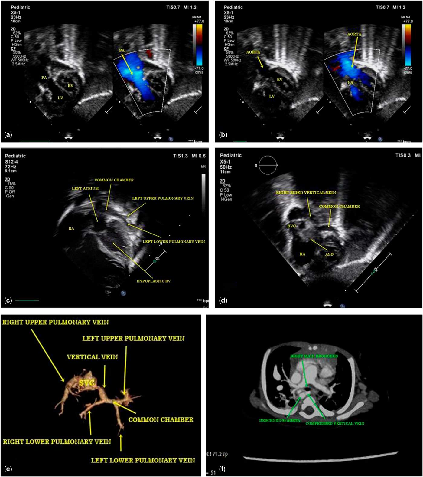
Transthoracic echocardiogram showed situs solitus, levocardia, corrected transposition of great vessels (atrioventricular discordance with ventriculoarterial discordance) (Fig 1a and b for showing ventriculoarterial discordance, Supplementary video 1), and borderline right ventricle size (tricuspid valve Z score was −2.8). Left-sided pulmonary veins were draining into a common chamber behind the left atrium (Fig 1c, Supplementary video 2). Right-sided pulmonary venous drainage was unclear on echocardiogram. A vertical venous channel arose from the right side of the common chamber that connected to the right-sided superior caval vein and finally drained into the right atrium (Fig 1d, Supplementary video 3). There was no significant pressure gradient in the vertical vein in its entire course.
(a) Transthoracic echocardiographic image in subcostal long axis view showing connection of right-sided left ventricle to pulmonary artery (ventriculo-arterial (VA) discordance). (b) Transthoracic echocardiographic image in subcostal long axis view showing connection of left-sided right ventricle to aorta (VA discordance). (c) Transthoracic echocardiographic image in apical four chamber view showing drainage of left-sided pulmonary veins into a common chamber, hypoplastic left-sided right ventricle, and dilated right atrium (RA). (d) Transthoracic echocardiographic image in subcostal short axis view showing drainage of right-sided vertical channel into RA via superior vena cava (SVC). (e) Three-dimensional reconstructed CT image showing drainage of dilated right upper pulmonary vein into SVC. (f) CT image showing compression of vertical vein between right main bronchus anteriorly and descending aorta posteriorly.

Small ostium secundum atrial septal defect (Fig 1d) was present, which was shunting right to left with severe pulmonary arterial hypertension. Interventricular septum was intact. Grade I diastolic dysfunction of the right ventricle was noted. There were competent tricuspid and mitral valves with right-sided aortic arch.
Three-dimensional volume-rendered CT was performed, which confirmed echocardiographic findings, as well as showed dilated right upper pulmonary vein, which was draining into the superior caval vein (Fig 1e). Left-sided pulmonary veins and right lower pulmonary vein were normal in size with no evidence of obstruction or narrowing. CT also showed vertical venous channel that was compressed between the right main bronchus anteriorly and descending thoracic aorta posteriorly (Fig 1f).
The right ventricle was of borderline size but was supporting systemic cardiac output; therefore, as of now only repair of total anomalous pulmonary venous connection was planned with the hope that the right ventricle may grow on follow-up for biventricular repair in the form of double-switch operation. In the operation theatre, pulmonary artery pressure was systemic. Total anomalous pulmonary venous connection was repaired with opening of the common chamber into the left atrium, resulting in right upper pulmonary vein drainage into the superior caval vein. A 3-mm atrial septal defect was left behind as a pop-off for the right ventricle as it had mild diastolic dysfunction. Vertical vein was ligated. Even after repair, pulmonary artery pressure was nearly systemic. We decided to shift the child with open chest with pulmonary artery pressure line in situ on inhaled nitric oxide support.
On the next day, pulmonary artery pressures were nearly systemic, and there was high dependency on nitric oxide; therefore, we decided to go for pulmonary artery banding. Loose pulmonary artery banding was performed, with a band gradient of 30 mmHg, as the patient went into bradycardia and hypotension on tight banding. Gradually, ventilatory support and nitric oxide requirement decreased and the patient was extubated on the 4th postoperative day with half systemic pulmonary pressures. The patient was discharged on pulmonary vasodilators in haemodynamically stable state with saturation of 94–95%. On follow-up, the patient remained stable with room air saturation of 94%.
Discussion
Corrected transposition of the great arteries is a rare defect representing ~0.5% of all CHD.Reference Tandon, Bose, Yoon and Schussler 1 It is characterised by the combination of atrioventricular and ventriculoarterial discordance. It may be present with or without associated anomalies. Merely 10% of the patients with corrected transposition of the great arteries do not have any associated anomalies such as ventricular septal defect, pulmonary artery stenosis, tricuspid valve abnormalities, and mitral valve abnormalities.Reference Zimmermann, Altman and Gantt 2
Darling’s study classified total anomalous pulmonary venous connection into four types, and the most common type is supracardiac.Reference Darling, Rothney and Craig 3 Total anomalous pulmonary venous connection is a rare congenital anomaly, corresponding to ~2% of all CHD.Reference Bharati and Lev 4 It consists of an abnormality of blood flow in which all four pulmonary veins drain into systemic veins or the right atrium with or without pulmonary venous obstruction.Reference Emmanouilides, Gutgesell, Riemenschneider and Allen 5
Echocardiography, cardiac CT, and cardiac MRI are successfully used for meticulous diagnosis.Reference Tandon, Bose, Yoon and Schussler 1
There are reports of association of corrected transposition of the great arteries with infradiaphragmatic total anomalous pulmonary venous connectionReference Hayashi, Hirata, Inuzuka and Hirata 6 and with systemic atriumReference Sharma, Talwar and Marwaha 7 in literature.
This is a very unusual association of corrected transposition of the great arteries with supracardiac total anomalous pulmonary venous connection via a right-sided vertical channel – that is, compressed between the bronchus and the descending aorta. Normally, the vertical channel runs between the bronchus and the pulmonary artery.
Treatment approach for corrected transposition of great vessels depends on symptoms and associated anomalies. When the ventricles are balanced without anatomical restriction, most surgeons agree that biventricular repair is the treatment of choice. When there is unfavourable anatomy, such as atrioventricular valve chordae straddling, multiple or inaccessible ventricular septal defect, borderline balanced ventricles, or inappropriate coronary artery anatomy, biventricular repair becomes difficult. Single ventricular repair, on the other hand, might be better under these circumstances.Reference Hsua, Changb, Huangb, Chenb and Chiub 8
The goal of surgery in total anomalous pulmonary venous connection is to redirect pulmonary veins to the left atrium through wide and non-restrictive connection. Careful intraoperative examination of the pulmonary venous drainage is mandatory for all patients. Precise technique for surgery adopted in an individual patient depends on the pattern of anatomic drainage, and an individualised surgical approach is recommended.
In this case, the right ventricle supported the systemic cardiac output even with borderline size, and thus we planned to only repair the total anomalous pulmonary venous connection with the hope that the right ventricle may grow in size for biventricular repair on follow-up. The common chamber was connected to the left atrium, resulting in drainage of the right upper pulmonary vein into the superior caval vein. Pulmonary artery banding was done as there was high dependency on nitric oxide with no significant fall in pulmonary artery pressure even after total anomalous pulmonary venous connection repair. Loose pulmonary artery banding was performed. If the right ventricle grows on follow-up, then the plan is to go for biventricular repair – double-switch operation with pulmonary artery debanding.
Supplementary material
To view supplementary material for this article, please visit https://doi.org/10.1017/S1047951118000677
Acknowledgements
None.
Financial Support
This research received no specific grant from any funding agency, commercial or not-for-profit sectors.
Conflicts of Interest
None.
Financial Support
This project was financed by a personal post-doctoral grant from EXTRA funds from the Norwegian Foundation for Health and Rehabilitation (grant no. 2009/2/0340).
Conflicts of Interest
None.
Ethical Standards
As this manuscript does not have any research over either human or animal, it does not need this statement as mentioned in author guidelines.