The β-thalassaemia syndromes collectively represent the most common monogenic disorders worldwide and are among the first diseases for which gene therapy was envisioned. These syndromes arise from autosomal recessive mutations that lead to the deficient or altered synthesis of the β-chain of haemoglobin. This in turn leads to an imbalance of α- and β-globin chains, resulting in the intracellular precipitation of excess α-globin chains and eventually ineffective erythropoiesis (Ref. Reference Stamatoyannopoulos1). Homozygotes or compound heterozygotes harbouring either null or very low expressing alleles present with profound anaemia that is lethal early in life if left untreated.
Mainstay treatment includes regular blood transfusions in combination with daily iron chelation, and has considerably extended the life expectancy of thalassaemic patients in the past 35 years. However, this lifelong treatment not only constitutes an enormous financial burden most challenging in developing countries (Ref. Reference Weatherall and Clegg2), but also severely compromises the quality of life of thalassaemic patients. Treatment noncompliance is common and many patients suffer from cardiac, hepatic and endocrine complications. Currently, allogeneic haematopoietic stem cell (HSC) transplantation is the only radical curative treatment available. A high cure rate (82–93%) can be achieved in thalassaemic class 1 and class 2 patients who are younger than 17 years of age using bone marrow transplantation from human leukocyte antigen (HLA)-matched sibling donors (Ref. Reference Lucarelli, Andreani and Angelucci3).
Despite this curative potential, HSC transplantation is associated with significant drawbacks, including limited availability of HLA-matched donors, the need for long-term immunosuppression to prevent or treat the graft-versus-host disease that is often associated with allogeneic HSC transplantation, narrow application to the youngest patients, and the increased risk of morbidity and mortality in older patients with organ damage (Lucarelli class 3 patients) due to immunological complications and nonrejection mortality (Refs Reference Gaziev4, Reference Lucarrelli5). Moreover, transplants from alternative donors, such as matched-unrelated or haploidentical donors, are associated with significantly lower disease-free survival (21–70%) and higher morbidity and mortality (25–30%) (Refs Reference La Nasa6, Reference La Nasa7, Reference Gaziev8, Reference Lucarelli and Gaziev9). As such, they remain largely investigational (Ref. Reference Lucarelli and Gaziev9).
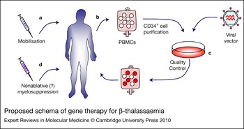
Given these limitations in treatment options, we and others have sought to develop an alternative treatment regimen for thalassaemia patients based on gene therapy. As shown in Figure 1, this approach involves the transfer of normal globin genes into patient-derived, autologous HSCs. These cells can then be used instead of allogeneic donor cells for transplantation. This approach carries the potential of a one-time permanent correction without the limitations of donor availability and the risks of immunological complications associated with allogeneic HSC transplantation. This includes the likely ability to use lower levels of myelosuppression as part of the transplantation regimen, and no post-transplant immunosuppression, which should in turn offer a substantial reduction in the morbidity and mortality associated with allogeneic HSC transplantation.
Proposed schema of gene therapy for β-thalassaemia. The basic schema proposed for the clinical gene therapy of β-thalassaemia is shown, with key advances or areas of research highlighted. (a) The patient is treated with granulocyte colony-stimulating factor (G-CSF) in order to mobilise haematopoietic stem cells into the peripheral blood. (b) Peripheral blood mononuclear cells (PBMCs) are collected and processed to isolate CD34+ haematopoietic stem cells. (c) These cells are cultured with a viral vector designed to express high levels of normal human β-globin (see Fig. 2 for vector details), and this is followed by quality control. (d) The patient is treated with moderate, nonablative myelosuppression, and then engrafted with the vector-modified cells (filled circles). See the text for details regarding these steps.

The complexity of globin gene regulation at ectopic chromatin sites and failure of vectors based on gammaretroviruses
As reviewed extensively elsewhere, most research efforts towards gene therapy for the β-thalassaemias to date have focused on the development of recombinant retroviral vectors as a means of gene delivery (Ref. Reference Lisowski and Sadelain10). These vectors, such as those based on gammaretroviruses or lentiviruses, are widely used in the setting of haematopoietic gene therapy because of their ability to efficiently integrate therapeutic genes into the target cell genome, resulting in long-term and stable gene transfer even after many rounds of cell division (Refs Reference Emery11, Reference Kohn12). However, vector integration occurs at sites throughout the target cell genome (Ref. Reference Berry13), rendering vector gene expression subject to a wide range of regulatory environments. This in turn can result in variation in vector gene expression and even overt vector silencing, a phenomenon known as chromosomal position effects (as reviewed in Ref. Reference Emery, Aker, Stamatoyannopoulos and Makrides14). Achieving a consistent and high-level expression of globin transgenes has further been complicated by the complexities of globin gene expression and regulation. These factors in turn can influence several challenges that must be met in order for gene therapy to become a realistic therapeutic approach for treating the β-thalassaemias, including the following: (Reference Stamatoyannopoulos1) efficient gene transfer into target HSCs; (Reference Weatherall and Clegg2) erythroid-lineage specific expression; (Reference Lucarelli, Andreani and Angelucci3) therapeutic levels of expression; and (Reference Gaziev4) vector safety.
The β-globin (HBB) locus has evolved to yield high-level and erythroid-specific expression. This developmental regulation is mediated in part by a group of elements collectively referred to as the locus control region (LCR) (Refs Reference Li15, Reference Grosveld16). The β-globin LCR includes at least seven separate elements defined by DNase hypersensitive sites (HS1–7) spread over a 25 kb segment upstream of the β-globin coding region. This region has been shown in several models to increase globin gene expression in a tissue-specific and developmentally specific fashion, and to shelter the rest of the β-globin locus from the effects of surrounding chromatin (e.g. Ref. Reference Blom van Assendelft17). In its native configuration, this LCR is far too large to include in retroviral vectors. Globin expression from first-generation gammaretroviral vectors that lacked LCR elements was minimal (Refs Reference Karlsson18, Reference Dzierzak, Papayannopoulou and Mulligan19). The first attempts to include LCR subfragments resulted in vectors that exhibited very low titres or genetic instability (Refs Reference Plavec20, Reference Novak21, Reference Emery22). Even when small segments of the key elements defined by HS2–4 were included without these problems, the resulting vectors exhibited modest and highly variable expression patterns, indicating sensitivity to position effects (Ref. Reference Sadelain23). Likewise, efforts to overcome retroviral vector instability or globin gene silencing or variable expression by the modification of globin gene sequences (Refs Reference Miller24, Reference Leboulch25), or incorporation of chromatin insulators (Ref. Reference Emery26), improved the expression and stability of gammaretroviral vectors, but expression still remained well below therapeutic levels. Collectively, efforts to achieve high-level β-globin expression by gammaretroviral vectors have met with limited success.
Lentiviral vectors and the revival of gene therapy for the β-thalassaemias
The failure in the 1980s and 1990s of gammaretroviral vectors to support the elements necessary to achieve high-level and consistent β-globin gene expression prompted investigations into alternative vector systems. In particular, lentiviral vectors, derived from human immunodeficiency virus 1 (HIV-1), raised great interest as a gene therapy vehicle due to their ability to transduce nondividing cells (Ref. Reference Miyoshi27), to hold larger inserts (~9–10 kb) than gammaretroviral vectors (Ref. Reference Kumar28), and to resist the genomic instability that sometimes plagued gammaretroviral vectors (Ref. Reference May29). Since their introduction, these vectors have undergone many improvements, including the reduction of HIV sequences and the separation of packaging genes onto independent plasmids so as to reduce the potential risk of generating replication-competent viruses (Ref. Reference Dull30). Most importantly, a self-inactivating (SIN) vector design was introduced that essentially eliminates expression of the viral sequences by deleting the U3 enhancer region of the viral long-terminal repeat (LTR) (Refs Reference Miyoshi31, Reference Zufferey32, Reference Naldini and Verma33, Reference Kay, Glorioso and Naldini34). With such a SIN vector design, vector expression can thus be driven exclusively by erythroid-specific enhancers and promoters. In turn, this allows for elevated and restricted globin gene expression, and an increased safety profile by reducing the potential for vector sequences to activate the expression of cellular genes flanking sites of vector integration in most tissues (Refs Reference Montini35, Reference Arumugam36).
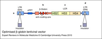
The past decade has seen many reports of lentiviral vectors carrying a variety of globin gene coding cassettes with elements from the β-globin LCR and either without (Refs Reference May29, Reference Pawliuk37, Reference Imren38, Reference Persons39, Reference Rivella40, Reference Miccio41, Reference Perumbeti42) or with (Refs Reference Arumugam36, Reference Puthenveetil43, Reference Aker44, Reference Arumugam45) chromatin insulators. Studies from the laboratory of Michel Sadelain were the first to demonstrate that these vectors could stably transmit and express therapeutic levels of human β-globin in a mouse model of β-thalassaemia intermedia (Ref. Reference May29). In this setting, vector expression increased haemoglobin levels from 8.0–8.5 g/dl in control mice to 11–13 g/dl, which was sufficient to correct both the anaemia and the extramedullary haematopoiesis, as well as to reduce hepatic iron accumulation. In addition, expression remained stable even after secondary transplantation (Ref. Reference May46). Subsequently, other groups also demonstrated long-term correction in murine β-thalassaemia intermedia models using lentiviral vectors carrying either the β-globin or the γ-globin gene (Refs Reference Imren38, Reference Persons39). Further studies from the laboratory of M. Sadelain also demonstrated that their vector could even support long-term survival in an embryonic-lethal mouse model of severe β-thalassaemia major (Cooley anaemia) (Ref. Reference Rivella40). In this model, fetal liver cells were used as a source of transplantable HSCs, which were transduced with the β-globin vector and transplanted into myeloablated adult recipients. Control mice receiving untransduced cells developed severe β-thalassaemia and died. In contrast, most vector-treated mice were converted after transplantation into a thalassaemia intermedia phenotype, whereas one achieved normal haemoglobin levels (12 g/dl). In support of the studies in mice, the group of Punam Malik demonstrated that transduction of bone marrow CD34+ cells from β-thalassaemia major patients with a lentiviral vector insulated by cHS4 (the 5′ DNase I HS4 of the chicken β-globin LCR) resulted in the phenotypic correction of erythroid cells following transplantation into immunodeficient mice (Ref. Reference Puthenveetil43). One recent publication reported that although the activity of a lentiviral vector encoding β-globin was effective at correcting both mild and severe β-thalassaemia in several settings, it was nevertheless dependent on the sites of vector integration (Ref. Reference Miccio41). This indicates that β-globin lentiviral vectors, even those that contain elements from the β-globin LCR, can remain sensitive to chromosomal position effects. This indication is bolstered by studies from our own laboratory and that of P. Malik demonstrating that the level and uniformity of expression from β-globin lentiviral vectors containing HS2, HS3 and HS4 can be improved by flanking these vectors with the cHS4 chromatin insulator (Refs Reference Aker44, Reference Arumugam45). A prototypical β-globin lentiviral vector is shown in Figure 2.
Optimised β-globin lentiviral vector. A prototypical state-of-the-art viral vector for human β-globin is shown, with key advances or areas of research highlighted. (a) The vector platform is based on a recombinant lentivirus that allows for stable and efficient gene transfer. (b) The β-globin coding cassette is optimised for size and expression, including the retention of intronic sequences that require the cassette to be included in the antisense direction relative to the vector transcription. (c) Extended regulatory elements from the β-globin locus control region (LCR), including DNase hypersensitive (HS) sites 2, 3 and 4, are included to achieve high-level expression. (d) Deletion of enhancers in the viral long-terminal repeats (LTRs) generates a ‘self-inactivating’ (SIN) vector design that allows for tighter regulation of the globin transgene and reduces the potential for activating proto-oncogenes that occasionally surround sites of vector integration. (e) The vector is flanked by chromatin insulators, in order to further improve vector expression and reduce the chance of activating surrounding cellular genes. This could include a single copy of the extended cHS4 insulator or alternative insulators under development. Other abbreviations: cPPT, central polypurine tract; enh–coding–pro, enhancer, coding sequence, promoter.

Insertional oncogenesis in gene therapy
The first unequivocal success of stem cell gene therapy was reported in 2000 from a trial involving children with X-linked severe combined immunodeficiency syndrome (X-SCID) (Ref. Reference Cavazzana-Calvo47). To date, 90% of patients with either X-SCID or a closely related disease, adenosine deaminase deficiency (ADA)-SCID, have been successfully treated in three separate gene therapy trials (Refs Reference Hacein-Bey-Abina48, Reference Gaspar49, Reference Aiuti50). In these trials, autologous HSCs were transduced with a gammaretroviral vector, and infused back into the patient with mild or no preconditioning. In most cases this resulted in the reconstitution of a functioning immune system that enabled the patients to live normal lives.
However, this great success was eventually overshadowed by subsequent reports that a total of five patients from the two trials for X-SCID developed a clonal T-cell proliferation and, in some cases, overt leukaemia (Refs Reference Hacein-Bey-Abina51, Reference Howe52). Molecular analysis revealed that these events were associated with specific instances where the vector provirus had integrated near a certain cellular proto-oncogene (LMO2) and had activated the inappropriate expression of this oncogene via the enhancer in the viral LTR (Ref. Reference Hacein-Bey-Abina53). Similar results were also seen in an unrelated trial for chronic granulomatous disease (CGD). In this case, mobilised peripheral blood HSCs were again transduced with a gammaretroviral vector. Initially, the life-threatening infections from which these patients suffer resolved, but once again an increase in the proportion of gene-modified myeloid cells was observed over time. This was found to be associated with the clonal expansion of cells with vector insertions associated with the proto-oncogene locus MDS1-Evi1 (MECOM) (Ref. Reference Ott54). In a study update (Ref. Reference Stein55), the two patients, 15 and 28 months after gene transfer, were found to have cytopaenias due to bone marrow myelodysplasia and they both exhibited loss of a single copy of chromosome 7 linked to a dominant MDS1-Evi1 clone.
Although insertional mutagenesis was the initiating event for the leukaemia developed in the X-SCID patient of the English trial, leukaemogenesis required the acquisition of secondary genetic lesions (Ref. Reference Howe52). It is presently unclear why there were several cases of vector-mediated oncogenesis in the X-SCID trials but not in the ADA-SCID trials. There is evidence that the X-SCID haematopoietic milieu or some disease-specific stimuli of the transgene may have been important for the high rate of transformation after an initiating event of insertional mutagenesis; aberrant restoration of interleukin 7 (IL-7)-dependent signalling, impaired antitumour immune surveillance due to very low numbers of natural killer (NK) cells, and constitutive expression of the therapeutic γc gene in thymocytes or thymocyte precursors are some aspects that may have contributed to the increased incidence of transformation (Refs Reference Howe52, Reference Aiuti56). In addition to the clinical trials just discussed, retroviral-vector-mediated oncogenesis has been observed in an increasing number of preclinical models, including studies in mice (Refs Reference Li57, Reference Shou58, Reference Kustikova59, Reference Li60) and nonhuman primates (Ref. Reference Seggewiss61).
Taken together, these events highlight the potential hazard of integrating gene therapy vectors in general, and in particular early generations of gammaretroviral vectors. Indeed, insertional mutagenesis represents an inescapable consequence of the transposition of genetic material (e.g. see Ref. Reference Sadelain62). When such transposition results in the integration of ectopic DNA sequences within or near cellular oncogenes or tumour suppressor genes, even if such events are rare, the potential exists that the event will lead to the activation or dysregulation of that gene. This is especially true if the vector contains strong enhancers or other potent regulatory elements. Initial estimations had assumed that the risk of insertional oncogenesis would be very low based on the assumption that proviral integration into the genome was random (Ref. Reference Baum63). However, subsequent investigations found that the insertion of retroviral vectors was, in fact, far from random. For example, large-scale studies of gammaretroviral integration sites essentially all point to a modest preference for integrations near the transcriptional start sites of transcriptionally active genes (Ref. Reference Berry13 and references therein). For example, our own studies found that 36% of all gammaretroviral vector integration sites in murine bone marrow progenitor cells resided within a ±5 kb window of a transcription start site, compared with only 14% for a simulated random control set (Ref. Reference Aker64). Unlike gammaretroviral vectors, vectors based on lentiviruses are less likely to integrate near transcription start sites, and instead show a preference for integration within the body of actively transcribed genes (Ref. Reference Berry13 and references therein). This preference appears to be directed at least in part through tethering of the lentiviral vector preintegration complex to a chromatin element mediated by the cellular protein LEDGF (PSIP1) (reviewed in Ref. Reference Poeschla65). However, it is important to note that even if vector integration was random, about 5% of all integration sites are still expected to reside within 100 kb of a known oncogene (Ref. Reference Aker64).
Safety features of globin lentiviral vectors
The lentiviral vectors for human β-globin that are currently considered for clinical trials have several features that should substantially reduce the risk of vector-mediated oncogenesis compared with the early-generation gammaretroviral vectors used in the SCID and other earlier gene therapy trials.
SIN vector configuration
The vector platform used for lentiviral vectors includes a SIN vector design. As described earlier, this involves deleting the viral enhancers in the vector LTRs. This not only minimises expression of the residual viral sequences, but also eliminates the potential for this enhancer to activate cellular genes flanking the site of vector integration. Indeed, active LTRs probably constitute the major determinants of genotoxicity (Ref. Reference Montini66).
Safer integration site profile of lentiviral vectors
The integration pattern of lentiviral vectors is thought to contribute to their overall safety, because they tend to integrate within the body of genes (Ref. Reference Schroder67) rather than near the promoters (like gammaretroviral vectors, Ref. Reference Wu68), where they are less likely to influence promoter activity.
Tissue-specific expression of globin vectors
The SIN lentiviral globin vectors contain only erythroid-specific regulatory elements, ensuring transcriptional activity only in the target-cell lineage (i.e. proerythroblasts). This lineage-restricted expression pattern minimises the risk of activating cellular oncogenes within all the other lineages, especially in primitive cells, with the high self-renewal capacity thought to be necessary for sustained malignant transformation (Ref. Reference Zychlinski69). Moreover, the β-globin vector transgene is activated only at late stages of erythroid differentiation, preceding naturally occurring enucleation, which essentially precludes the potential for oncogenic transformation (Ref. Reference Chang and Sadelain70).
Disease-associated oncogenic potential
Thalassaemia per se is not associated with the sort of elevated oncogenic risk known to be linked with X-SCID. As such, the possibility of acquiring additional genetic ‘hits’ before or after gene transfer is low. This is augmented by the fact that the selective pressure on genetically corrected cells in β-thalassaemia is very modest compared with the selective pressure exerted on corrected lymphoid cells in the setting of X-SCID. Indeed, one recently reported study used an ex vivo immortalisation assay to demonstrate that a typical SIN-lentiviral vector for β-globin exhibits a very low genotoxic potential (Ref. Reference Arumugam36). However, two other in vivo studies demonstrate that such vectors still retain the ability to activate genes flanking sites of vector integration (Ref. Reference Hargrove71) or to exhibit an integration site profile that suggests the presence of an in vivo selective pressure for clones containing a vector provirus more favourable to high levels of vector-derived expression (Ref. Reference Miccio41).
Chromatin insulators
To further augment the safety of humans globin vectors, some investigators have incorporated chromatin insulator elements in the vector design. This is an appealing strategy for potentially diminishing vector-mediated proto-oncogene activation. Chromatin insulators are DNA elements that help form functional boundaries between adjacent chromatin domains (reviewed in Refs Reference Emery, Aker, Stamatoyannopoulos and Makrides14, Reference Gaszner and Felsenfeld72, Reference Wallace and Felsenfeld73). They have been reported in species as diverse as yeast and humans, and have been shown to have a key role in global gene regulation. There are two basic classes of chromatin insulators: enhancer-blocking insulators and barrier insulators. Enhancer-blocking insulators prevent enhancer-mediated transcriptional activation of adjacent promoters, whereas barrier insulators block the encroachment of silencing heterochromatin into adjoining regions of open chromatin that are otherwise transcriptionally permissive. The best-studied vertebrate element is a 1.2-kb fragment containing cHS4 of the chicken β-globin LCR (Ref. Reference Chung, Bell and Felsenfeld74). Several studies have demonstrated the ability of this element to protect the expression of viral gene therapy vectors from silencing chromosomal position effects (e.g. see Refs Reference Emery26, Reference Rivella75, Reference Emery76, Reference Yannaki77). This has even been the case for globin lentiviral vectors containing extended regions of the β-globin LCR (Refs Reference Arumugam36, Reference Aker44, Reference Arumugam45). In this capacity, the cHS4 insulator (or other barrier insulators) provides an indirect means of reducing the risk of vector-mediated oncogenesis by reducing the vector titre dose needed to achieve therapeutic levels of expression. In addition, several lines of evidence, including studies using an in vivo tumour model (Ref. Reference Li60), suggest that the enhancer-blocking activity of the cHS4 insulator might also directly reduce the rate of vector-mediated oncogenesis (Refs Reference Evans-Galea78, Reference Ramezani, Hawley and Hawley79). However, it is important to note that it has yet to be directly demonstrated that chromatin insulators can improve the safety of globin lentiviral vectors, especially in the setting of therapeutic animal models.
The need for bone marrow conditioning in thalassaemia gene therapy
Several lines of evidence indicate that successful β-globin gene therapy will result in the selective survival of genetically correct erythrocytes (e.g. Ref. Reference Nishino80 and references therein). However, such selective pressure will not extend to the primitive haematopoietic stem/progenitor cells that give rise to long-term stable engraftment. As a consequence, the long-term engraftment of gene-corrected cells in this setting will likely require either an in vivo selection strategy (Refs Reference Neff81, Reference Ragg82, Reference Jin83, Reference Emery84, Reference Zhao85) or bone marrow conditioning, in order to allow the genetically modified HSCs to reach therapeutically relevant frequencies. Indeed, preclinical gene transfer studies in animal models and clinical studies with allogeneic bone marrow transplantation suggest that a level of gene-corrected HSCs in excess of 20% will be required for the treatment of β-thalassaemia (discussed below), well beyond the 0.01% expected in the absence of selection or bone marrow conditioning.
In the setting of allogeneic HSC transplantation for haematological disease, ablative bone marrow conditioning serves predominantly to attenuate the immunological barriers between the host and the donor, although it also serves to open up space in the bone marrow ‘niche’ by depleting the host HSC pool and to eradicate disease. In the autologous HSC transplantation setting, where immunological barriers do not exist, ablation serves only to eradicate disease burden and to open up the bone marrow niche. Myeloablation, however, is accompanied by significant levels of treatment-related morbidity and mortality, which are highly undesirable in patients with nonmalignant diseases.
As an alternative, much effort over recent years has been directed towards developing less toxic, nonmyeloablative conditioning regimens (reviewed in Ref. Reference Burroughs and Storb86). In addition to their application in the setting of allogeneic transplantation for haematological disease, these approaches are also being applied in HSC gene therapy (Refs Reference Gaspar49, Reference Aiuti87). In this latter setting, nonmyeloablative, low-intensity conditioning facilitates engraftment with substantially reduced treatment-related toxicity.
Reports of nonmyeloablative conditioning in gene marking studies of mice and nonhuman primates (Refs Reference Mardiney and Malech88, Reference Rosenzwieg89) have provided direct evidence for facilitated engraftment of transduced cells. Such regimens are also proving effective in a number of clinical gene therapy trials (Refs Reference Gaspar49, Reference Aiuti50, Reference Ott54). Indeed, earlier attempts to correct ADA-SCID, CGD, Fanconi anaemia and Gaucher disease by gene therapy without preceding conditioning were, in general, disappointing (Refs Reference Bordignon90, Reference Kohn91, Reference Malech92, Reference Kohn93, Reference Dunbar94, Reference Schuening95, Reference Kelly96). The proof of principle for bone marrow conditioning as a means of facilitating engraftment of gene-corrected cells was clearly provided in recent ADA-SCID and CGD trials (Refs Reference Aiuti50, Reference Ott54, Reference Kang97), in which transplantation of gene-modified cells after a partially myeloablative conditioning regimen resulted in correction or improvement of the genetic diseases. In particular, conditioning based on busulfan (4 mg/kg) or melphalan (140 mg/m2) was shown to effectively enhance engraftment of gene-modified HSCs in ADA-SCID (Refs Reference Aiuti50, Reference Gaspar98). However, in the setting of CGD, where there is no selective advantage for the transduced HSCs, a higher degree of ablation (8–10 mg/kg busulfan) was needed. Nevertheless, this dose was well tolerated, allowing for haematopoietic reconstitution within 21–28 days (Refs Reference Ott54, Reference Kang97).
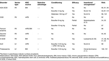
Full myeloablation has been considered in the gene therapy of genetic diseases lacking a proliferative advantage of corrected cells. In the first clinical trials of stem cell gene therapy involving the use of lentiviral vectors, including one trial for adrenoleukodystrophy (ALD) (Ref. Reference Cartier99) and one trial for β-thalassaemia (Refs Reference Bank, Dorazio and Leboulch100, Reference Kaiser101), a fully myeloablative conditioning regimen with busulfan and cyclophosphamide or busulfan alone was used. Table 1 summarises the clinical experience in HSC gene therapy for monogenic disorders.
Summary of the stem cell gene therapy clinical trials for monogenic disorders

aNumbers in parentheses indicate numbers of patients.
Abbreviations: ADA-SCID, ADA-deficient SCID; ALD, adrenoleukodystrophy; BM, bone marrow; CGD, chronic granulomatous disease; CPM, cyclophosphamide; HSC, haematopoietic stem cell; LV, lentiviral; mPB, mobilised peripheral blood; RV, retroviral; SCID, severe combined immunodeficiency; X-SCID, X-linked SCID.
It is likely that a substantial degree of conditioning will be necessary in gene therapy for the β-thalassaemias. In this setting, patients often exhibit massive expansion of the haematopoietic compartment and extensive extramedullary haematopoiesis that could substantially dilute the therapeutic effect of infused corrected cells in an unconditioned host. However, the degree of bone marrow ablation needed to ensure adequate engraftment of gene-modified cells without unacceptable toxicity under these settings is presently unknown.
Studies involving allogeneic HSC transplantation in patients with β-thalassaemia and sickle cell disease indicated that even small levels of donor chimerism can render recipients transfusion independent (Refs Reference Andreani102, Reference Iannone103), supporting the concept that even low levels of engraftment with gene-modified autologous HSCs could provide significant benefit. However, the threshold level of normal HSCs and erythroid chimerism required for clinical improvement has not ultimately been defined. Gene transfer studies in a mouse model of β-thalassaemia suggest that an HSC chimerism level of 20–30% and a transgene expression level in excess of 15% of total α-globin will be necessary to ameliorate disease and achieve transfusion independence in patients with β-thalassaemia (Ref. Reference Persons104). As mentioned above, this therapeutic potential is dependent in part on the extended survival of genetically correct erythrocytes, which translates into a selective advantage at the level of circulating red blood cells (e.g. Ref. Reference Nishino80 and references therein). In addition, ineffective erythropoiesis in the diseased bone marrow will provide a competitive advantage to the corrected erythroid progenitors. Indeed, a therapeutic amplification at the level of genetically corrected mature erythroblasts was shown at an HSC chimerism of 30–50% (Ref. Reference Miccio41).
Based on these observations, it is predicted that full myeloablation might not be necessary to achieve significant therapeutic benefit in the setting of gene therapy for β-thalassaemia. We expect that 8 mg/kg busulfan or 140 mg/m2 melphalan could provide adequate ablation to allow transduced HSCs to reach clinically meaningful levels, while maintaining an adequate level of safety commensurate with a Phase I clinical trial. However, it would be important to counterbalance the limited engraftment that would be expected under these conditions by using methods that ensure high levels of gene transfer and high doses of gene-modified cells.
Obtaining target cells for thalassaemia gene therapy
Given the absence of a disease-associated growth advantage at the level of stem/progenitor cells and the preference for a reduced intensity conditioning regimen, it will be necessary to start with a very large number of transplantable HSCs in order to achieve adequate levels of engraftment with gene-modified cells in patients with β-thalassaemia. Obtaining a sufficient number of cells represents a major challenge in this setting. Granulocyte colony-stimulating factor [G-CSF (CSF3)]-mobilised peripheral blood currently represents the most widely used source of HSCs for allogeneic and autologous transplantation (Ref. Reference Gratwohl105), because of the high yield of CD34+ cells, a shorter time to engraftment and the less invasive procedure, compared with bone marrow harvest (Ref. Reference To106). However, the optimal source of HSCs for transduction has not yet been established.
The gene transfer rates obtained with mobilised HSCs might be higher than steady-state bone marrow, although this is still an issue of debate (Refs Reference Dunbar107, Reference Horn108, Reference Thomasson109). The highest levels of in vivo gene marking were shown by the cytokine combination of G-CSF and stem cell factor [SCF (KITLG)] (Refs Reference Dunbar107, Reference Hematti110, Reference Hematti111, Reference Bodine112), but SCF is not available for clinical use. However, there is a lack of studies directly comparing G-CSF-mobilised peripheral blood versus bone marrow or showing a clear superiority in the actual repopulating cell transduction of bone marrow over G-CSF-mobilised cells (Ref. Reference Hematti111). In practice, both sources, bone marrow (Refs Reference Cavazzana-Calvo47, Reference Aiuti50) and G-CSF-mobilised blood (Refs Reference Ott54, Reference Cartier99), have been successfully used in clinical gene therapy trials. Because G-CSF mobilisation is the preferred method for obtaining the high numbers of HSCs required for the gene therapy of β-thalassaemia, we believe it remains the best option for this purpose until proven otherwise in the clinical setting.
Mobilisation with G-CSF, in normal donors and in patients with haematological malignancies, is generally well tolerated with only mild side effects. However, rare events of splenic rupture, in healthy donors and in patients undergoing autologous cell transplantation, have been reported (Refs Reference O'Malley, Whalen and Banks113, Reference Brown and Dale114, Reference Becker115). Of more concern, G-CSF has been shown to precipitate severe sickle cell crises in patients with sickle cell disease, including one death (Refs Reference Abboud, Laver and Blau116, Reference Adler117). For this population, the options for collecting HSCs for gene transfer purposes are thus restricted to conventional bone marrow harvest under local or general anaesthesia with careful perioperative management, or alternative mobilisation procedures (e.g. Plerixafor, formerly known as AMD3100) (Refs Reference Broxmeyer118, Reference Larochelle119). In contrast, G-CSF mobilisation is not expected to be associated with serious side effects in patients with β-thalassaemia owing to the distinctive pathophysiology of the disease compared with sickle cell disease. This is supported by one study that found G-CSF mobilisation to be safe and effective in a mostly paediatric cohort of β-thalassaemia patients (Ref. Reference Li120). However, in adult β-thalassaemia patients, the evolving disease sequelae might present unique challenges. In particular, both the impaired organ function associated with accumulated iron and the as yet unknown effects of chronic infusions and chelation on the bone marrow reservoir might adversely affect both the safety and efficacy of G-CSF mobilisation. In addition, the chronic splenomegaly often found in adult patients with severe β-thalassaemia might either reduce the efficiency of G-CSF mobilisation by sequestering the mobilised HSCs or even increase the probability of splenic rupture. Another possibility is that G-CSF could also exacerbate the leukocytosis often associated with β-thalassaemia patients who have undergone splenectomy, leading to an increased risk of thrombosis. Our recent studies in a mouse model of β-thalassaemia give experimental credence to these concerns (Ref. Reference Yannaki121).
To address these issues, we recently initiated a clinical trial designed to assess the safety and efficacy of G-CSF mobilisation in adult patients with β-thalassaemia (http://clinicaltrials.gov/ct2/show/NCT00336362). This includes an assessment of whether pretreatment with hydroxyurea (HU) can reduce the potential risks associated with G-CSF in this patient population. We hypothesised that in nonsplenectomised patients HU pretreatment would reduce extramedullary haematopoiesis and thereby reduce the size of the spleen and potentially the risk for rupture, and in splenectomised patients HU would reduce the high level of white blood cells and platelets and thereby reduce the risk of thrombosis (Ref. Reference Yannaki and Stamatoyannopoulos122).
Thalassaemia is the first clinical entity to be treated with gene therapy in which the bone marrow is directly affected by the disease. It is currently unknown how chronic transfusions and chelation may have affected the quantity and quality of HSCs and their amenability to gene transfer. Only clinical studies could address whether HSCs harvested from a hypercellular bone marrow with endless intramedullary cell destruction or cytokine-mobilised HSCs will be superior in terms of yield and gene transfer rates in a gene therapy setting.
Clinical applications
Spurred by success in preclinical models, the first clinical trial directed towards the use of a β-globin lentiviral vector for the treatment of patients with β-thalassaemia was initiated in 2007 in France (Refs Reference Bank, Dorazio and Leboulch100, Reference Kaiser101). In addition, at least two other groups have announced plans to carry out similar trials (http://oba.od.nih.gov/oba/rac/PROTOCOL.pdf; Refs Reference Sadelain123, Reference Persons124). In the 2007 trial initiated in France, Philippe Leboulch and colleagues treated two patients with β-thalassaemia using CD34+ cells transduced with a lentiviral vector encoding the β-globin gene mutated at amino acid 87 (Refs Reference Bank, Dorazio and Leboulch100, Reference Persons124). Although the amino acid substitution was originally incorporated to prevent globin polymerisation in the setting of sickle cell disease, this variant also provides a means of distinguishing between endogenous and vector-derived globin molecules. In addition, this vector was flanked with a truncated version of the cHS4 chromatin insulator. Unfortunately, the 250 bp fragment chosen for this application is missing key elements needed for both optimum barrier activity (Ref. Reference Aker44) and enhancer-blocking activity (Ref. Reference Chung, Bell and Felsenfeld74). Furthermore, this element was used as a doublet, even though such duplicated sequences are well known to frequently recombine in retroviral vectors (Ref. Reference Pathak and Temin125). Autologous cells were harvested from bone marrow, and the recipients were conditioned with full myeloablation with busulfan. Owing to delayed haematological recovery and cytopaenia-related toxicity, the first patient received unmanipulated back-up cells for rescue and was reconstituted by unmodified thalassaemic marrow cells. The second patient, an 18-year-old individual with severe βE/β0-thalassaemia (HbE/β-thalassaemia), who was dependent on monthly transfusions, received ex vivo transduced HSCs with the aforementioned vector and conditioning regimen. Haematopoietic recovery was again delayed but uneventful with approximately 10% engraftment of gene-modified HSCs. The patient maintains haemoglobin levels of 9.5 g/dl, and remained transfusion independent for more than one year. The contribution of vector-encoded β-globin to total haemoglobin was estimated to be 3 g/dl, consistent with preclinical mouse studies. This was accompanied by a similar increase in HbF (fetal haemoglobin), an observation that has yet to be explained. However, vector integration site analysis revealed the emergence of a partially dominant haematopoietic clone harbouring a vector insertion in the HMGA2 proto-oncogene (Refs Reference Kaiser101, Reference Persons124). The clinical relevance of this finding remains unclear. It is possible the clone simply arose through a stochastic process inherent to engraftment with limited numbers of HSCs, in which case the clonal frequency should stabilise or may even recede as a result of clonal exhaustion (with a commensurate loss of the high-level vector expression associated with this clone). In contrast, this clone may continue to expand, suggesting an underlying premalignant growth perturbation. Careful, long-term monitoring of the patient will be necessary to distinguish between these possibilities.
Research in progress and outstanding research questions
The experience with gene therapy for X-SCID, ADA-SCID, CGD and, more recently, X-linked ALD serves as clear proof of principle for the use of HSC gene therapy in the treatment of monogenic disorders. Gene therapy for β-thalassaemia is now on the way, as demonstrated by the preliminary success of the second patient in the French thalassaemia trial. Whether the state-of-the-art erythroid-specific SIN lentiviral vectors prove safer than more conventional gammaretroviral vectors will require longer follow-up and more patients.
In addition, the unique pathology of β-thalassaemia presents several unique challenges that will likely require additional attention. First, the optimal level of ablation in the hypercellular thalassaemic bone marrow needs to be determined so as to ensure effective engraftment of gene-engineered cells, while minimising both haematopoietic and nonhaematopoietic toxicity. Second, the optimal source of HSCs must be addressed, especially for patients with sickle cell disease for whom G-CSF cannot be used. It is also possible that patients with β-thalassaemia may respond differently to G-CSF mobilisation compared with normal donors or patients with haematological malignancies, depending on the degree of splenomegaly or the absence of the spleen. Bone marrow harvest or alternative mobilisation agents, such as Plerixafor/AMD3100 (Refs Reference Broxmeyer118, Reference Larochelle119), may prove to be necessary for collecting HSCs from patients with haemoglobinopathies. Third, the optimal cell dose needs to be assessed. High numbers of gene-modified cells will probably be needed in order to compete with residual endogenous HSCs that remain in the setting of sublethal preparative regimens. However, there are both known and unknown risks associated with collecting HSCs before vector transduction, which would be compounded by the need for excessively high numbers of HSCs. In addition, there are likely dose-dependent risks associated with the number of gene-modified cells returned to the patient, because the likelihood of vector integration within or near proto-oncogenes increases proportionately to the number of integrated vectors. Thus, it will be necessary to determine the minimum number of input cells required to achieve the durable engraftment of a sufficient number of gene-modified cells in order to achieve a clinically relevant threshold. Fourth, it is likely that the majority of patients enrolled in the first trials for haemoglobin disorders will be adults, in contrast to the paediatric populations that have been the focus of the most successful HSC gene therapy trials reported to date. Adult patients with thalassaemia or sickle cell disease often present with accumulated organ damage due to iron overload or repeated vassocclusive crises, respectively, an unknown bone marrow stem cell reservoir, and ‘older’ stem cells that may be less amenable to gene transfer compared with those of young patients. Fifth, the risk of insertional oncogenesis in stem cell gene therapy with lentiviral vectors needs to be addressed and vector configurations for increasing biosafety need to be defined. So far, there is preclinical evidence of a safer integration pattern with lentiviral vectors as compared with the hitherto clinically used gammaretroviral vectors. Existing knowledge in humans is limited and inconclusive: in the two ALD patients treated with a lentiviral vector, the therapeutic effect was accompanied by highly polyclonal haematopoiesis without clustering of vector insertions in growth-related genes, whereas the thalassaemic patient in the French trial developed a partially dominant haematopoietic clone associated with a lentiviral vector insertion in the HMGA2 proto-oncogene.
Induced pluripotent stem (iPS) cells: beyond gene therapy
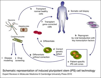
The demonstration that differentiated somatic cells can be reprogrammed into a primitive state resembling embryonic stem cells, so-called iPS cells, has opened new avenues of research and potential for therapeutic intervention (reviewed in Refs Reference Patel and Yang126, Reference Rashid and Vallier127). This technology circumvents most ethical concerns associated with the use of embryonic stem cells as well as the technical limitations associated with a related technology, somatic cell nuclear transfer. As first described, this technology relied on the use of viral vectors to transfer key transcription factors required for establishing and maintaining pluripotency (Refs Reference Takahashi and Yamanaka128, Reference Yu129) into a patient's somatic cells. Alternative strategies quickly evolved, including the use of episomal vectors and direct delivery of recombinant proteins (Refs Reference Yu130, Reference Kim131). These cells have already proved useful in experimental applications, including the modelling of human disease (Refs Reference Ebert132, Reference Lee133) and as a tool for drug screening (Refs Reference Kiskinis and Eggan134, Reference Park135). Although much work lies ahead before it is possible to rigorously produce and characterise clinical-grade iPS cells, such clinical applications are clearly on the horizon, including cell replacement for degenerative diseases and the treatment of genetic diseases in combination with gene therapy. The clinical relevance of iPS technology is shown in Figure 3.
Schematic representation of induced pluripotent stem (iPS) cell technology. (a) Somatic cells obtained from a patient are (b) directly reprogrammed through transduction with viral vectors expressing key transcription factors associated with pluripotency to (c) result in patient-derived iPS cell clones. In the case of an inherited monogenic disorder, these could then be (d) modified by gene correction, (e) induced to undergo differentiation into the desired cell type and (f) transplanted into the patient. In the case of an acquired disease, the patient-derived iPS cells could be (g) induced to differentiate, without further genetic manipulation, into a healthy version of the cell types that are damaged in the patient and then (h) transplanted to replace damaged or lost cells. These differentiated cells can also be used (i) in disease modelling for molecular studies or (j) for screening drug candidates in otherwise difficult to study diseases.

The screening of vector integration sites before infusion of gene-modified cells and the administration of cells with known, ‘harmless’ insertions to the patient would be a major safety precaution in HSC gene therapy. However, this is not currently feasible owing to the inability to readily expand HSCs in vitro. In contrast, patient-specific iPS cells could be either screened for safe integration sites or even corrected by gene targeting, and then expanded in vitro and differentiated into transplantable HSCs. The proof of principle for the feasibility of this strategy was provided in a humanised sickle cell anaemia mouse model (Ref. Reference Hanna136) In this study, autologous iPS cells were generated from skin fibroblasts, and reprogrammed in vitro by retroviral transduction with a cocktail of four transcription factors [Oct4 (POU5F1), SOX2, KLF4 and c-Myc (MYC)]. Following gene correction of the sickle cell mutation by homologous recombination, these cells were then forced to differentiate into HSCs through exposure to cytokines and the HOXB4 transcription factor, and were finally transferred back into diseased donor animals. The result was a correction in both clinical and laboratory parameters. In a more recent report, genetic correction of somatic cells from Fanconi anaemia patients with a lentiviral vector allowed the cells to be reprogrammed into iPS cells. These cells could then be redifferentiated into phenotypically normal myeloid and erythroid haematopoietic progenitor cells with the same efficiency as iPS cells derived from non-Fanconi donors (Ref. Reference Rega137). The successful reprogramming of skin fibroblasts to iPS cells from a patient with β-thalassaemia and their differentiation into cells producing haemoglobin have also been recently reported (Ref. Reference Ye138).
Current barriers tempering clinical utility of iPS cells
Before nuclear reprogramming can fulfil its potential for clinical applications, substantial additional advances will be necessary. These include improving systems for both reprogramming adult cells to a pluripotent state, and then redifferentiating these cells to the clinically relevant state. In addition, it will be necessary to eliminate the potential for generating tumourigenic cells, and to develop preclinical models that can help ensure high-quality clinical studies. This includes the development of preclinical models that can take into account all the variables often associated with human disease, including genetic, epigenetic, environmental and age-related factors (Refs Reference Rashid and Vallier127, Reference Okita and Yamanaka139, Reference Yamanaka and Blau140). With regard to thalassaemia and other haematological inherited or acquired diseases, robust evidence that human iPS cells could be reliably and reproducibly differentiated into haematopoietic lineages is still needed.
Conclusions
The successful preclinical studies in thalassaemia mouse models, the accumulating data on lentiviral-vector-mediated HSC transduction and the anticipated increased safety of lineage-restricted globin SIN lentiviral vectors strongly support the initiation of Phase I gene therapy clinical trials in β-thalassaemia. However, questions remain regarding the optimal vector design, the optimal means of acquiring and engrafting HSCs in patients with β-thalassaemia and potential complications associated with an adult thalassaemia patient population. These questions must be addressed before the enormous potential for gene therapy in the treatment of the thalassaemias can be realised. We believe this goal can best be met through rationally designed clinical trials.
Acknowledgements and funding
We thank the reviewers for their insightful comments. This work was supported by NIH Grant 2U01HL066947.