Introduction
Olfactory dysfunction is a common condition with a reported prevalence of between 4 and 25 per cent.Reference Vennemann, Hummel and Berger1–Reference Bramerson, Johansson, Ek, Nordin and Bende4 The prevalence increases dramatically with age, with more than half of those aged 65 to 80 years having evidence of major olfactory impairment.Reference Murphy, Schubert, Cruickshanks, Klein, Klein and Nondahl3, Reference Doty, Shaman, Applebaum, Giberson, Siksorski and Rosenberg5
The ability to smell is a vitally important sense. When absent or altered, it can result in significant disability and reduction in a person's quality of life.Reference Miwa, Furukawa, Tsukatani, Costanzo, DiNardo and Reiter6 Furthermore, derangement in olfaction can have life-threatening consequences; for example, missing gas leaks in the home or eating spoiled food.
There are several classifications of olfactory disturbance, with the main groups being anosmia, complete loss of smell, hyposmia, reduced ability to smell and cacosmia, distortion in the perception of smell. Disturbances in olfaction can result from pathological processes at any level along the olfactory pathway. Overall, the most common causes of olfactory dysfunction are nasal and/or sinus disease, viral upper respiratory tract infection and head trauma.Reference Allis and Leopold7 A diagnosis of nasal or sinus disease, such as chronic rhinosinusitis with or without nasal polyps, can be easily confirmed by the associated symptoms and nasal examination (e.g. nasal endoscopy) findings.Reference Fokkens, Lund, Mullol, Bachert, Alobid and Baroody8
Patients presenting with an isolated symptom of olfactory dysfunction, with negative nasal endoscopy findings, represent a diagnostic dilemma. Therefore, many patients with olfactory dysfunction undergo imaging of the olfactory tract, using computed tomography or magnetic resonance imaging (MRI). These imaging studies are usually conducted to rule out structural abnormalities, in particular lesions such as olfactory neuroblastomas. However, there is almost no published work on the identification rates, using cross-sectional imaging, of treatable pathology in olfactory dysfunction. Furthermore, olfactory lesions are very rare; only 1000 cases of olfactory neuroblastoma have been reported in the literature since Berger and Luc first described the condition in 1924.Reference Broich, Pagliari and Ottaviani9
We aimed to investigate the value of scanning patients with olfactory dysfunction to determine whether scanning can identify abnormalities that may cause the olfactory dysfunction, and whether the findings of the imaging studies alter the management of these patients.
Materials and methods
We identified 100 consecutive patients who had undergone an MRI scan for the primary complaint of altered sense of smell. Magnetic resonance imaging is the standard investigation for olfactory dysfunction within our unit. We retrospectively identified all relevant MRI scans performed in our unit, working back from November 2013. An audit tool was used to scan the free text of the electronic MRI request form for the part-word ‘osmia’ (as in ‘anosmia’ for example). We then searched through the corresponding electronic clinical notes and MRI reports to identify those patients with a primary presentation of olfactory dysfunction. We excluded all patients diagnosed with chronic rhinosinusitis, with or without nasal polyps, as this is a known cause of olfactory dysfunction (chronic rhinosinusitis is clearly identifiable from patient history and physical examination findings). The demographic details and MRI findings of those patients identified for inclusion in the study were collected, and initial and subsequent management plans were recorded.
Results
All patients had undergone MRI at the Freeman Hospital, Newcastle upon Tyne, on either a 1.5T Avanto or Symphony machine (Siemens, Erlangen, Germany). A standard protocol for scanning was used: axial T2-weighted whole brain sequences, and coronal T1-weighted, T2-weighted and fluid attenuated inversion recovery (‘FLAIR’) sequences through the olfactory bulbs and tracts and sinonasal tract. The imaging reports had been written by the same specialist head and neck consultant radiologist.
A hundred consecutive patients with olfactory dysfunction were scanned over a 48-month study period, between September 2009 and November 2013. The vast majority of imaging studies took place towards the end of this period when the demand for MRI of patients with anosmia increased significantly. Our cohort included 38 males and 62 females, with a mean age of 52 years (range, 6–82 years). Of these, 75 had anosmia, 10 had hyposmia and 15 had cacosmia, based on clinical reports. No patients underwent physiological measurements of smell; diagnosis was made entirely on the basis of clinical history in all patients.
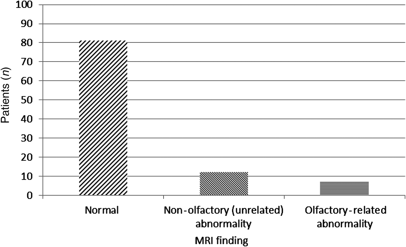
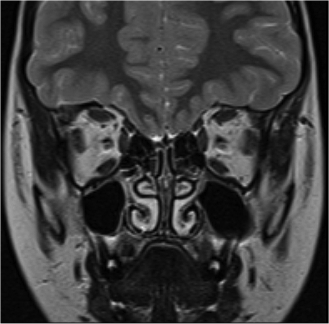
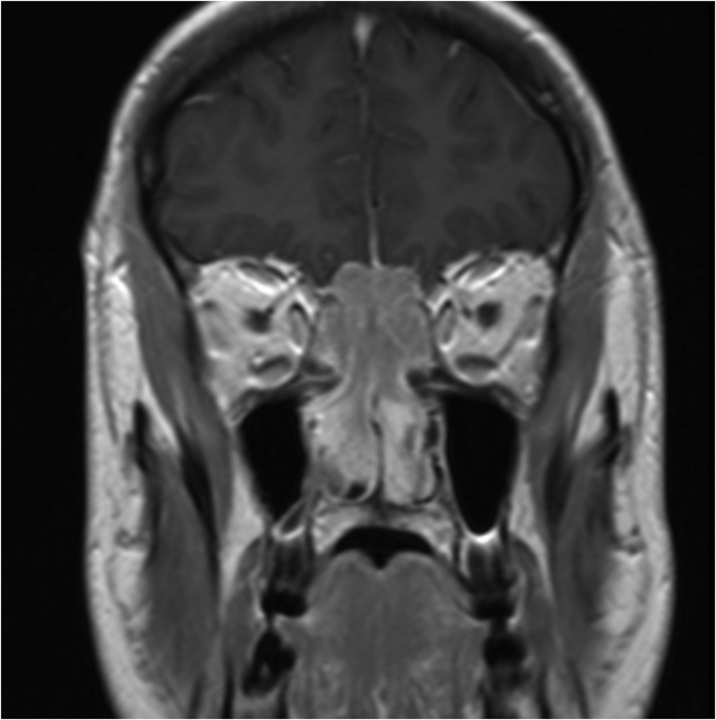
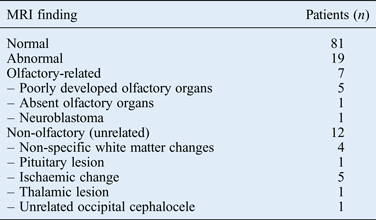
Magnetic resonance imaging revealed abnormalities in 19 patients (19 per cent). Of these, only seven patients (7 per cent) had abnormalities that were considered clinically relevant to the presentation of olfactory dysfunction (Table I and Figures 1–4).
Magnetic resonance imaging (MRI) findings for 100 consecutive patients who presented to the Freeman Hospital with olfactory dysfunction symptoms.

Coronal, fluid attenuated inversion recovery (‘FLAIR’) magnetic resonance image demonstrating normal olfactory bulbs (arrow).
Coronal, T2-weighted magnetic resonance image demonstrating absent olfactory apparatus.
Coronal, post-contrast, T1-weighted magnetic resonance image showing nasal and extradural components of an olfactory neuroblastoma.
Magnetic resonance imaging findings*
*For 100 consecutive patients who presented to the Freeman Hospital with symptoms of olfactory dysfunction and underwent magnetic resonance imaging (MRI).
A comparison of the findings for children (aged less than 16 years, n = 5) with those for adults (equal to or more than 16 years old, n = 95) revealed some interesting findings. The MRI results for over half (60 per cent) of the children in our cohort indicated a reason for the olfactory dysfunction (all had poorly developed or completely absent olfactory organs). In the adult cohort, however, only 4 per cent had olfactory-related abnormalities as identified by scanning. Three patients had poorly developed or completely absent olfactory organs and one patient had an olfactory neuroblastoma.
We subsequently assessed whether the MRI scan results altered management practice. Out of 100 patients scanned, only 1 patient (with olfactory neuroblastoma) had any alteration in management after scanning. In fact, only 37 per cent of patients were seen back in the clinic; most were sent a letter informing them of normal scan findings, and no follow up was arranged.
In our cohort, a likely aetiology was documented for 49 per cent of patients; probable causes included a post-viral condition (39 per cent), trauma (5 per cent) and medication (3 per cent). All patients had normal physical examination findings, with normal nasal endoscopy and flexible laryngoscopy findings, except for the patient with olfactory neuroblastoma. The case note review for this patient described a mass in the nasal cavity and post-nasal space, as observed on rigid endoscopy, without any other nasal symptoms.
We subsequently searched our database to identify the number of patients with olfactory neuroblastoma (detected by MRI) who reported olfactory dysfunction. We identified another three patients who presented over the last six years. One presented with a unilateral nasal polyp on repeat endoscopy. The other two patients did not have any associated nasal or neurosurgical symptoms or signs. These patients were all younger (less than 50 years old) when compared with our cohort; patient history and clinical examination did not determine the cause of the olfactory dysfunction.
Discussion
Of the patients who presented with olfactory dysfunction and underwent imaging of the olfactory tract, 99 per cent had no abnormality found that altered their management. Within that 99 per cent, six patients had abnormalities related to the olfactory tract that were benign and untreatable. However, those findings did not alter the management of the olfactory dysfunction, but simply provided the patients with an explanation for their symptoms. We also found that children with olfactory dysfunction were far more likely to have an identifiable cause for their symptoms on MRI scanning, such as an underdeveloped olfactory tract.
Many patients provided a likely cause for the olfactory dysfunction, such as post-viral anosmia, which may negate the need for imaging of the olfactory tract. Furthermore, endoscopic examination findings were normal in all our cohort patients, except for the olfactory neuroblastoma patient. A search of our database for olfactory neuroblastoma patients over the last six years revealed that half had physical signs on endoscopic examination. However, two patients with olfactory neuroblastoma had no signs or symptoms. Nevertheless, these patients were both young (aged less than 50 years) compared with our cohort, and their history revealed no cause for the olfactory dysfunction.
Olfactory disturbance is a serious condition that can have a major impact on patients' lives. While the reassurance associated with normal scan findings can provide comfort for the clinician and patient, it does come at some cost, averaging at around £200 per MRI scan. Most departments are under pressure to deliver targets, and scanning patients in cases where imaging will not alter management should be discouraged. Furthermore, the MRI procedure itself can be quite unpleasant, particularly for claustrophobic individuals.
Clinical decisions should ultimately rest on a clinician's judgement. However, we suggest that many patients are being scanned unnecessarily. As this study demonstrates, the imaging results do not significantly alter the management of affected patients, which is mainly focused on olfactory rehabilitation advice such as food safety and the fitting of gas alarms.Reference Geyer and Nilssen10 Given the characteristics of the olfactory dysfunction patients in our study, particularly the age distribution, we feel that decisions regarding patient selection for scanning could be more rational. For example, many of our patients were elderly and it is known that these patients are more likely to have olfactory dysfunction of unknown or benign aetiology than younger people. The oldest patient with olfactory-related pathology in our series was 42 years old. Our findings also indicated that the likelihood of having olfactory pathology with olfactory dysfunction is far greater in children than adults. Over half of the children in our study had olfactory pathology as observed on MRI scans, compared with only 4 per cent of the adults.
• Magnetic resonance imaging (MRI) scanners and other imaging equipment are expensive and in high demand
• Olfactory lesions and treatable causes of olfactory dysfunction are rare
• For 99 per cent of the olfactory dysfunction patients in this study, olfactory tract MRI revealed no abnormality that altered clinical management
• In 6 per cent of patients, olfactory tract-related abnormalities were identified but these findings did not alter management
• Imaging was more likely to determine the cause of olfactory dysfunction symptoms in children than adults
• Aetiology was indicated by patient history in almost half (49 per cent) of the cases, without the need for imaging
In the most recent review of the literature on olfactory dysfunction, by Hong et al., it was suggested that imaging may not be necessary in patients where the aetiology is clear (e.g. a post-viral condition) and the physical examination findings are normal.Reference Hong, Holbrook, Leopold and Hummel11 Our study findings strongly support this rationale. However, the authors of that review do recommend that if the history and physical examination offer no clear cause, MRI scanning should be performed to rule out a central mass lesion or neurodegenerative disorder. Our findings are also in line with those of Busaba.Reference Busaba12 Following a smaller study of 28 patients with olfactory dysfunction, the author concluded that scanning of the olfactory tract did not add to the clinical information obtained from patient history and physical examination findings.
Our study has clear limitations in that it comprises a relatively small cohort from only one centre. Furthermore, the conclusions made rely on data from clinical letters and MRI scan reports. However, there is limited published work available on the topic addressed in this study. We believe that large-scale, national and international studies are needed to fully assess the identification rates of treatable pathology revealed by cross-sectional imaging examinations in those presenting with olfactory dysfunction. This will allow for greater evidence-based risk stratification and management of olfactory dysfunction, and may reduce costs for healthcare providers.