Introduction
Allergic rhinitis is one of the most common allergic diseases, and it is believed that the pathogenesis of allergic rhinitis may be the result of environmental, immunological and genetic factors.Reference Greiner, Hellings, Rotiroti and Scadding 1 – Reference Rosenwasser 4 Till now, many molecules have been shown to participate in the initiation and progression of allergic rhinitis.
MicroRNA (miRNA) is a small single-stranded RNA with full length, identifying the 3'- untranslated region of target messenger RNA, and plays an important role in gene expression.Reference Ambros 5 – Reference Bartel 7 A number of studies have demonstrated that miRNA is involved in the development of tumours, autoimmune diseases and other systemic diseases.Reference Calin, Dumitru, Shimizu, Bichi, Zupo and Noch 8 , Reference Yang, Li and Padgett 9 Our previous microarray showed that miRNA let-7a was up-regulated in upper respiratory tract disease (H Yu, unpublished data), but its role in allergic rhinitis is unclear.
Osteopontin (OPN) is expressed by macrophages, smooth muscle cells and T lymphocytes. It is an early T cell activating factor, and plays an important role in cell chemotaxis and adhesion, cytokine expression, immune response, and inflammation.Reference Liu, Lu, Yu, Hua, Cui and Huang 10 Many pro-inflammatory factors, such as interleukin 1 and tumour necrosis factor-α, can promote OPN transcription.Reference Liu, Lu, Yu, Hua, Cui and Huang 10 Previously, our group reported that OPN expression was up-regulated in the mucosa of an allergic rhinitis model and allergic rhinitis patients, and could be modulated by many kinds of cytokines.Reference Liu, Lu, Yu, Hua, Cui and Huang 10
This study aimed to investigate the role and significance of miRNA let-7a in mice with experimental allergic rhinitis, and examine whether miRNA let-7a has any effect on OPN expression.
Materials and methods
Mice
Thirty-two mature female C57BL/6 mice, aged eight weeks and weighing 20–25 g, were selected (purchased from the experimental animal centre of Huazhong University of Science and Technology), and raised in a specific pathogen-free facility. They were randomly divided into four groups, with eight mice in each group.
MicroRNA synthesis
MiRNA let-7a was synthesised by Shanghai Genechem (Shanghai, China), with mature chain and minor chain respectively: 5'-UGAGGUAGUAGGUUGUAUAGUU-3' 5'-CUAUACAAUCUACUGUCUUUCC-3', and miRNA let-7a control: 5'-AUCCGCGCGAUAGUACGUAUU-3' 5'-AATACGTACTATCGCGCGGAT-3', which were designed according to a previous paper.Reference Makino, Jinnin, Hirano, Yamane, Eto and Kusano 11
Allergic rhinitis model
As per previous reports,Reference Liu, Yu, Wang, Zhang, Huang and Cui 12 , Reference Kim, Yang, Park, Ahn, Son and Kim 13 the miRNA let-7a group, miRNA control group and ovalbumin (OVA) group mice were treated with a subcutaneous injection of 40 g aluminium hydroxide as adjuvant and 2 mg OVA (Sigma-Aldrich, St Louis, Missouri, USA) diluted in 400 ml phosphate-buffered saline on day 0 and day 7, once daily. For phosphate-buffered saline group mice, OVA was replaced with phosphate-buffered saline.
After the 14th day, the miRNA let-7a group, miRNA control group and OVA group were challenged with 10 per cent OVA diluted in phosphate-buffered saline into the nose, once a day for 5 continuous days. For phosphate-buffered saline group mice, OVA was replaced with phosphate-buffered saline. For miRNA let-7a group, mice were given miRNA let-7a and Lipofectamine 2000 reagent (Sigma-Aldrich, USA) mixture (40 M miRNA let-7a diluted in 40 ml phosphate-buffered saline) half an hour before the first, second and third challenge. The control group was given miRNA let-7a control and 2000 liposome mixture (40 M diluted in 40 ml phosphate-buffered saline). Mice were sacrificed on day 19.
Mouse behaviour detection
Twenty-four hours after the last intranasal phosphate-buffered saline or OVA challenge, each mouse was challenged with intranasal OVA. Immediately after OVA challenge, the number of nose-scratching events in the 10 minutes after the last challenge was counted independently by two observers, as reported elsewhere.Reference Kim, Yang, Park, Ahn, Son and Kim 13
Evaluation of goblet cells in nasal mucosa
Twenty-four hours after the last challenge, mice were sacrificed and the heads were taken. They were prepared for conventional fixed paraffin embedded sections. The sections were stained with periodic acid-Schiff stain for the observation of goblet cells.
Nasal lavage fluid collection
Mice were sacrificed by dislocation and intratracheally injected with 1 ml saline. Nasal lavage fluid collection was acquired and the fluid was centrifuged. The collection of cell debris and nasal lavage fluid were stored in the refrigerator at −20 °C for other tests.
Nasal lavage fluid eosinophil counting
The cells isolated from the nasal cavity were diluted with 1 ml saline, and 10 ml was applied to the carrier plate, fixed by acetone for 15 minutes and then stored at −20 °C. Eosinophils were counted by routine haematoxylin and eosin staining.
Detection of interleukins 5 and 13, interferon-γ protein
The levels of interleukin (IL)-5, IL-13 and interferon-γ in mouse nasal lavage fluid were detected according to the steps of the enzyme-linked immunosorbent assay kit (Wuhan Boster Biological, Hubei, China).
Real-time polymerase chain reaction
Total RNA of mouse nasal mucosa was extracted and subjected to reverse transcription. The expression of related genes was analysed by fluorescence quantitative polymerase chain reaction. The primers of polymerase chain reaction for OPN and GADPH (glyceraldehyde 3-phosphate dehydrogenase) were: (sense primer) 5’-CGGATTTGGCCGTATTGGG-3’ (antisense primer) 5’-CTCGCTCCTGGAAGATGG-3’, OPN (sense primer) 5’-GCCTGTTTGGCATTGCCTCCTC-3’ (antisense primer) 5’-CACAGCATTCTGTGGCGCAAGG-3’, as reported in our previous study.Reference Liu, Lu, Yu, Hua, Cui and Huang 10
Statistical analysis
The experimental data were analysed using SPSS® statistical software, version 15.0. The original data were analysed using the Mann–Whitney U test. The difference was statistically significant when p < 0.05.
Results
Nose-scratching events
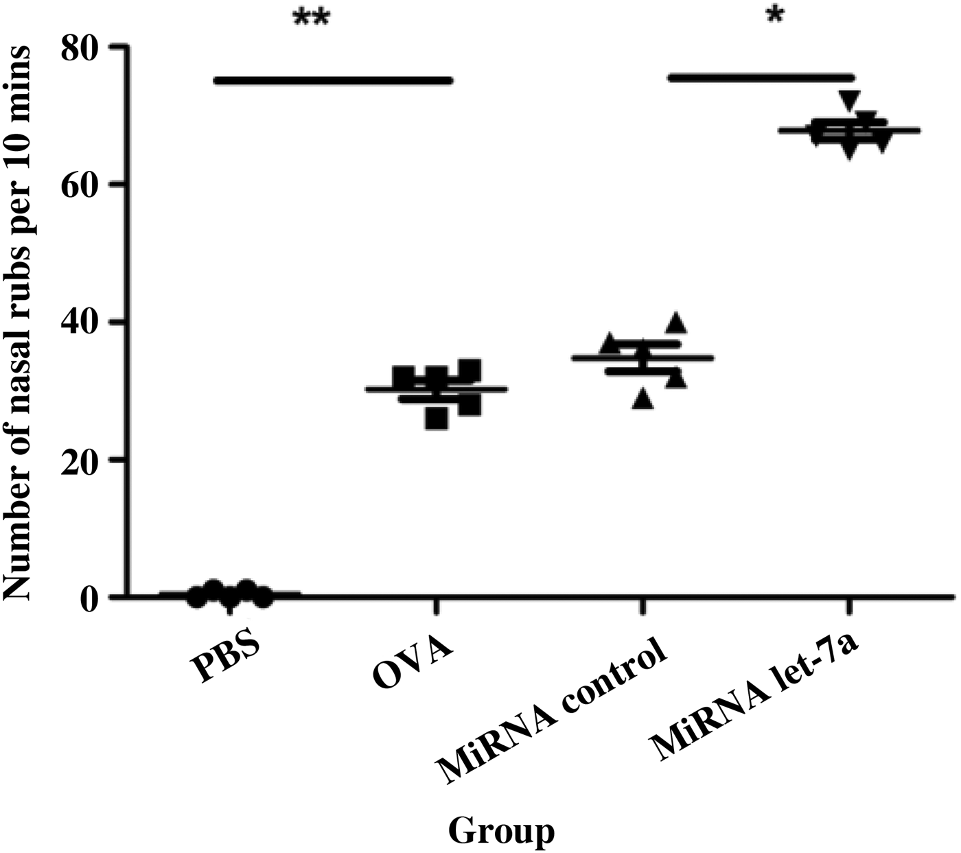
The number of nose-scratching events in the OVA group was higher than that in the phosphate-buffered saline group (p < 0.05). After miRNA let-7a treatment, nose-scratching events were significantly higher than in the miRNA control group (p < 0.05) (Figure 1).
Intranasal microRNA (miRNA) let-7a treatment increased nose-scratching events. The number of nasal rubbing movements was counted for 10 minutes immediately after the last nasal challenge. Data are indicated as mean ± standard deviation of five mice per group. Data are representative of three independent experiments with similar results. *p < 0.05; **p < 0.01. Mins = minutes; PBS = phosphate-buffered saline

Eosinophils in nasal mucosa
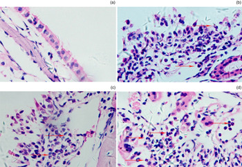
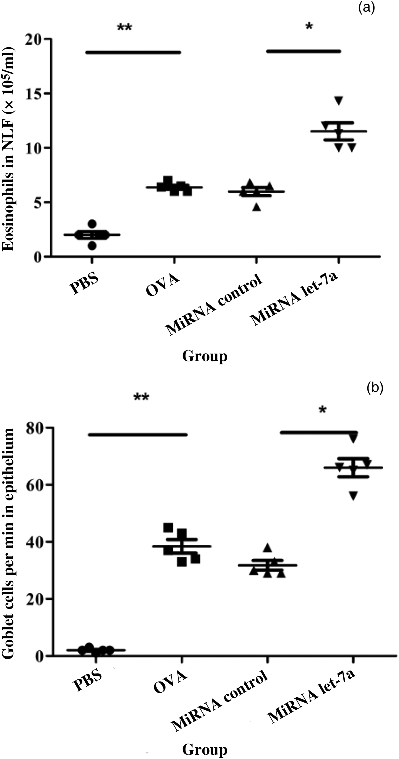
Haematoxylin and eosin staining showed the inflammatory cells and eosinophils in the nasal mucosa of the allergic rhinitis mice (Figure 2). Analysis of the nasal lavage fluid cells showed that eosinophil numbers in the nasal lavage fluid of OVA mice were significantly higher than in the normal control group (p < 0.05). After miRNA let-7a treatment, eosinophil numbers in the miRNA let-7a group were significantly higher than in the miRNA control group (p < 0.05) (Figure 3a).
Photomicrographs showing staining of nasal mucosa are presented for each group: (a) phosphate-buffered saline (PBS), (b) OVA, (c) microRNA (miRNA) control and (d) miRNA let-7a. Red arrows indicate eosinophils in nasal mucosa. (H&E; ×200)

Intranasal microRNA (miRNA) let-7a administration increased the numbers of (a) eosinophils in nasal lavage fluid (NLF) and (b) goblet cells in the nasal mucosa in allergic rhinitis mice. Data are indicated as mean ± standard deviation of five mice per group. Data are representative of three independent experiments with similar results. *p < 0.05; **p < 0.01. Min = minute

Goblet cells in nasal mucosa
The number of goblet cells in the nasal mucosa of the OVA group was significantly higher than in the normal control group (p < 0.05). The number of goblet cells in the nasal mucosa of the miRNA let-7a group was significantly higher than in the miRNA control group (p < 0.05) after treatment with miRNA let-7a (Figure 3b).
Interleukins 5 and 13 and interferon-γ
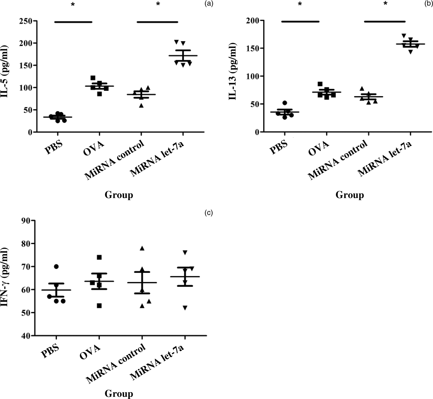
Compared with the miRNA control group, the interleukin (IL)-5 and IL-13 levels were significantly higher (p < 0.05) in the miRNA let-7a group. The difference in interferon-γ levels was not statistically significant (p > 0.05) (Figure 4).
Intranasal microRNA (miRNA) let-7a administration enhanced type 2 helper T cell related cytokine levels. The nasal lavage fluid levels of (a) interleukin (IL)-5, (b) IL-13 and (c) interferon (IFN)-γ were measured by enzyme-linked immunosorbent assay. Data are indicated as mean ± standard deviation of five mice per group. Data are representative of three independent experiments with similar results. *p < 0.05. PBS = phosphate-buffered saline

OPN in nasal mucosa
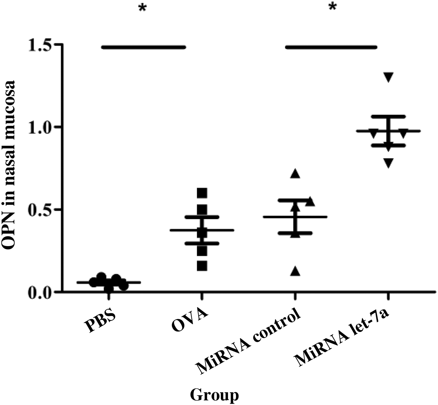
Compared with the miRNA control group, the OPN levels were significantly higher (p < 0.05) in the miRNA let-7a group (Figure 5).
Intranasal microRNA (miRNA) let-7a administration increased OPN expression in nasal mucosa. OPN messenger RNA relative expression was measured by polymerase chain reaction. Data are indicated as mean ± standard deviation of five mice per group. Data are representative of three independent experiments with similar results. *p < 0.05. PBS = phosphate-buffered saline

Discussion
In this study, we found that miRNA let-7a treated mice had a significantly increased number of nose-scratching events, nasal mucosal eosinophilia and goblet cell hyperplasia. These findings suggest that miRNA let-7a plays an important role in the setting of allergic rhinitis. To the best of our knowledge, this is the first study investigating miRNA in allergic rhinitis.
MicroRNA is a class of highly conserved, small molecule, non-coding RNA, with a length of about 22 nucleotides, which has the function of regulating post-transcriptional gene expression. It has been demonstrated that miRNA is involved in the development of tumours, autoimmune diseases and other systemic diseases.Reference Calin, Dumitru, Shimizu, Bichi, Zupo and Noch 8 , Reference Yang, Li and Padgett 9 , Reference Roggli, Britan, Gattesco, Lin-Marq, Abderrahmani and Meda 14 In recent years, more and more miRNA have been shown to play an important role in allergic diseases.Reference Lu, Munitz and Rothenberg 15 , Reference Lu, Hartner, Lim, Fabry, Mingler and Cole 16 A variety of miRNAs have been implicated in a series of pathophysiological processes, including inflammatory cell infiltration, cytokine secretion, airway hyper-responsiveness, smooth muscle cell proliferation and phenotypic changes in allergic airway diseases.Reference Lu, Munitz and Rothenberg 15 , Reference Lu, Hartner, Lim, Fabry, Mingler and Cole 16
MicroRNA let-7a is a member of the miRNA let-7 family. Previous studies have shown that miRNA let-7a plays an important role in tumour diseases. MicroRNA let-7a is a new tumour suppressor gene candidate for lung cancer, colorectal cancer, and so on.Reference Liu, Yu, Wang, Zhang, Huang and Cui 12 , Reference Shell, Park, Radjdai, Schickel, Kistner and Jewell 17 A study of a murine asthma model, using an miRNA chip to detect miRNA expression in the lungs of mice, found that the miRNA let-7 family accounted for a larger proportion of miRNA, and miRNA let-7f expression was the highest.
The miRNA let-7 family may play an important role in the pathogenesis of asthma, suggesting that miRNA is involved in the occurrence and development of bronchial asthma, and plays an important role in the regulation of allergic diseases.Reference Polikepahad, Knight, Naghavi, Oplt, Creighton and Shaw 18 In recent years, studies have shown that MiRNA let-7a may play an important role in allergic diseases.Reference Polikepahad, Knight, Naghavi, Oplt, Creighton and Shaw 18 Our previous miRNA microarray showed that miRNA let-7a was up-regulated in upper respiratory tract disease. In order to study its role in allergic rhinitis, we observed the effect of miRNA let-7a on allergic rhinitis in mice. We found that after transfection of miRNA let-7a, clinical symptoms of the mice were significantly enhanced, mainly nose-scratching events and sneezing symptoms. In addition, inflammatory cell infiltration and goblet cell hyperplasia in the nasal mucosa were significantly higher in the miRNA let-7a group than in the miRNA let-7a control group. These results indicate that miRNA let-7a can aggravate the clinical symptoms and pathological severity of allergic rhinitis mice.
The pathogenesis of allergic rhinitis is complicated, and the imbalance of type 1/type 2 helper T cells plays a key role in the pathogenesis of allergic rhinitis. Under normal circumstances, the differentiation and function of type 1/type 2 helper T cells in the body are in a state of equilibrium; when the type 2 helper T cytokines predominate, allergic rhinitis may occur.Reference Rosenwasser 4 We found that the levels of interleukin (IL)-5 and IL-13 in nasal lavage fluid were significantly higher than in the non-transfection group, but there was no significant difference in interferon-γ. These findings suggest that miRNA let-7a is involved in the regulation of type 2 helper T cells. Studies have shown that the target gene of miRNA let-7a may be IL-13 in allergic diseases.Reference Shell, Park, Radjdai, Schickel, Kistner and Jewell 17 , Reference Polikepahad, Knight, Naghavi, Oplt, Creighton and Shaw 18 However, it is necessary to further verify whether miRNA let-7a has any other target genes in allergic diseases.
-
• MicroRNA (miRNA) let-7a increased the number of nose-scratching events in allergic rhinitis mice
-
• It also promoted nasal mucosal eosinophilia and goblet cell hyperplasia in allergic rhinitis mice
-
• In addition, it increased interleukins 5 and 13 levels in nasal mucosa
-
• The miRNA let-7a treated mice had significantly enhanced OPN expression in nasal mucosa
-
• MicroRNA let-7a can promote the development of allergic rhinitis partly by regulating OPN expression
OPN, together with calcium ions (CaReference Bousquet, Khaltaev, Cruz, Denburg, Fokkens and Togias 2 +) and nitric oxide, are second messengers. OPN participates in tumour cell transformation, invasion and metastasis, smooth muscle cell proliferation, and phenotype transformation. Furthermore, it is closely related to atherosclerosis, infection, and the occurrence and development of a variety of physiological and pathological phenomena.Reference Cantor and Shinohara 19 Therefore, it is a kind of new cell signalling molecule. Many pro-inflammatory factors, such as IL-1 and tumour necrosis factor-α, can promote OPN transcription.Reference Liu, Lu, Yu, Hua, Cui and Huang 10 , Reference Cantor and Shinohara 19 Previously, our group reported that OPN expression was up-regulated in the mucosa of an allergic rhinitis model and allergic rhinitis patients, and could be modulated by many kinds of cytokines.Reference Liu, Lu, Yu, Hua, Cui and Huang 10 In this study, we found that miRNA let-7a treatment could significantly decrease OPN expression in the nasal mucosa of allergic rhinitis mice, indicating that miRNA let-7a might regulate OPN expression in the setting of allergic rhinitis. This is the first investigation to report that miRNA let-7a might modulate OPN production in allergic diseases. Previously, we found that OPN was directly regulated by Clara cell 10-kDa protein; however, whether miRNA let-7a modulates OPN expression in a direct or indirect way should be further elucidated in future studies.
In this study, we explored the role of miRNA let-7a in allergic diseases using an allergic rhinitis model. The findings suggested that miRNA let-7a is closely related to the pathogenesis of allergic rhinitis. Although the exact mechanism of miRNA let-7a in allergic diseases requires further exploration, this study provides additional evidence for the role of miRNAs in allergic diseases. With further studies on the regulation of miRNA let-7a expression, the mechanism of allergic rhinitis and other allergic diseases will become clear, which may provide a new strategy for the diagnosis and treatment of such diseases.
Acknowledgements
This study was supported by a National Natural Science Foundation of China grant (number 81300812) and a grant from the Ministry of Education of China (Program for Changjiang Scholars and Innovative Research Team in University (IRT_14R20)).